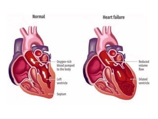
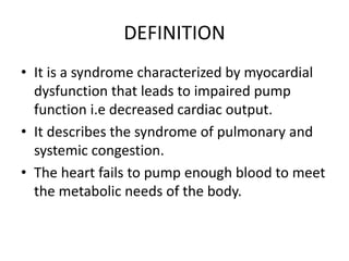
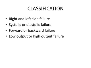
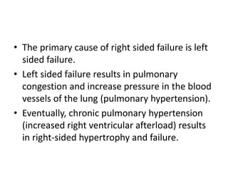
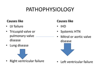
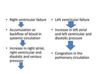
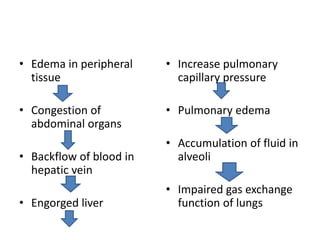
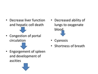
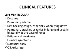
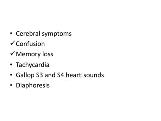
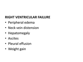
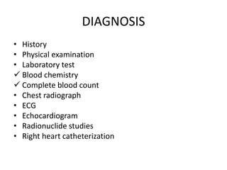
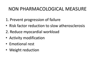
CONGESTIVE HEART FAILURE
Neelu Aryal
Lecturer
DEFINITION
• It is a syndrome characterized by myocardial
dysfunction that leads to impaired pump
function i.e decreased cardiac output.
• It describes the syndrome of pulmonary and
systemic congestion.
• The heart fails to pump enough blood to meet
the metabolic needs of the body.
CLASSIFICATION
• Right and left side failure
• Systolic or diastolic failure
• Forward or backward failure
• Low output or high output failure
RIGHT AND LEFT SIDED HEART FAILURE
• Right side heart failure refers to signs and
symptoms of increased pressure and congestion
in the systemic veins and capillaries.
• Left sided heart failure refers to sign and
symptoms of elevated pressure and congestion in
pulmonary veins and capillaries.
NOTE: Failure of 1 ventricle often produces changes
in the other ventricles, leading to biventricular
failure.
LEFT SIDED HEART FAILURE
• The most common form of Heart failure is left
sided heart failure.
• Left sided failure results from left ventricular
dysfunction, which prevents normal blood
flow and causes blood to back up into the left
atrium and into the pulmonary veins.
• The increase pulmonary pressure causes fluids
extravasation (leakage) from pulmonary
capillary bed into the interstitium and then
the alveoli, which is manifested as pulmonary
congestion and edema.
RIGHT SIDED HEART FAILURE
• Right sided failure causes a back up of blood into
the right atrium and venous circulation.
• This leads to venous congestion in the systemic
circulation and results in:
Jugular venous distention
Hepatomegaly
Splenomegaly
Vascular congestion of GI tract
Peripheral edema
• The primary cause of right sided failure is left
sided failure.
• Left sided failure results in pulmonary
congestion and increase pressure in the blood
vessels of the lung (pulmonary hypertension).
• Eventually, chronic pulmonary hypertension
(increased right ventricular afterload) results
in right-sided hypertrophy and failure.
FORWARD VERSUS BACKWARD HEART FAILURE
(DIRECTION OF BLOOD FLOW)
FORWARD FAILURE
• Inadequate output of affected ventricle causes
decreased perfusion of vital organs, resulting
in symptoms such as mental confusion,
muscle weakness and altered kidney function.
BACKWARD FAILURE
• Damming of the blood behind the affected
ventricle causes increased pressure in the atrium
behind the affected ventricle, increased pressure
in the venous and capillary system behind the
atria, and movement of fluid from capillary into
the interstitium, resulting in symptoms such as
pulmonary edema.
• Both backward and forward failure occur in most
patient with CHF
LOW OUTPUT AND HIGH OUTPUT FAILURE
LOW OUTPUT HEART FAILURE
• Most heart failure is low output
• Not enough cardiac output is available to
meet usual needs.
• The person shows the evidence of impaired
circulation and vasoconstriction with cool,
pale extremities.
HIGH OUTPUT FAILURE
• It occurs when a condition causes the heart to
work harder to meet increased needs (eg:
sepsis, anemia)
• The person appears warm and flushed.
SYSTOLIC AND DIASTOLIC HEART FAILURE
SYSTOLIC FAILURE
• Is an abnormality in systolic function leading
to a problem with contraction and ejection of
the blood (eg: dilated cardiomyopathy)
DIASTOLIC FAILURE
• Abnormality in diastolic function leading to a
problem with heart relaxing and filling with
blood. (eg: hypertrophic cardiomyopathy)
RISK FACTORS AND ETOLOGY
A) Underlying causes of HF
1) Intrinsic myocardial disease
• Cardiomyopathy
• Coronary artery disease
2) Excess volume load
• Aortic regurgitation
• Mitral regurgitation
• Tricuspid regurgitation
• Left and right shunt
3) Excess resistance to ejection
• Hypertension
• Aortic stenosis
• Pulmonic stenosis
• Hypertrophic cardiomyopathy
4) Increased body demand
• Thyrotoxicosis
• Anemia
• Pregnancy
5) Iatrogenic myocardial damage
• Drugs
• Radiation
B) MAJOR ETIOLOGIES
• Coronary artery disease
• MI
• Primary cardiomyopathy
• Hypertension
• Vascular heart disease, particularly aortic
C) PRECIPITATING FACTOS
• Dysrhythmias
• Cardiac Infection (increases myocardial oxygen
demand)
• Pulmonary embolism
• Physical and emotional exertion
• Uncontrolled hypertension
• MI
• Medications
β blockers
Estrogens
Chemotherapy agents
• Renal disease
• Excessive transfusion or intravenous fluid
PATHOPHYSIOLOGY
Causes like
• LV failure
• Tricuspid valve or
pulmonary valve
disease
• Lung disease
• Right ventricular failure
Causes like
• IHD
• Systemic HTN
• Mitral or aortic valve
disease
• Left ventricular failure
• Right ventricular failure
• Accumulation or
backflow of blood in
systemic circulation
• Increase in right atrial,
right ventricular and
diastolic and venous
pressure
• Left ventricular failure
• Increase in left atrial
and left ventricular and
diastolic pressure
• Congestion in the
pulmonary circulation
• Edema in peripheral
tissue
• Congestion of
abdominal organs
• Backflow of blood in
hepatic vein
• Engorged liver
• Increase pulmonary
capillary pressure
• Pulmonary edema
• Accumulation of fluid in
alveoli
• Impaired gas exchange
function of lungs
• Decrease liver function
and hepatic cell death
• Congestion of portal
circulation
• Engorgement of spleen
and development of
ascities
• Decreased ability of
lungs to oxygenate
blood
• Cyanosis
• Shortness of breath
CLINICAL FEATURES
LEFT VENTRICULAR
• Dyspnea
• Pulmonary edema
• Dry, hacking cough, especially when lying down
• Pulmonary crackles or rayles in lung field usually
bilaterally at the base of lungs
• Fatigue and weakness
• Urinary symptoms
Nocturia: early
Oliguria: late
• Cerebral symptoms
Confusion
Memory loss
• Tachycardia
• Gallop S3 and S4 heart sounds
• Diaphoresis
RIGHT VENTRICULAR FAILURE
• Peripheral edema
• Neck vein distension
• Hepatomegaly
• Ascites
• Pleural effusion
• Weight gain
DIAGNOSIS
• History
• Physical examination
• Laboratory test
 Blood chemistry
 Complete blood count
• Chest radiograph
• ECG
• Echocardiogram
• Radionuclide studies
• Right heart catheterization
CLINICAL MANAGEMENT
• Non pharmacological measures
• Pharmacological interventions
• Surgical management
NON PHARMACOLOGICAL MEASURE
1. Prevent progression of failure
• Risk factor reduction to slow atherosclerosis
2. Reduce myocardial workload
• Activity modification
• Emotional rest
• Weight reduction
3. Control excess salt and water retension
• Dietary salt restriction
• Fluid restriction
PHARMACOLOGICAL INTERVENTIONS
• Angiotensin converting enzyme (ACE
inhibitors)
Captopril
Enalapril
(decrease BP, decrease afterload)
• Angiotensin receptor blockers
Losartan
(decrease BP, decrease afterload)
• Beta blockers
Metoprolol
Atenolol
( dilates blood vessels and decrease afterload)
• Diuretics
Loop diuretics : Furosemide
(decrease fluid volume overload)
• Digitalis
Digoxin
(improves contractility)
• Calcium channel blockers
Amlodipin
Nefidipin
(vasodilation and reduction of systemic
vascular resistance)
SURGICAL MANAGEMENT
• Intra aortic balloon pump
• Ventricular assist device
• Cardiomyoplasty
• Cardiac transplantation
COMPLICATION
• Acute pulmonary edema
• Death
NURSING MANAGEMENT
• Decreased cardiac output related to inability
of the heart to pump effectively
• Fluid volume excess related to decreased renal
blood flow, causing increase level of
aldosterone and antidiuretic hormones
THANK YOU

Congestive heart failure

  • 1.
  • 3.
    DEFINITION • It isa syndrome characterized by myocardial dysfunction that leads to impaired pump function i.e decreased cardiac output. • It describes the syndrome of pulmonary and systemic congestion. • The heart fails to pump enough blood to meet the metabolic needs of the body.
  • 4.
    CLASSIFICATION • Right andleft side failure • Systolic or diastolic failure • Forward or backward failure • Low output or high output failure
  • 5.
    RIGHT AND LEFTSIDED HEART FAILURE • Right side heart failure refers to signs and symptoms of increased pressure and congestion in the systemic veins and capillaries. • Left sided heart failure refers to sign and symptoms of elevated pressure and congestion in pulmonary veins and capillaries. NOTE: Failure of 1 ventricle often produces changes in the other ventricles, leading to biventricular failure.
  • 6.
    LEFT SIDED HEARTFAILURE • The most common form of Heart failure is left sided heart failure. • Left sided failure results from left ventricular dysfunction, which prevents normal blood flow and causes blood to back up into the left atrium and into the pulmonary veins.
  • 7.
    • The increasepulmonary pressure causes fluids extravasation (leakage) from pulmonary capillary bed into the interstitium and then the alveoli, which is manifested as pulmonary congestion and edema.
  • 8.
    RIGHT SIDED HEARTFAILURE • Right sided failure causes a back up of blood into the right atrium and venous circulation. • This leads to venous congestion in the systemic circulation and results in: Jugular venous distention Hepatomegaly Splenomegaly Vascular congestion of GI tract Peripheral edema
  • 9.
    • The primarycause of right sided failure is left sided failure. • Left sided failure results in pulmonary congestion and increase pressure in the blood vessels of the lung (pulmonary hypertension). • Eventually, chronic pulmonary hypertension (increased right ventricular afterload) results in right-sided hypertrophy and failure.
  • 10.
    FORWARD VERSUS BACKWARDHEART FAILURE (DIRECTION OF BLOOD FLOW)
  • 11.
    FORWARD FAILURE • Inadequateoutput of affected ventricle causes decreased perfusion of vital organs, resulting in symptoms such as mental confusion, muscle weakness and altered kidney function.
  • 12.
    BACKWARD FAILURE • Dammingof the blood behind the affected ventricle causes increased pressure in the atrium behind the affected ventricle, increased pressure in the venous and capillary system behind the atria, and movement of fluid from capillary into the interstitium, resulting in symptoms such as pulmonary edema. • Both backward and forward failure occur in most patient with CHF
  • 13.
    LOW OUTPUT ANDHIGH OUTPUT FAILURE
  • 14.
    LOW OUTPUT HEARTFAILURE • Most heart failure is low output • Not enough cardiac output is available to meet usual needs. • The person shows the evidence of impaired circulation and vasoconstriction with cool, pale extremities.
  • 15.
    HIGH OUTPUT FAILURE •It occurs when a condition causes the heart to work harder to meet increased needs (eg: sepsis, anemia) • The person appears warm and flushed.
  • 16.
  • 17.
    SYSTOLIC FAILURE • Isan abnormality in systolic function leading to a problem with contraction and ejection of the blood (eg: dilated cardiomyopathy)
  • 19.
    DIASTOLIC FAILURE • Abnormalityin diastolic function leading to a problem with heart relaxing and filling with blood. (eg: hypertrophic cardiomyopathy)
  • 21.
    RISK FACTORS ANDETOLOGY A) Underlying causes of HF 1) Intrinsic myocardial disease • Cardiomyopathy • Coronary artery disease
  • 22.
    2) Excess volumeload • Aortic regurgitation • Mitral regurgitation • Tricuspid regurgitation • Left and right shunt
  • 23.
    3) Excess resistanceto ejection • Hypertension • Aortic stenosis • Pulmonic stenosis • Hypertrophic cardiomyopathy
  • 24.
    4) Increased bodydemand • Thyrotoxicosis • Anemia • Pregnancy
  • 25.
    5) Iatrogenic myocardialdamage • Drugs • Radiation
  • 26.
    B) MAJOR ETIOLOGIES •Coronary artery disease • MI • Primary cardiomyopathy • Hypertension • Vascular heart disease, particularly aortic
  • 27.
    C) PRECIPITATING FACTOS •Dysrhythmias • Cardiac Infection (increases myocardial oxygen demand) • Pulmonary embolism • Physical and emotional exertion • Uncontrolled hypertension
  • 28.
    • MI • Medications βblockers Estrogens Chemotherapy agents • Renal disease • Excessive transfusion or intravenous fluid
  • 29.
    PATHOPHYSIOLOGY Causes like • LVfailure • Tricuspid valve or pulmonary valve disease • Lung disease • Right ventricular failure Causes like • IHD • Systemic HTN • Mitral or aortic valve disease • Left ventricular failure
  • 30.
    • Right ventricularfailure • Accumulation or backflow of blood in systemic circulation • Increase in right atrial, right ventricular and diastolic and venous pressure • Left ventricular failure • Increase in left atrial and left ventricular and diastolic pressure • Congestion in the pulmonary circulation
  • 31.
    • Edema inperipheral tissue • Congestion of abdominal organs • Backflow of blood in hepatic vein • Engorged liver • Increase pulmonary capillary pressure • Pulmonary edema • Accumulation of fluid in alveoli • Impaired gas exchange function of lungs
  • 32.
    • Decrease liverfunction and hepatic cell death • Congestion of portal circulation • Engorgement of spleen and development of ascities • Decreased ability of lungs to oxygenate blood • Cyanosis • Shortness of breath
  • 33.
    CLINICAL FEATURES LEFT VENTRICULAR •Dyspnea • Pulmonary edema • Dry, hacking cough, especially when lying down • Pulmonary crackles or rayles in lung field usually bilaterally at the base of lungs • Fatigue and weakness • Urinary symptoms Nocturia: early Oliguria: late
  • 34.
    • Cerebral symptoms Confusion Memoryloss • Tachycardia • Gallop S3 and S4 heart sounds • Diaphoresis
  • 35.
    RIGHT VENTRICULAR FAILURE •Peripheral edema • Neck vein distension • Hepatomegaly • Ascites • Pleural effusion • Weight gain
  • 36.
    DIAGNOSIS • History • Physicalexamination • Laboratory test  Blood chemistry  Complete blood count • Chest radiograph • ECG • Echocardiogram • Radionuclide studies • Right heart catheterization
  • 37.
    CLINICAL MANAGEMENT • Nonpharmacological measures • Pharmacological interventions • Surgical management
  • 38.
    NON PHARMACOLOGICAL MEASURE 1.Prevent progression of failure • Risk factor reduction to slow atherosclerosis 2. Reduce myocardial workload • Activity modification • Emotional rest • Weight reduction
  • 39.
    3. Control excesssalt and water retension • Dietary salt restriction • Fluid restriction
  • 40.
    PHARMACOLOGICAL INTERVENTIONS • Angiotensinconverting enzyme (ACE inhibitors) Captopril Enalapril (decrease BP, decrease afterload)
  • 41.
    • Angiotensin receptorblockers Losartan (decrease BP, decrease afterload) • Beta blockers Metoprolol Atenolol ( dilates blood vessels and decrease afterload)
  • 42.
    • Diuretics Loop diuretics: Furosemide (decrease fluid volume overload) • Digitalis Digoxin (improves contractility)
  • 43.
    • Calcium channelblockers Amlodipin Nefidipin (vasodilation and reduction of systemic vascular resistance)
  • 44.
    SURGICAL MANAGEMENT • Intraaortic balloon pump • Ventricular assist device • Cardiomyoplasty • Cardiac transplantation
  • 45.
  • 46.
    NURSING MANAGEMENT • Decreasedcardiac output related to inability of the heart to pump effectively • Fluid volume excess related to decreased renal blood flow, causing increase level of aldosterone and antidiuretic hormones
  • 47.