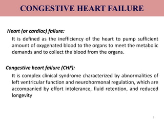
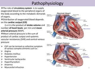
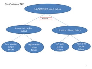
2. CONGESTIVE HEART FAILURE
Heart (or cardiac) failure:
It is defined as the inefficiency of the heart to pump sufficient
amount of oxygenated blood to the organs to meet the metabolic
demands and to collect the blood from the organs.
Congestive heart failure (CHF):
It is complex clinical syndrome characterized by abnormalities of
left ventricular function and neurohormonal regulation, which are
accompanied by effort intolerance, fluid retention, and reduced
longevity
2
3. Pathophysiology
The role of circulatory system is to supply
oxygenated blood to the peripheral organs of
the body according to the metabolic demands
of organs.
Distribution of oxygenated blood depends
on the cardiac output (CO)
(i.e it is the product of stroke volume and
number of heart beats per min and mean
arterial pressure (MAP).
Mean arterial pressure is the sum of
product of cardiac output and systemic
vascular resistances (SVR) and central venous
pressure
CHF can be termed as collective symptom
of various complex ailments such as :
Angina
Diabetes mellitus
Hypertension
Ventricular tachycardia
Hyperthyroidism
Anaemia
Myocardial infraction
3
4. Congestive heart failure
BASED ON
Amount of cardiac
output Position of heart failure
High cardiac
output
failure
Low cardiac
output
failure
Left side
cardiac
failure
Right side
Cardiac
failure
Classification of CHF
4
5. Based on amount of cardiac output
Low-cardiac output failure
• It is most common congestive heart failure.
• The metabolic demands of the body organs are normal with in limits but the heart fails
to pump sufficient amount of oxygenated blood to the organs of the body.
• The primary cause of LCOF is the ventricular systolic dysfunction and ventricular
diastolic dysfunction.
Ventricular systolic dysfunction
• Myocardial infarction weakens the muscles of ventricles and make them inefficient to
pump the required volume of blood.
• Thus results in low cardiac output and low ejection fraction.
Ventricular diastolic dysfunction
• Hypertrophy is responsible for the stiffening of heart muscle
• The stiffened muscle of the ventricles fails to relax during diastolic and thus cannot
collect sufficent amount of blood.
• This ultimately results in low cardiac output.
5
6. High cardiac output failure
•It occurs very rarely.
•Hyperthyroidism , anaemia & arteriovenous shunt, enhances the metabolic
demands of the body for myocardial oxygen ,which cannot be met even by the
increased pumping action of the heart.
Low cardiac output failure High cardiac output failure
Most frequent Very rarely
Metabolic demands of the body organs for
oxygen are normal and within limits
Metabolic demands of the body for oxygen
is very high
Myocardial fraction is prominent factor
leading to the failure of systolic & diastolic
function of the ventricles, ultimetly results
in low cardiac output failure
Hyperthyroidism, anaemia, arteriovenous
shunt causes high cardiac output failure.
6
7. Based on the position of heart failure
• The failure of either right side or left side of the heart to pump the blood
leads to the failure of the other side and ultimately results in the failure of
the both sides of the heart.
Left side cardiac failure
• The failure of the left ventricle to pump the entire blood present in it during
systole results in retention of some amount of blood after every systole
• Thus blood is accumulated in the left ventricle after few systole of the heart.
• The left ventricle fails to accept the blood from auricles and lungs thus the
uncollected blood due to back-up pressure remains in the lungs resulting in
pulmonary oedema.
7
8. Right side cardiac failure
• The failure of right ventricle to pump the entire blood present in it during
systole results in retention of some amount of blood after every systole.
• Thus blood is accumulated in right ventricle after few systoles.
• The left ventricle fails to accept the blood from peripheral organs and
ultimately results in generalized systemic oedema or peripheral oedema.
Left side cardiac failure Right side cardiac failure
Is the result of right side cardiac failure Is the result of left side cardiac failure
Inefficent pumping action of left
ventricle is responsible for the
accumulation of blood in the ventricles
Inefficient pumping action of right ventricle
is responsible for the accumulation of
blood in right ventricle
Left ventricle fails to accept/collect the
blood from lungs due to back pressure
Right ventricle fails to accept/collect the
blood from peripheral organs.
Pulmonary congestion/oedema is the
final result
Peripheral generalized oedema is the
final result
8
9. Pathophysiology
• Normal filling capacity of left ventricle is about 130 ml, out of which about 70ml
undergoes ejection, while the remaining volume persist in the ventricles.
• The volume of blood ejected from the left ventricle reduces to about 55ml
In condition of left ventricular systolic dysfunctioning.
• Any factor that tends to increase the stress on the heart or lead to myocardial
infraction results in left ventricular systolic dysfunction.
(LVSD).
• The eventual consequences is an impairment in the systolic contraction or diastolic
relaxation or both.
• Imapariment in the contracting ability of the heart results in systolic dysfunction
,due to this ejection faction tends to get lowered.
• The diastolic function is concerned with the filling of the ventricles, such filling is
governed by the venous return and adequate dilation of the ventricles.
• In case of diastolic dysfunction, the ventricles do not dilate properly resulting in
relatively less filling.
• If the diastolic dysfunction persists for longer periods, it result in systolic
dysfunction and remodelling of the ventricles. 9
10. Clinical condition that results in systolic and systolic dysfunctioning
include:
• Hypertension
• Aortic stenosis
• Valvular defects
• Acute myocardial infraction
• Cardiomyopathies
• Thus the ventricles fails to fill in the required volume of blood and also fails in its
subsequent ejection.
• This results in in the drastic reduction in the stroke volume and cardiac output.
• A decrease in cardiac ouput leads to reduced perfusion of essential organs.
• The body tries to compensate for the reduced cardiac output by stimulating the
functioning of various compensatory mechanisims
10
11. Compensatory mechanisms of congestive heart
failure
• To enhances the cardiac output, body compensates for the intrinsic cardiac effects
in the following manner.
1. Increased sympathetic discharge
• To compensate for the decreased B.P, baroreceptors located in the arch of aorta
carotid sinuses and walls of the heart get stimulated and causes activation of
beta-adrenergic receptors leading to an increase in rate and force of contraction
of heart.
• An increase in venous return (preload) is also seen due to the activation of alpha-adrenergic
receptors.
• Increased rate and force of contraction together with the increased preload
results in an initial increase in the cardiac output.
• Vasoconstriction of the arteries due to alpha stimulation also causes an increase
in after load, leading to fall in ejection fraction.
• As a result the cardiac output decreases.
11
12. Activation of Renin- Angiotensin
Aldosterone (RAA)
• Fall in the cardiac output decreases the renal perfusion rate, as a result the RAA
system gets activated .
• Angiotensin 2 causes vasoconstriction and an increase in the peripheral vascular
resistance(PVR).
• while aldosterone leads to increased retention of sodium and water, there by
increasing the blood volume.
• PVR effects the after load during which the heart is unable to pump the extra
blood volume.
• This leads to the development of back up pressure causing pulmonary congestion
and peripheral oedema.
12
13. Cardiac Remodeling
• It is most important mechanisum by which body compensated for the intrinsic cardiac
effects.
• It involves changes in the shape of the heart (from normal to spherical) due to
myocardial hypertrophyt.
• During cardiac remodelling, the connective tissue cells as well as the abnormal
myocardial cells undergoes proliferation and dilation instead of steching under the
influences of angiotensin 2.
• In the early stages, the remodelled heart maintains the cardiac performances.
• But later on ,hypertrophy may exert certain adverse effects like ischaemic changes,
decrease in the rate and force of contraction of heart.
• After certain period of time the compensatory mechanisms get exhausted and worsen
the cardiac performances.
• The stress on heart increase and a stage is reached where these mechanisms fails to
maintain the adequate cardiac output.
The heart produce signs &symptoms like:
• Dyspnoes, hypoxia
• Pulmonary & peripheral oedema
• Hepatic congestion and enlarged liver
• Enlarged heart
• Decreased exercise tolerance due to decrease in cardiac output
• Decreased urine formation due to decrease in renal perfusion rate.
13
15. Clinical manifestations/signs and symptoms
• Fluid retention
• Pulmonary congestion
• Dyspnoea & orthopnoea
CVS MANIFESTATIONS
• Resting tachycardia
• Ventricular arrhythmias
• Enlargement of heart
RENAL MANIFESTATIONS
• Nocturia
• Oliguria
OTHER MANIFESTATIONS
• Reduced cardiac output lead to poor perfusion of skeletal muscle resulting in
fatigue.
• Reduced perfusion to brain results in altered mental states & confusion.
• Reduced perfusion may also causes the patient to appear pale with cold and
sweaty hands.
15
17. TREATMENT OF CHF
• There are two distinct goals of drug therapy
in CHF:
Relief of congestion/low cardiac output
symptoms & restoration of cardiac
performance:
o Inotropic drugs-digoxin,
dobutamine,amrinone/milrinone.
o Diuretics: furosemide, thiazides.
o Vasodilators: ACE inhibitors/AT1 antagonist,
hydralazine, nitrate.
o BETA blocker: metoprolol,bisprolol,carvedilol
Arrest/reversal of disease progression &
prolongation of survival
ACE inhibitors/AT1antagonist (ARBs).
Beta-blockers
Aldosterone antagonist-spironolactone..
17
19. Thiazide Diuretics
Hydrochlorothiazide, metolazone, chlorthalidone
• Thiazide diuretics inhibit the Na-Cl symporter.
• This is a channel present on the luminal side of the distal
convoluted tubule of the nephron and functions via secondary
active transport.
• Sodium enters the cell down it concentration gradient
(“downhill”), while chlorine goes up its electrical gradient
(inside of the cell is negative).
• Chlorine is able to travel “uphill” without direct usage of energy
because it uses the energy that sodium dissipates when it goes
down its concentration gradient.
• Once these two ions enter the cell, the sodium is shuttled
across the basolateral side of the cell via Na/K ATPase (primary
active transport) and chlorine travels across a chlorine channel
(facilitated diffusion). (RFUMS, 2006)
Mechanism of Action of Thiazide Diuretics:
• Individuals that suffer from high blood pressure are typically
given thiazide diuretics. It is believed that thiazide inhibits the
Na-Cl symporter by binding to the Cl- site of the transporter.
This shuts down the pump and leads to excretion of sodium and
chloride. By inhibiting these solutes to be reabsorbed into the
plasma, they will cause the urine to have a greater
concentration (increased amount of osmotically active solutes)
and this will give water the gradient to remain in the filtrate.
Also water will not be reabsorbed in the collecting tubule
because the plasma osmolality will be reduced, so it will need to
leave the plasma because the extracellular fluid osmolality will
be low.
• thiazide diuretics are successful at reducing blood pressure
because they reduce the plasma volume. It is possible that
thiazide diuretics also act to relax the smooth muscle of blood
vessels to reduce the resistance to blood flow. Patients that take
these medications will tend to have a greater frequency of
urination accompanied by increased solute and water loss.
19
20. Loop Diuretics
Furosemide, Bumetanide , Torsemid
• The Na+, K+, 2Cl- symporter, a carrier-mediated process,.
• It is the major reabsorptive mechanism in the thick ascending limb (TAL).
• All four ions are transported by secondary active transport into the TAL epithelial cells, at their apical surface,
using the energy derived from the Na+/K+-ATPase co-transporter, also a carrier mediated mechanism.
Mechanism of Action of Loop Diuretics:
• Loop diuretics act on the Na-K-2Cl symporter in the thick ascending limb of the loop of Henle to inhibit sodium
and chloride reabsorption. Because magnesium and calcium reabsorption in the thick ascending limb is
dependent on sodium and chloride concentrations.
• loop diuretics also inhibit their reabsorption. By disrupting the reabsorption of these ions, loop diuretics
prevent the urine from becoming dilute and disrupt the generation of a hypertonic renal medulla. Without such
a concentrated medulla, water has less of an osmotic driving force to leave the collecting duct system,
ultimately resulting in increased urine production. This diuresis leaves less water to be reabsorbed into the
blood, resulting in a decrease in blood volume.
• Loop diuretics cause vasodilation of the veins and of the kidney's blood vessels, mechanically causing a
decrease in blood pressure. The collective effects of decreased blood volume and vasodilation decrease blood
pressure.
Adverse reaction:
•pre-renal azotemia
•Hypokalemia
•Skin rash
•ototoxicity
20
21. Potassium-Sparing Diuretics
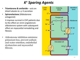
• The K-sparing diuretics are weak diuretics alone.
• They are primarily used as adjuncts to thiazides and loop diuretics or for potassium and
magnesium spacing. Instead of using thiazides alone for hypertension ,triamterene is also
used by combination.
• Amiloride can be used for magnesium deficiency because it increases renal reabsorption.
• If a patient who has hypomagnesemia, and you can't give them enough magnesium
orally, because of laxative action, give amiloride.
• Also, amiloride is useful for patients taking lithium who have polyuria and complain of
having to get up three or four times at night. At a dose of 5 mg bid, amiloride reduces
urine volume by 30%.
• "Don't use any K-sparing diuretics with angiotensin-converting enzyme
inhibitors, angiotensin II receptor blockers [or] nonsteroidals.
• Be cautioned against using them when serum creatinine levels are above 2 mg/dL.
Specific side effects seen with K-sparing diuretics include
• Hyperchloremic acidosis;
• Hyperkalemia, especially if administered with an ACE inhibitor, angiotensin II receptor
blocker or in patients with diabetes;
• Gynecomastia, impotence in men or irregular menstrual cycles in women (only with use
of spironolactone);
• Folic acid deficiency (with chronic use of triamterene); or acute renal failure (with
triamterene when used with indomethacin [Indocin]).
21
22. K+ Sparing Agents
• Triamterene & amiloride – acts on
distal tubules to ↓ K secretion
• Spironolactone (Aldosterone
antagonist)
it improve survival in CHF patients due
to the effect on renin-angiotensin-aldosterone
system with subsequent
effect on myocardial remodeling and
fibrosis
• Aldosterone inhibition minimize
potassium loss, prevent sodium
and water retention, endothelial
dysfunction and myocardial
fibrosis.
22
23. Renin–Angiotensin- System
• The renin-angiotensin system (RAS) or the renin-angiotensin-aldosterone
system (RAAS) is a hormone system that regulates blood pressure and water (fluid)
balance.
• When blood volume is low, juxtaglomerular cellsin the kidneys secrete renin directly
into circulation.
• Plasma renin then carries out the conversion of angiotensinogen released by the liver
to angiotensin I.
• Angiotensin I is subsequently converted to angiotensin II by the enzyme angiotensin
converting enzyme found in the lungs.
• Angiotensin II is a potent vaso-active peptide that causes blood vessels to constrict,
resulting in increased blood pressure.
• Angiotensin II also stimulates the secretion of the hormone aldosterone from
the adrenal cortex
• Aldosterone causes the tubules of the kidneys to increase the reabsorption of sodium
and water into the blood. This increases the volume of fluid in the body, which also
increases blood pressure.
• If the renin-angiotensin-aldosterone system is too active, blood pressure will be too
high.
• There are many drugs that interrupt different steps in this system to lower blood
pressure. These drugs are one of the main ways to control high blood pressure
(hypertension),hear failure,kidney failure, and harmful effects of diabetes. 23
26. Angiotensin Converting Enzyme (ACE)
Inhibitors
• Captopril, Lisinopril, Enalapril, Ramipril, Quinapril.
Mode of action:
• Angiotensin 1 Angiotensin 2
ACE Inhibitors
Hences, they inhibit the generation of angiotensin 2,a potent
vasoconstrictor.
They also inhibit the release of aldosterone & vasopressin,
thereby inhibiting fluid and slat retention thus decreasing
the preload.
Elevate the levels of bradykinin, vasodilator thus enhancing
renal & cardiac perfusion.
26
27. Scope for ACE Inhibitors…..
Drug
reactions
•Hypotension
•Dry cough
•Renal
dysfunction
•Proteinuria
•Dizziness
•Joint pain
27
28. Angiotensin Receptor AT-1 blockers
(ARB)
Losartan ,candesartan,valsartan
• Angiotensin 2 ,a vasocontrictor is concerned with ventricular remodelling and fluid
retention.
• These drugs inhibit the binding of angiotensin 2 to its AT₁ receptor.
• Thus they preclude the a bove mentioned effects of angiotensin 2.
• These agents do not exert any action on bradykinin and thus do not produce
cough.
• Has comparable effect to ACE I
• Can be used in certain conditions when ACE I are contraindicated
Adverse drug reactions
•Hypotension
•Impariment of renal functioning
Dose
Candesartan
•Initial: 4-8mg
•Targeted dose -32mg
Valsartan
•Initial:40mg
•Targeted dose -160mg
28
29. ACE-Inhibitors and ARB effects
• Vasodilation
• Decreased fluid retention (afterload & preload)
• Reduction in aldosterone secretion
• Inhibition of cardiac and vascular remodeling
29
30. Inotropes
• Increase force of contraction
• All increase intracellular cardiac Ca++ concentration
• Eg:
– Digitalis (cardiac glycoside)
– Dobutamine (β-adrenergic recepter agonist)
– Amrinone (PDE inhibitor)
30
31. Cardiac glycosides : Digoxin (DIGITALIS)
inhibit Na +,K + ATPase , pump which
It inhibits the
Functions in the exchange of Na⁺ for k⁺ ions.
Such blockage results in intracellular
accumulation of Na⁺ ions .
These ions are then exchanged with Ca₂⁺ ions
through Na⁺ - Ca₂⁺ exchange carries.
These ca₂⁺ ions increase the contractility of the
myocardium which is beneficial to the failing
heart.
Digoxin enhances the cholinergic activity which
reduces the HR and AV conduction .
Due to this the time required for diastolic filling
gets enhanced while the myocardial o2
consumption is retarted.
The sympathetic outflow comprising renin,
aldosterone is also decreased by dioxin
31
32. Drug reaction
• Bradycardia
• Nausea
• Vomiting
• Visual disturbances
• Non paroxysomal junctional
tachycardia
• Supraventricular
tachycardia
• Sexual dysfunction
• Neuralgic pain
USES:
• For tachyarrhythmias
• For ventricular arrhythmias
32
33. β -Adrenergic Agonists
Dopamine
• Dopamine acts at a variety of receptors (dose dependant)
• Rapid elimination- can only be administered as a continuous infusion
Dobutamine
•Stimulates beta-adrenergic receptors and produces a positive inotropic
response.
•Unlike the vasoconstriction seen with high doses of dopamine,
dobutamine produces a mild vasodilatation
33
35. BIPYRIDINES
phosphodiesterase inhibitors
• Targets PDE -3 (found in cardiac and smooth muscle)
• Ex. Inamrinone , milrinone
alter the intracellular
movements of calcium by
influencing the sarcoplasmic
reticulum
increasing inward
calcium flux
in the heart during
the
action potential
increase myocardial contractility
Inhibition of
PDE3
Increase in cAMP
the conversion of inactive protein
kinase to active form
Protein kinases are responsible for
phosphorylation of Ca channels
increased Ca entry into the cell
↑ Vascular Permeability leads to
↓ in intravascular fluid Volume
increase in
contractility
vasodilation
35
36. β1 Blockers
MOA
bisoprolol, carvedilol , metoprolol
Heart failure is accompanied by an
increase activation of sympathetic
nervous system.
• This brings about structural &
functional modification in the
myocardium.
• β Blockers inhibit the sympathetic
outflow of norepinephrine and
counteract the changes produced.
The ventricular remodelling in heart
failure is also reversed by
β Blockers
• Increases beta receptor sensitivity.
Adverse drug reaction
• Hypotension
• Bradycardia
• Worsening of CHFsymptoms.
36
37. Vasodilators
• Isosorbide dinitrate, isosorbide mononitrate, and hydralazine also used
specially in patients who cannot tolerate ACE inhibitors.
37
38. Vasodilator(Hydralazine)
• It directly relaxes the arterioles & arteries reducing the peripheral vascular
reesistances & preload.
• It also help to reduce after load.
Adverse drug reaction:
• Nausea
• Palpitation
• Tachycardia
• Salt & water retention on prolong therapy.
38
39. NITRATES & NITRITES
Nitroglycerin is denitrated by
glutathione S -transferase in smooth muscle
Free nitrite ion is released, which is then converted to Nitric
Oxide
activation of guanylyl cyclase enzyme
increase in cGMP
dephosphorylation of myosin light chain , preventing the
interaction of myosin with actin(Myosin light chain
kinase essential for smooth muscle contraction).
Results in vasodilation
39
40. Calcium Channel Blockers for Vasodialation
Nisoldipine , Isradipine
bind more effectively to open channels and inactivated channels
(inner side of the membrane)
reduces the frequency of opening in response to depolarization
marked decrease in transmembrane calcium current
activation of myosin light chain kinase
Vascular smooth muscle (the most sensitive
long-lasting relaxation
40
41. NISIRITIDE(BNP)
• Brain (B-type) natriuretic peptide (BNP) is secreted constitutively by
ventricular myocytes in response to stretch
• Niseritide = recombinant human BNP
• Naturally occurring atrial natriuretic peptide may vascular
permeability may reduce intravascular volume)
Main Side Effect:
• hypotension
41
42. Human BNP binds to the particulate guanylate cyclase receptor
of vascular smooth muscle and endothelial
intracellular concentrations (cGMP) ↑
smooth muscle cell relaxation
dilate veins and arteries
systemic and pulmonary vascular resistances ↑
Indirect ↑ in cardiac output and diuresis.
Effective in HF because preload and afterload↓
42