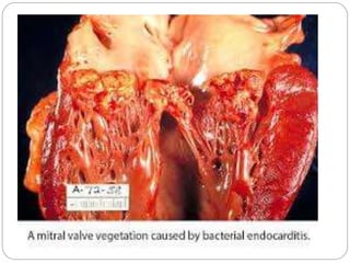
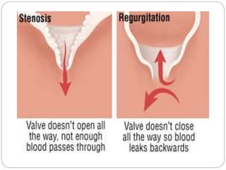
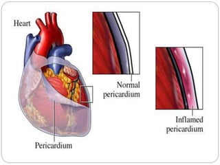
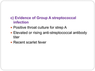
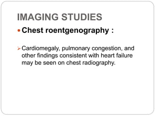
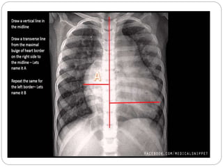
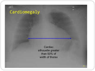
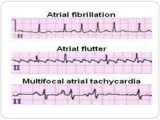
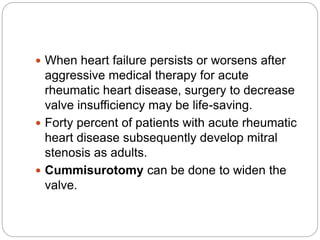
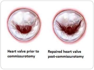
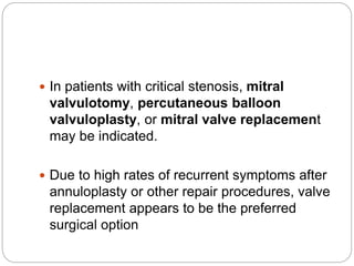
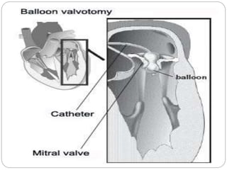
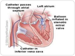
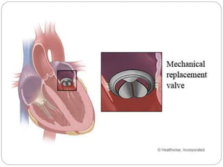
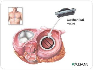
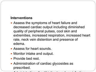
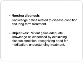
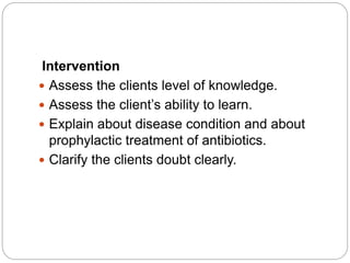
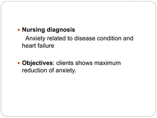
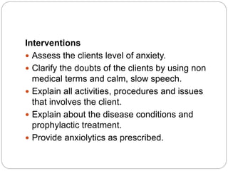
Neelu Aryal
Msc Nursing 2nd year
RHEUMATIC HEART
DISEASE
INTRODUCTION
 Rheumatic fever is a diffuse inflammatory
disease characterized by a delayed response
to an infection by group A beta-hemolytic
streptococci (GAS) in the tonsilopharyngeal
area, affecting the heart, joints, central
nervous system, skin and subcutaneous
tissues.
 It is thought that 40-60% of patients with ARF
will go on to developing RHD.
DEFINITION
 Rheumatic heart disease is a chronic
condition resulting from rheumatic fever which
involves all the layers of the heart (i.e.
pancarditis) and is characterized by scarring
and deformity of the heart valves.
 The commonest valves affecting are the mitral
and aortic, in that order. However all four
valves can be affected.
INCIDENCE
 Rheumatic fever is principally a disease of
childhood, with a median age of 10 years,
although it also occurs in adults (20% of
cases).
 Rheumatic fever occurs in equal numbers in
males and females, but the prognosis is
worse for females than for males.
 The disease is seen more commonly in poor
socio-economic strata of the society living in
damp and overcrowded place.
INCIDENCE(contd…)
 Common in the developing countries like
India, Pakistan.
 The incidence of RF in Developing countries
is 27-100/1 lac /yr (G.S.Sainani 2006)
 The incidence of rheumatic fever (RF) varies
from 0.2 to 0.75/1000/ year in school
children 5–15 years of age (2001 Govt.
Census) (Anil Grover,Padamavati S et al,
et.al INJ 2002).
ETIOLOGY
 Group A beta-hemolytic streptococcus.
 Rheumatic fever
RISK FACTORS
 Poor socio-economic status: People who
are poor and belongs to low socio-economic
conditions are prone to get Rheumatic heart
disease.
 Over-crowding: People who are living in a
slum or damp area are more prone to get
Rheumatic heart disease.
 Age: It appears most commonly in children
between the age of 5 to 15 years.
RISK FACTORS(contd…)
 Climate and season: It occurs more in the
rainy season and in the cold climate.
 Upper respiratory tract infection:
Rheumatic fever is an outcome of upper
respiratory tract infection with group A beta-
hemolytic streptococcus.
RISK FACTORS(contd…)
 Previous history of Rheumatic fever: The
client with previous history of Rheumatic fever
are at high risk to develop Rheumatic heart
disease.
 Genetic predisposition: Rheumatic heart
disease shows familier tendancy.
PATHOPHYSIOLOGY
Causative agent (Group A
Beta-hemolytic streptococci)
Untreated Strep throat
Rheumatic fever
All layers of the heart and the
mitral valve become inflamed
Vegetation forms
Valvular regurgitations and
stenosis
Heart failure
CLINICAL
MANIFESTATIONS
Major manifestations
 Carditis
 Polyarthritis
 Chorea
 Erythema marginatum
 Subcutaneous nodules
MAJOR
MANIFESTATIONS(contd…)
 Carditis
MAJOR
MANIFESTATIONS(contd…)
 Arthritis
MAJOR
MANIFESTATIONS(contd…)
 Chorea
MAJOR
MANIFESTATIONS(contd…)
 Erythema marginatum
MAJOR
MANIFESTATIONS(contd…)
 Subcutaneous nodules
Minor manifestations include:
 Fever associated with weakness, malaise,
weight loss and anorexia
 Arthralgia
Laboratory findings
 Positive throat culture for group A beta-
hemolytic streptococci.
Laboratory
findings(contd…)
 Elevated acute phase reactants:
a) Erythrocyte sedimentation rate
b) C-reactive protein
c) Leukocytosis
Laboratory
findings(contd…)
• Prolonged P-R interval
DIAGNOSTIC EVALUATIONS
 A diagnosis of rheumatic heart disease is
made after confirming antecedent rheumatic
fever.
 The modified Jones criteria (revised in 1992)
provide guidelines for the diagnosis of
rheumatic fever.
JONES CRITERIA
 2 major or
1 major and 2 minor
Jones’ criteria for the diagnosis of
Rheumatic fever
Major manifestations
 Carditis
 Polyarthritis
 Chorea
 Erythema marginatum
 Subcutaneous nodules
Minor manifestations
a) Clinical findings
 Previous rheumatic fever or rheumatic heart
disease.
 Arthralgia
 Fever associated with weakness, malaise,
weight loss and anorexia
b) Laboratory findings
 Elevated ESR, C-reactive protein and
Leukocytosis
 ECG and echocardiogram to confirm rhythm
problems and structural changes (prolonged
P-R interval).
 Chest X-ray shows enlarged heart.
c) Evidence of Group A streptococcal
infection
 Positive throat culture for strep A
 Elevated or rising anti-streptococcal antibody
titer
 Recent scarlet fever
IMAGING STUDIES
 Chest roentgenography :
Cardiomegaly, pulmonary congestion, and
other findings consistent with heart failure
may be seen on chest radiography.
 Doppler-echocardiogram
In acute rheumatic heart disease, Doppler-
echocardiography identifies and quantitates
valve insufficiency and ventricular dysfunction.
 In chronic rheumatic heart disease,
echocardiography may be used to track the
progression of valve stenosis and may help
determine the time for surgical intervention.
HEART CATHETERIZATION
 In acute rheumatic heart disease, this
procedure is not indicated.
 With chronic disease, heart catheterization
has been performed to evaluate mitral and
aortic valve disease and to balloon stenotic
mitral valves.
ON ECG
 Sinus tachycardia most frequently
accompanies acute rheumatic heart disease.
Alternatively, some children develop sinus
bradycardia from increased vagal tone.
 Patients with rheumatic heart disease also
may develop atrial flutter, multifocal atrial
tachycardia, or atrial fibrillation from chronic
mitral valve disease and atrial dilation.
HISTOLOGIC FINDINGS
 Pathologic examination of the insufficient
valves may reveal verrucous lesions at the
line of closure.
 Aschoff bodies (perivascular foci of
eosinophilic collagen surrounded by
lymphocytes, plasma cells, and macrophages)
are found in the pericardium, perivascular
regions of the myocardium, and endocardium.
 Anitschkow cells are plump macrophages
within Aschoff bodies.
 In the pericardium, fibrinous and serofibrinous
exudates may produce an appearance of
"bread and butter" pericarditis.
MEDICAL MANAGEMENT
1. Eradicate infection
 Preventive and prophylactic therapy is indicated after
rheumatic fever and acute rheumatic heart disease to
prevent further damage to valves.
 Primary prophylaxis (initial course of antibiotics
administered to eradicate the streptococcal infection)
also serves as the first course of secondary
prophylaxis (prevention of recurrent rheumatic fever
and rheumatic heart disease).
 An injection of 0.6-1.2 million units of benzathine
penicillin G intramuscularly every 4 weeks is the
recommended regimen for secondary prophylaxis for
most US patients.
 Administer the same dosage every 3 weeks in
areas where rheumatic fever is endemic, in
patients with residual carditis, and in high-risk
patients.
 Continue antibiotic prophylaxis indefinitely for
patients at high risk (eg, health care workers,
teachers, daycare workers) for recurrent GABHS
infection.
 Patients with rheumatic fever with carditis and
valve disease should receive antibiotics for at
least 10 years or until age 40 years.
 Patients with rheumatic heart disease and valve
damage require a single dose of antibiotics 1
hour before surgical and dental procedures to
help prevent bacterial endocarditis.
 Patients who had rheumatic fever without valve
damage do not need endocarditis prophylaxis.
 Do not use penicillin, ampicillin, or amoxicillin for
endocarditis prophylaxis in patients already
receiving penicillin for secondary rheumatic fever
prophylaxis (relative resistance of PO
streptococci to penicillin and aminopenicillins.
 Alternate drugs recommended by the American
Heart Association for these patients include PO
clindamycin (20 mg/kg in children, 600 mg in
adults) and PO azithromycin or clarithromycin (15
mg/kg in children, 500 mg in adults).
2. Maximize cardiac output
 Corticosteroids are used to treat carditis,
especially if heart failure is evident.
 If heart failure develops, treatment, including ACE
inhibitors, beta blockers and diuretics, is effective.
3. Promote comfort
 Client with arthritic manifestations obtain relief
with salicylates.
 Bed rest is usually prescribed to reduce cardiac
effort until evidence of inflammation has
subsided.
SURGICAL
MANAGEMENT
 When heart failure persists or worsens after
aggressive medical therapy for acute
rheumatic heart disease, surgery to decrease
valve insufficiency may be life-saving.
 Forty percent of patients with acute rheumatic
heart disease subsequently develop mitral
stenosis as adults.
 Cummisurotomy can be done to widen the
valve.
 In patients with critical stenosis, mitral
valvulotomy, percutaneous balloon
valvuloplasty, or mitral valve replacement
may be indicated.
 Due to high rates of recurrent symptoms after
annuloplasty or other repair procedures, valve
replacement appears to be the preferred
surgical option
NURSING MANAGEMENT
 Nursing diagnosis
Pain related to inflammatory response in
the joints.
 Objectives: The client verbalizes increased
comfort as evidenced by reports of reduced
discomfort, expression of joint pain reduction,
relaxed body posture and a calm facial
expression.
Interventions
 Assess the level of pain, duration, intensity
and frequency of pain.
 Complete bed rest and provide comfortable
position.
 Provide diversional therapy and psychological
support.
 Administer analgesics as needed.
 Nursing diagnosis
Decreased cardiac output related to valve
dysfunction or HF.
 Objectives: client increases cardiac output as
evidenced by regular cardiac rhythm, heart
rate, blood pressure, respiration and urine
output within normal limit.
Interventions
 Assess the symptoms of heart failure and
decreased cardiac output including diminished
quality of peripheral pulses, cool skin and
extremities, increased respiration, increased heart
rate, neck vein distention and presence of
edema.
 Assess for heart sounds.
 Monitor intake and output.
 Provide bed rest.
 Administration of cardiac glycosides as
prescribed.
 Nursing diagnosis
Knowledge deficit related to disease condition
and long term treatment.
 Objectives: Patient gains adequate
knowledge as evidenced by explaining
disease condition, recognizing need for
medication, understanding treatment.
Intervention
 Assess the clients level of knowledge.
 Assess the client’s ability to learn.
 Explain about disease condition and about
prophylactic treatment of antibiotics.
 Clarify the clients doubt clearly.
 Nursing diagnosis
Anxiety related to disease condition and
heart failure
 Objectives: clients shows maximum
reduction of anxiety.
Interventions
 Assess the clients level of anxiety.
 Clarify the doubts of the clients by using non
medical terms and calm, slow speech.
 Explain all activities, procedures and issues
that involves the client.
 Explain about the disease conditions and
prophylactic treatment.
 Provide anxiolytics as prescribed.
BIBLIOGRAPHY
 Black JM, Hawks JH. Medical surgical nursing. 8th
ed.vol-2. Elsevier; p.1396-1401
 Lewis, Heitkemper, Dirksen O’Brien, Bucher. Medical
surgical nursing.7th ed.New Delhi.Elsevier;p. 875-82
 URL:https://en.wikipedia.org/wiki/Rheumatic_fever
 URL:http://www.slideshare.net/miel9156/rheumatic-
heart-disease-3264045
 URL:http://www.powershow.com/view/3d4797-
MWI0M/Rheumatic_heart_disease_RHD_powerpoint
_ppt_presentation
 URL:http://gmch.gov.in/estudy/e%20lectures/Patholog
y/cvs5%20RF.pdf
THANK YOU

Rheumatic Heart Disease

  • 1.
    Neelu Aryal Msc Nursing2nd year RHEUMATIC HEART DISEASE
  • 2.
    INTRODUCTION  Rheumatic feveris a diffuse inflammatory disease characterized by a delayed response to an infection by group A beta-hemolytic streptococci (GAS) in the tonsilopharyngeal area, affecting the heart, joints, central nervous system, skin and subcutaneous tissues.  It is thought that 40-60% of patients with ARF will go on to developing RHD.
  • 3.
    DEFINITION  Rheumatic heartdisease is a chronic condition resulting from rheumatic fever which involves all the layers of the heart (i.e. pancarditis) and is characterized by scarring and deformity of the heart valves.  The commonest valves affecting are the mitral and aortic, in that order. However all four valves can be affected.
  • 4.
    INCIDENCE  Rheumatic feveris principally a disease of childhood, with a median age of 10 years, although it also occurs in adults (20% of cases).  Rheumatic fever occurs in equal numbers in males and females, but the prognosis is worse for females than for males.  The disease is seen more commonly in poor socio-economic strata of the society living in damp and overcrowded place.
  • 5.
    INCIDENCE(contd…)  Common inthe developing countries like India, Pakistan.  The incidence of RF in Developing countries is 27-100/1 lac /yr (G.S.Sainani 2006)  The incidence of rheumatic fever (RF) varies from 0.2 to 0.75/1000/ year in school children 5–15 years of age (2001 Govt. Census) (Anil Grover,Padamavati S et al, et.al INJ 2002).
  • 6.
    ETIOLOGY  Group Abeta-hemolytic streptococcus.  Rheumatic fever
  • 7.
    RISK FACTORS  Poorsocio-economic status: People who are poor and belongs to low socio-economic conditions are prone to get Rheumatic heart disease.  Over-crowding: People who are living in a slum or damp area are more prone to get Rheumatic heart disease.  Age: It appears most commonly in children between the age of 5 to 15 years.
  • 8.
    RISK FACTORS(contd…)  Climateand season: It occurs more in the rainy season and in the cold climate.  Upper respiratory tract infection: Rheumatic fever is an outcome of upper respiratory tract infection with group A beta- hemolytic streptococcus.
  • 9.
    RISK FACTORS(contd…)  Previoushistory of Rheumatic fever: The client with previous history of Rheumatic fever are at high risk to develop Rheumatic heart disease.  Genetic predisposition: Rheumatic heart disease shows familier tendancy.
  • 10.
    PATHOPHYSIOLOGY Causative agent (GroupA Beta-hemolytic streptococci) Untreated Strep throat Rheumatic fever
  • 11.
    All layers ofthe heart and the mitral valve become inflamed Vegetation forms Valvular regurgitations and stenosis Heart failure
  • 16.
    CLINICAL MANIFESTATIONS Major manifestations  Carditis Polyarthritis  Chorea  Erythema marginatum  Subcutaneous nodules
  • 17.
  • 19.
  • 20.
  • 21.
  • 22.
  • 23.
    Minor manifestations include: Fever associated with weakness, malaise, weight loss and anorexia  Arthralgia
  • 24.
    Laboratory findings  Positivethroat culture for group A beta- hemolytic streptococci.
  • 25.
    Laboratory findings(contd…)  Elevated acutephase reactants: a) Erythrocyte sedimentation rate b) C-reactive protein c) Leukocytosis
  • 26.
  • 27.
    DIAGNOSTIC EVALUATIONS  Adiagnosis of rheumatic heart disease is made after confirming antecedent rheumatic fever.  The modified Jones criteria (revised in 1992) provide guidelines for the diagnosis of rheumatic fever.
  • 28.
    JONES CRITERIA  2major or 1 major and 2 minor
  • 29.
    Jones’ criteria forthe diagnosis of Rheumatic fever Major manifestations  Carditis  Polyarthritis  Chorea  Erythema marginatum  Subcutaneous nodules
  • 30.
    Minor manifestations a) Clinicalfindings  Previous rheumatic fever or rheumatic heart disease.  Arthralgia  Fever associated with weakness, malaise, weight loss and anorexia
  • 31.
    b) Laboratory findings Elevated ESR, C-reactive protein and Leukocytosis  ECG and echocardiogram to confirm rhythm problems and structural changes (prolonged P-R interval).  Chest X-ray shows enlarged heart.
  • 32.
    c) Evidence ofGroup A streptococcal infection  Positive throat culture for strep A  Elevated or rising anti-streptococcal antibody titer  Recent scarlet fever
  • 33.
    IMAGING STUDIES  Chestroentgenography : Cardiomegaly, pulmonary congestion, and other findings consistent with heart failure may be seen on chest radiography.
  • 37.
     Doppler-echocardiogram In acuterheumatic heart disease, Doppler- echocardiography identifies and quantitates valve insufficiency and ventricular dysfunction.
  • 38.
     In chronicrheumatic heart disease, echocardiography may be used to track the progression of valve stenosis and may help determine the time for surgical intervention.
  • 40.
    HEART CATHETERIZATION  Inacute rheumatic heart disease, this procedure is not indicated.  With chronic disease, heart catheterization has been performed to evaluate mitral and aortic valve disease and to balloon stenotic mitral valves.
  • 42.
    ON ECG  Sinustachycardia most frequently accompanies acute rheumatic heart disease. Alternatively, some children develop sinus bradycardia from increased vagal tone.  Patients with rheumatic heart disease also may develop atrial flutter, multifocal atrial tachycardia, or atrial fibrillation from chronic mitral valve disease and atrial dilation.
  • 47.
    HISTOLOGIC FINDINGS  Pathologicexamination of the insufficient valves may reveal verrucous lesions at the line of closure.  Aschoff bodies (perivascular foci of eosinophilic collagen surrounded by lymphocytes, plasma cells, and macrophages) are found in the pericardium, perivascular regions of the myocardium, and endocardium.
  • 48.
     Anitschkow cellsare plump macrophages within Aschoff bodies.  In the pericardium, fibrinous and serofibrinous exudates may produce an appearance of "bread and butter" pericarditis.
  • 51.
    MEDICAL MANAGEMENT 1. Eradicateinfection  Preventive and prophylactic therapy is indicated after rheumatic fever and acute rheumatic heart disease to prevent further damage to valves.  Primary prophylaxis (initial course of antibiotics administered to eradicate the streptococcal infection) also serves as the first course of secondary prophylaxis (prevention of recurrent rheumatic fever and rheumatic heart disease).  An injection of 0.6-1.2 million units of benzathine penicillin G intramuscularly every 4 weeks is the recommended regimen for secondary prophylaxis for most US patients.
  • 52.
     Administer thesame dosage every 3 weeks in areas where rheumatic fever is endemic, in patients with residual carditis, and in high-risk patients.  Continue antibiotic prophylaxis indefinitely for patients at high risk (eg, health care workers, teachers, daycare workers) for recurrent GABHS infection.  Patients with rheumatic fever with carditis and valve disease should receive antibiotics for at least 10 years or until age 40 years.
  • 53.
     Patients withrheumatic heart disease and valve damage require a single dose of antibiotics 1 hour before surgical and dental procedures to help prevent bacterial endocarditis.  Patients who had rheumatic fever without valve damage do not need endocarditis prophylaxis.  Do not use penicillin, ampicillin, or amoxicillin for endocarditis prophylaxis in patients already receiving penicillin for secondary rheumatic fever prophylaxis (relative resistance of PO streptococci to penicillin and aminopenicillins.
  • 54.
     Alternate drugsrecommended by the American Heart Association for these patients include PO clindamycin (20 mg/kg in children, 600 mg in adults) and PO azithromycin or clarithromycin (15 mg/kg in children, 500 mg in adults).
  • 55.
    2. Maximize cardiacoutput  Corticosteroids are used to treat carditis, especially if heart failure is evident.  If heart failure develops, treatment, including ACE inhibitors, beta blockers and diuretics, is effective.
  • 56.
    3. Promote comfort Client with arthritic manifestations obtain relief with salicylates.  Bed rest is usually prescribed to reduce cardiac effort until evidence of inflammation has subsided.
  • 57.
  • 58.
     When heartfailure persists or worsens after aggressive medical therapy for acute rheumatic heart disease, surgery to decrease valve insufficiency may be life-saving.  Forty percent of patients with acute rheumatic heart disease subsequently develop mitral stenosis as adults.  Cummisurotomy can be done to widen the valve.
  • 60.
     In patientswith critical stenosis, mitral valvulotomy, percutaneous balloon valvuloplasty, or mitral valve replacement may be indicated.  Due to high rates of recurrent symptoms after annuloplasty or other repair procedures, valve replacement appears to be the preferred surgical option
  • 65.
  • 66.
     Nursing diagnosis Painrelated to inflammatory response in the joints.  Objectives: The client verbalizes increased comfort as evidenced by reports of reduced discomfort, expression of joint pain reduction, relaxed body posture and a calm facial expression.
  • 67.
    Interventions  Assess thelevel of pain, duration, intensity and frequency of pain.  Complete bed rest and provide comfortable position.  Provide diversional therapy and psychological support.  Administer analgesics as needed.
  • 68.
     Nursing diagnosis Decreasedcardiac output related to valve dysfunction or HF.  Objectives: client increases cardiac output as evidenced by regular cardiac rhythm, heart rate, blood pressure, respiration and urine output within normal limit.
  • 69.
    Interventions  Assess thesymptoms of heart failure and decreased cardiac output including diminished quality of peripheral pulses, cool skin and extremities, increased respiration, increased heart rate, neck vein distention and presence of edema.  Assess for heart sounds.  Monitor intake and output.  Provide bed rest.  Administration of cardiac glycosides as prescribed.
  • 70.
     Nursing diagnosis Knowledgedeficit related to disease condition and long term treatment.  Objectives: Patient gains adequate knowledge as evidenced by explaining disease condition, recognizing need for medication, understanding treatment.
  • 71.
    Intervention  Assess theclients level of knowledge.  Assess the client’s ability to learn.  Explain about disease condition and about prophylactic treatment of antibiotics.  Clarify the clients doubt clearly.
  • 72.
     Nursing diagnosis Anxietyrelated to disease condition and heart failure  Objectives: clients shows maximum reduction of anxiety.
  • 73.
    Interventions  Assess theclients level of anxiety.  Clarify the doubts of the clients by using non medical terms and calm, slow speech.  Explain all activities, procedures and issues that involves the client.  Explain about the disease conditions and prophylactic treatment.  Provide anxiolytics as prescribed.
  • 74.
    BIBLIOGRAPHY  Black JM,Hawks JH. Medical surgical nursing. 8th ed.vol-2. Elsevier; p.1396-1401  Lewis, Heitkemper, Dirksen O’Brien, Bucher. Medical surgical nursing.7th ed.New Delhi.Elsevier;p. 875-82  URL:https://en.wikipedia.org/wiki/Rheumatic_fever  URL:http://www.slideshare.net/miel9156/rheumatic- heart-disease-3264045  URL:http://www.powershow.com/view/3d4797- MWI0M/Rheumatic_heart_disease_RHD_powerpoint _ppt_presentation  URL:http://gmch.gov.in/estudy/e%20lectures/Patholog y/cvs5%20RF.pdf
  • 75.