Downloaded 107 times


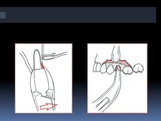


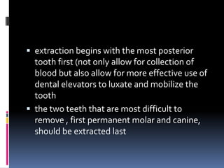


The document outlines the process and considerations for complicated exodontia in oral and maxillofacial surgery, focusing on patient classification by the ASA system to assess health risks, and extraction techniques for both closed and open procedures. It details surgical principles including flap design, suturing techniques, and specific methods for removing single-rooted and multi-rooted teeth, emphasizing the importance of careful assessment and technique selection based on individual patient needs. Additionally, it highlights the policy for managing root fragments and guidelines for performing multiple extractions efficiently.


