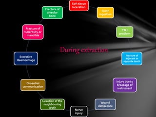
All the mistakes are rectified.Complete and precise knowledge about EXODONTIA .I would like to again focus on compatibility of this ppt;some pictures differ from original one.Animations and Transitions added are not visible .Good for beginners to understand and remember.Images give you better way to grasp.Enjoy and have fun watching this ppt.
3. • Since the earliest period of history; extraction of the tooth
has been considered a very formidable procedure by the
layman.
• It is because of the horrifying experiences associated with
the tooth extraction in the past.
• Even today the removal of a tooth is dreaded by a patient
almost more than any other surgical procedure.
• Many patient suffer from extraction phobia and are often
difficult to care for.
• Despite modern methods of anesthesia many dentist still
believe that speed is essential when extracting the teeth.
Introduction
4. The ideal tooth extraction is -
the painless removal of the whole tooth, or root, with minimal trauma to the investing tissues, so
that the wound heals uneventfully and no post operative prosthetic problem is created
- Geoffrey L. Howe
Definition
5. 1st dentist ever was an EGYPTIAN
- HESI RE (3100-2181 BC)
The history of dental extraction
forceps is very old and goes back to the
time of ARISTOTLE (384 – 322 BC).
He described the mechanics of oral
surgery forceps.
This was 100 years before
ARCHIMEDES studied and discussed
the principles of the lever.
HISTORY
6. Dental history Arabic dentist cauterizing dental
pulp
Curing a tooth with the fumes from
henbane seeds
The martyrdom of St. Apollonia, shows the
tortuous extraction of teeth
German travelling dentist
7. Travelling dentist in a Dutch
The Italian ‘oral surgeon' that effortlessly
removes jaw bones
8. Dental pelican , which was invented in the 14th century by GUY
DE CHAULIAC and used until the late 18th century
9. Indication
THERAPEUTIC INDICATIONS:-
As a general rule:
Any time surgery is considered there has to be a relevant indication, i.e.
whenever a tooth is to be extracted. Extracting a tooth without the relevant
indication is unsound
- (Gaisbauer 1997)
PROPHYLACTIC INDICATION:-
Prophylactic extraction of teeth that may cause infectious complications or
bodily harm constituting an unnecessary risk to therapy if the teeth are not
extracted
10. Teeth located within the area of the
tumor
Extraction of teeth not worth
preserving.
Teeth severely damaged by caries.
11. Retained roots
Teeth with peri-apical infections
that cannot be preserved.
Teeth with apical changes.
12. Teeth with apical changes after
root treatment.
Teeth damaged by trauma
• multiple fractures,
• longitudinal fracture,
• extensive bony defect in the
periodontal region
13. Teeth severely damaged by
periodontal
Disease extraction of impacted and
dislocated teeth
14. In order to maintain normal occlusion
(elongated or heavily tilted teeth,
particularly with previous contacts)
Teeth not worth being preserved from a
prosthetic point of view.
Over erupted tooth without antagonist prior
to extensive prosthodontic treatment.
In case of crowding or impeded
eruption, milk teeth and permanent
teeth should be extracted for
orthodontic purpose.
PROSTHODONTIC INDICATIONS:- ORTHODONTIC INDICATIONS:-
18. Risk of caries in adjacent teeth due to
crowded, partially impacted teeth which
cannot be cleaned adequately
Resorption of the alveolar bone due to
repeated infection around partially
impacted teeth.
If there is the risk of root resorption of
adjacent teeth due to direct contact
with impacted teeth
Preventive measures against relapse
due crowding of anterior teeth
30. also known as ‘forceps
extraction’
It consists of removing teeth
with the use of forceps/elevators.
The blades of these
instruments are forced down the
periodontal membrane between the
tooth root and bony socket.
Intra-alveolar
31. Also known as
‘surgical extraction’
This method involves the
dissection of the tooth or
root from its bony
attachments.
This separation is
achieved by removal of
some of the bone
investing the roots which
are then extracted by the
use of forceps or elevators
Trans-alveolar
32. Maxillary extraction Mandibular extraction
Chair position
•Maxillary occlusal plane at 60⁰ to the
floor.
•Height of dental chair is at 8cm below
the shoulder level of operator
•Pt. is positioned upright and
mandibular occlusal plane is parallel to
the floor.
•Height of dental chair is at 16 cm below
the shoulder level of operator
35. LEVER PRINCIPLES:-
eg. Elevators (which is a lever of
the first order)
It is the simple machine to lift heavy
objects by exerting small amount of
force
Thus elevators includes
An effort arm which is the handle.
Fulcrum is the crest of the alveolar
bone
Resistance arm which exerts the
pressure on the tooth which is the
resistance here.
Maximum mechanical advantage is
gained by keeping the effort arm
longer than the resistance arm.
36. WEDGE PRINCIPLE:-
Eg. apexo elevator, Warnick James
elevators , straight elevator, dental
forceps
This is done by wedging the
elevators or beaks of the forceps
between the tooth and socket in
periodontal ligament space .
This displaces the tooth
occlusally and the instrument can
further be pushed into the socket
to displace the tooth further till it
can be removed completely
Mechanical advantage of wedge
is 2.5;calculated by dividing the
length of the slope by the wedge’s
width
37. WHEEL AND AXLE PRINCIPLE:-
Eg. Cryer’s elevator and cross bar
Resembles the wheel of a vehicle
attached to an axle or a central pole
around which the wheel moves.
The wheel and axle principle is
actually a modified form of lever
principle, but it can move a load
farther than a lever can
The handle serve as the wheel and
blade engages the tooth .
When the handle is rotated the
force created on the blade of the
elevator is multiplied creating a
greater mechanical advantage to
elevate tooth out of its socket.
Mechanical advantage is 4.6
38. Expansion of bony socket by the use of wedge shaped beaks of forceps.
Removal of teeth from the socket
PRINCIPLES OF FORCEP USE:-
39. Principles of extraction
1 Expansion of bony
socket.
Bony is relatively elastic
so it is possible to expand
the bony socket and
remove the teeth.
This is done by holding
the tooth in the apical
end and moved with the
help of forceps causing
the expansion of the
bony socket
40. 2. Loosening of soft tissue
attachment around the tooth
3. Luxation of teeth using
dental elevator
It is done using the No. 9
periosteal elevator.
The instrument has sharp
pointed end and broader rounded
end.
The pointed end is used to begin
periosteal reflection and reflect
dental papilla.
The pointed end is used in prying
motion to elevate dental papilla
between the teeth or attached
gingiva around the tooth
Usually done with straight elevator or
No. molt elevator .
The straight elevator is inserted
perpendicular to the interdental space
after reflection of interdental papillae.
The elevator is then turned in such a
way that the inferior position of blade
rests on alveolar bone and the tooth to be
extracted.
Strong, slow, steady, forceful turning of
the handle moves the tooth in a posterior
direction, which results in some
expansion of alveolar bone and tearing of
periodontal ligament
41. 4 Luxation of teeth with forceps
The major portion of the force is directed
towards the thinnest and therefore weakest
bone’
Thus in maxilla and all but the molar teeth
in the mandible is labial and buccal.
The surgeon uses slow steady force to
displace the tooth buccally.
The tooth is then moved again towards
the opposite direction with slow deliberate
strong pressure .
Buccal and lingual pressure continue to
expand the alveolar socket.
42. 5 Adaptation of forceps to the teeth
Beaks of forceps are adapted
automatically to the tooth apical to
the cervical line that is to the root
surface so that the beaks grasps the
root underneath the loosened soft
tissue.
Lingual beak is usually seated first.
Beak must be held parallel to the
long axis .
The beaks acts as wedges to dilate
the crestal bone on the buccal and
lingual aspects
43. Once the alveolar bone has expanded sufficiently and the
tooth has been luxated a slight tractional force usually
directed buccally can be used
Tooth is delivered out of socket.
6 Removal of teeth from the socket
44. Reflection of soft tissue
Protection of other teeth
Supporting and stabilizing the
mandible
Deliver the whole tooth, root,
dislodged fillings.
Stabilization of patients head
Support of alveolar bone
Tactile information
Compress socket
Role of opposite hand
45. 3 fundamental requirement:-
1. Adequate access and visualization of field of surgery.
2. An unimpeded pathway for removal f tooth.
3. Use of controlled force to luxate and remove the teeth.
Requirement for closed extraction
46. 1. Positioning of the pt.
2. Administration of local anesthesia.
3. Loosening and luxation of the soft tissue attachment from the tooth.
4. Adaptations of forceps to the teeth.
5. Luxation of the teeth by forceps.
6. Removal of teeth
Procedures for extraction
47. Pre-extraction treatment planning
- dentures , soft tissue surgery , implant .
EXTRACTION SEQUENCE :-
MAXILLARY TEETH FIRST
- infiltration anesthetic :more rapid
- debris may fall into the empty sockets
- with mainly buccal force
THE MOST POSTERIORTEETH FIRST
THE MOST DIFFICULT (MOLAR AND CANINE ) LAST.
(8-7-5-4-6-2-1-3)
MULTIPLE EXTRACTION
(Reference from oral and maxillofacial surgery textbook (4th edi.) by NEELIMA MALIK)
48. 1. Apical grip.
2. Labial movement.
3. Palatal movement.
4. Rotational movement.
5. Dilatation of the socket.
6. Final delivery in labial
direction.
Extraction of maxillary anteriors
49. Extraction of maxillary premolars
1. Apical grip.
2. Labial movement.
3. Palatal movement.
4. Dilatation of the socket.
5. Final delivery in buccal
direction.
50. 1. Apical grip.
2. Labial movement.
3. Palatal movement.
4. Dilatation of the socket.
5. Final delivery in buccal
direction.
Extraction of maxillary molars
51. Extraction of mandibular anterior
1. Apical grip.
2. Labial movement.
3. Lingual movement.
4. Rotational movement.
5. Dilatation of the socket.
6. Final delivery in labial
direction.
52. Extraction of mandibular premolar
1. Apical grip.
2. Labial movement.
3. Lingual movement.
4. Primary rotatory
movement in 2nd
premolar only.
5. Dilatation of the socket.
6. Final delivery in buccal
direction.
53. Extraction of mandibular molar
1. Apical grip.
2. Labial movement.
3. Lingual movement.
4. Dilatation of the
socket.
5. Final delivery in
buccal direction.
54. 1. Infection
2. Size Of Wound
3. Blood Supply
4. Resting Of Part
5. Foreign Bodies
6. General Condition Of The Patient
FACTORS INFLUENCING THE HEALING
57. Avoid use of other gauze
Bite gently
Take medicines timely
Gargle with lukewarm saline
water after 24hrs.
Avoid use of straws
Avoid hot food; take soft ,
cold and fluids.
Avoid Smoking, Tobacco,
Alcohol
Use ice packs in case of pain
and swelling
60. Soft tissue
laceration
Tooth
ingestion
TMJ
problem
Fracture of
adjacent or
opposite tooth
Injury due to
breakage of
instrument
Wound
dehiscence
Nerve
injury
Luxation of the
neighbouring
tooth
Oroantral
communication
Excessive
Haemorrhage
Fracture of
tuberosity or
mandible
Fracture of
alveolar
bone
During extraction