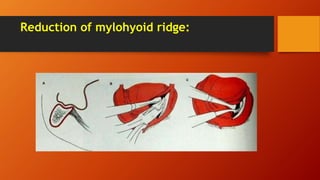
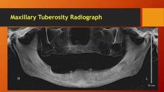
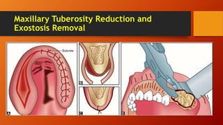
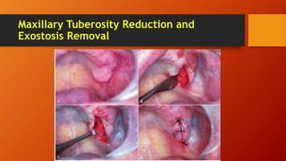
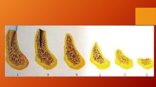
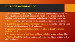
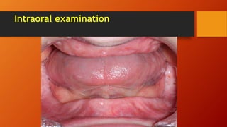
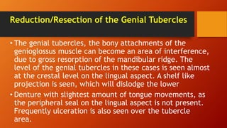
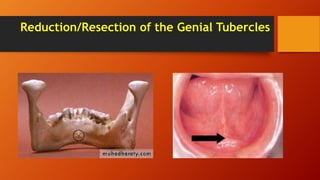
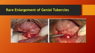
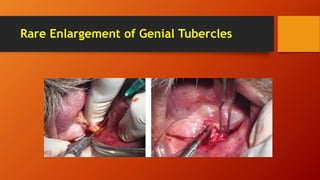
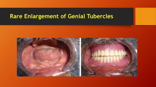
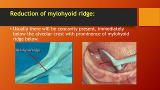
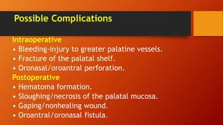
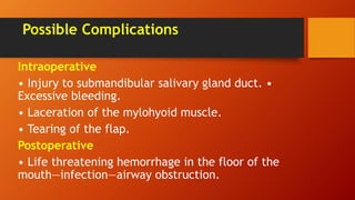
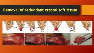
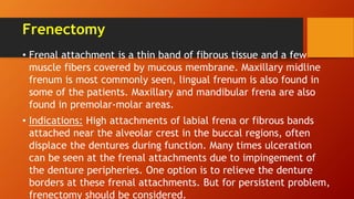
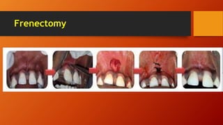
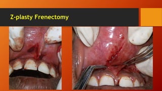
Preprosthetic Surgery
Dr. Haydar Munir Salih Alnamer
BDS, PhD (BOARD CERTIFIED)
pathophysiology of edentulous Bone loss
• Complete denture retention and stability is dependent on the
well adapted fit, hydrostatic pressure and physical features of
the alveolar process such as contour and height. Over the years,
the progressive bone loss, which is seen in the individuals, can
negatively impact prosthetic stability, retention and
serviceability.
• Causes of Bone Loss
• Physiologic
• Environmental
• Pathologic or
• Combination of above causes.
Metabolic factors
• Aging: Continuous resorption of the alveolar ridges,
after teeth extractions over the years.
• Trauma: Bone loss secondary to trauma (during
extraction or otherwise)
• Periodontal disease: Generalized bone loss is seen due
to extensive periodontal problems.
• Long term denture usage: It is a known fact, that
tension forces result in bone opposition and
compressive forces result in bone resorption
Patterns of Bone Loss
• Most of the bone loss occurs in the first year of denture
wearing and it is ten times greater, than the loss seen in the
following years
• Four times more bone loss in the mandible, than in the
maxilla over the years (maxilla distributes the compressive
forces over a wider surface area).
• Extractions of teeth done at different times with long-time
gaps will exhibit irregular bony ridge pattern.
Aims of preprosthetic surgery
1. Provide adequate bony tissue support for the placement of RPD/CD—
removable partial denture or complete denture (optimum ridge, height and
width and contour)
2. Provide adequate soft tissue support. Optimum vestibular depth.
3. Elimination of pre-existing bony deformities, e.g. tori, prominent
mylohyoid ridge, genial tubercle.
4. Correction of maxillary and mandibular ridge relationship.
5. Elimination of pre-existing soft tissue deformities, e.g. epulis, flabby
ridges, hyperplastic tissues.
6. Relocation of frenal/muscle attachments.
7. Relocation of mental nerve.
8. Establishment of correct vestibular depth.
Intraoral examination
Examination of the alveolar ridges, both maxillary and mandibular should be
carried out along with the soft tissue examination of the entire oral cavity
including posterior pharynx. Inspection and palpation should be carried out.
1. Ridge form should be inspected for the amount and contour of the bone.
2. Quality and quantity of the overlying soft tissues of the denture bearing areas
to be ascertained (vestibular depth and area).
3. Location of frenal/muscle attachments in relation to the alveolar crest
should be noted.
4. Presence or absence of soft tissue and bony pathology should be looked for.
5. Relationship of the maxillary alveolar arch to the mandibular alveolar arch in
all three planes.
Intraoral examination
Radiological evaluation
• Radiological assessment should include orthopantograph or
panoramic and lateral cephalometric radiographs.
• In difficult cases, advanced imaging techniques such as
computed tomography— dental CT scan can be used. 3D CT scan
be used, if cost permits.
• The radiographs should be studied to detect any presence of
bony pathological lesions, presence of impacted teeth, cysts,
tumors, root pieces, etc. Bony trabecular pattern, distance of
the neurovascular bundle from the alveolar crest, level of
mental foramen, the size and pneumatization of the maxillary
sinus can be also scrutinized from the X-rays.
Radiological evaluation
Diagnostic model
Alveoloplasty
• Alveoloplasty refers to surgical recontouring of the
alveolar process. This contouring is done with the
purpose to take care of bony projections, sharp crestal
bone or undercuts. Primary alveoloplasty is always
done at the time of multiple extraction or single
extraction. Conservation is the key factor in this
procedure.
• After simple extraction, digital compression of the
alveolar cortices is done immediately. This procedure
is referred as simple alveoloplasty
Alveoloplasty
Simple Conservative Alveoloplasty with
Multiple Extractions
If multiple adjacent teeth are to be extracted in a
single sitting, the simple alveoloplasty technique is
advocated, if there are no other bony irregularities
present. Immediately after extractions, the buccal and
lingual plates should be compressed with firm digital
pressure and the gingival tissue is repositioned and the
entire ridge is palpated for locating sharp bony spicules
or undercuts. These should be trimmed with rongeur
and the edges smoothened with bone file
Alveoloplasty
Alveoloplasty after the Post extraction
Healing
• Many times, multiple extractions are carried out at different
times, which results in the irregular ridge. The ridge may be
knife edged or multiple areas will show sharp bony spicules,
which are very painful to touch. Here, crestal incision is taken
and mucoperiosteal flap is reflected judiciously. Care is taken
not to tear the flap, as at the sharp points, the separation
becomes difficult. The side ways separation with periosteal
elevator will help the smooth reflection. Sometimes releasing
incisions may be needed. The sharp areas or large undercuts
should be trimmed with rongeur or round bur and suturing
done.
Alveoloplasty after the Post extraction Healing &
Elimination of Unfavorable Undercuts
Reduction/Resection of the Genial Tubercles
• The genial tubercles, the bony attachments of the
genioglossus muscle can become an area of interference,
due to gross resorption of the mandibular ridge. The
level of the genial tubercles in these cases is seen almost
at the crestal level on the lingual aspect. A shelf like
projection is seen, which will dislodge the lower
• Denture with slightest amount of tongue movements, as
the peripheral seal on the lingual aspect is not present.
Frequently ulceration is also seen over the tubercle
area.
Reduction/Resection of the Genial Tubercles
Rare Enlargement of Genial Tubercles
Reduction of mylohyoid ridge:
• Usually there will be concavity present, immediately
below the alveolar crest with prominence of mylohyoid
ridge below.
Reduction of mylohyoid ridge:
Excision of Tori
In maxilla: Usually seen in midline of the palate.
Incidence:20 percent in females, 10 percent in
male patients. Multiple shapes and configurations
can be seen. It will vary from single smooth
elevation to multiloculated pedunculated bony
masses.
Excision of Tori
In mandible: Found in 8 percent of population, with
same incidence in male and females. Usually bilateral
tori in the premolar region on the lingual aspect are
seen. May be single or multiple or multilobulated. When
the teeth are present, tori are of no consequence and
usually there are no speech problems associated with
them. But after extraction, the upper denture
construction or lower partial/full denture construction
is not possible, unless tori are excised.
Indications for Reduction/Removal/Excision
of Tori
1. An extremely large torus, filling the palatal vault.
2. A large torus, that may extend beyond the post-dam area.
3. Ulceration/traumatization/hyperkeratinization of the
overlying mucosa.
4. Deep bony undercuts.
5.Interference with the function—speech-deglutition.
6. Psychological consideration—malignancy/cancer phobia.
7. Food lodgement under the folds and projection of the tori.
Torus palatino
Torus palatino removal
Removal of large tori by Division of the
torus into multiple segments should be
done with the bur (vertical and horizontal
multiple cuts).
Torus palatino removal
Removal of small tori
Torus palatino removal
Possible Complications
Intraoperative
• Bleeding-injury to greater palatine vessels.
• Fracture of the palatal shelf.
• Oronasal/oroantral perforation.
Postoperative
• Hematoma formation.
• Sloughing/necrosis of the palatal mucosa.
• Gaping/nonhealing wound.
• Oroantral/oronasal fistula.
Torus mandibularis
Torus mandibularis Removal
Possible Complications
Intraoperative
• Injury to submandibular salivary gland duct. •
Excessive bleeding.
• Laceration of the mylohyoid muscle.
• Tearing of the flap.
Postoperative
• Life threatening hemorrhage in the floor of the
mouth—infection—airway obstruction.
Maxillary Tuberosity Reduction and Exostosis
Removal
Excess horizontal and vertical bony/soft tissue in the maxillary
tuberosity region may interfere with denture construction.
• Excessive bony undercut/mobile or redundant soft
tissue/hyperplastic fibrous tissue may be present (or both).
• The excess tissue protrudes into the intermaxillary space to a
degree that, there is insufficient space for lower dentures.
Radiographs are often necessary—OPG or intraoral periapical view:
• To determine the extent of the bone and soft tissue in the
enlarged tuberosity region.
• To locate the level of the floor of the maxillary sinus.
Maxillary Tuberosity Radiograph
Maxillary Tuberosity Reduction and
Exostosis Removal
Maxillary Tuberosity Reduction and
Exostosis Removal
Soft Tissue Surgeries for the Correction of
Alveolar Ridge: Removal of redundant crestal
soft tissue
Presence of fibrous, hyperplastic tissue often gives
rise to flabby, wobbly ridge form. Dense,
fibromatous or the softer and redundant type of
tissue results in an unstable base for dentures.
• In maxilla—enlarged tuberosity.
• In mandible—enlarged retromolar pad.
Removal of redundant crestal soft tissue
Denture granuloma or hyperplasia:
• It is seen in the palatal region or at the vestibular depth,
obliterating the sulcus or sometimes on lingual aspect of the
lower dentures. The tissue is inflamed, fibrous and
hyperplastic.
• epulis fissurata: These are the benign, pedunculated lesions
present as excessive or redundant tissue of the vestibule,
frequently associated with over extension of the denture
border or ill-fitting dentures
• Palatal papillary hyperplasia: It is due to chronic denture
irritation, under an ill-fitting dentures.
Denture granuloma or hyperplasia & epulis
fissurata
Treatment
Frenectomy
• Frenal attachment is a thin band of fibrous tissue and a few
muscle fibers covered by mucous membrane. Maxillary midline
frenum is most commonly seen, lingual frenum is also found in
some of the patients. Maxillary and mandibular frena are also
found in premolar-molar areas.
• Indications: High attachments of labial frena or fibrous bands
attached near the alveolar crest in the buccal regions, often
displace the dentures during function. Many times ulceration
can be seen at the frenal attachments due to impingement of
the denture peripheries. One option is to relieve the denture
borders at these frenal attachments. But for persistent problem,
frenectomy should be considered.
Frenectomy
Z-plasty Frenectomy
Z-plasty Frenectomy
The Z plasty procedure can be used, when the frenum is broad and the vestibule is short.
These type of procedures can be used for eliminating the frenum, as well as for deepening
the vestibule (some amount of vertical lengthening can be obtained). It also lessens the
tension of the scar band.
Lingual Frenectomy
Lingual frenum is attached to the crest of the alveolar
ridge and it connects to the tongue, below the tip of the
tongue in edentulous patient. In dentulous patient, it is
attached to the lingual gingiva, behind the mandibular
incisors. This condition is also known as tongue tie or
ankyloglossia.
Aim of Surgery
• To correct speech
• Prior to denture construction
• To improve the tongue mobility
Lingual Frenectomy
Ridge extension procedure (Vestibuloplasty)
• Whenever there is an inadequate vestibular depth
present, (due to mandibular atrophy and high muscle and
soft tissue attachments) to increase the retention and
stability of the denture, deepening of the vestibule is
considered
• Vestibuloplasty can be done in the maxilla or in the
mandible or in both the jaws.
• Mandibular techniques are further divided into two
categories:
• 1. Those done on the labial side
• 2. Those done on the lingual side.
labial vestibular procedures
• The procedure will be known as transpositional flap
vestibuloplasty or lip switch procedure, when the soft
tissues from the inner aspect of the lip is shifted to a
favorable zone on the alveolar bone, so that the increase in
the denture bearing area is achieved.
• This method effectively increases the vestibular depth in
the mandible, when the patient has a bone height of 15
mm or more in the anterior region. Implants or bone grafts
should be considered in the patients having less than 15
mm of bone height in the anterior region. The mucosa must
be healthy and exhibit no fibrosis, scarring or hyperplasia.
lip switch procedure
Submucosal Vestibuloplasty Technique
Pre prosthetic surgery

Pre prosthetic surgery

  • 1.
    Preprosthetic Surgery Dr. HaydarMunir Salih Alnamer BDS, PhD (BOARD CERTIFIED)
  • 2.
    pathophysiology of edentulousBone loss • Complete denture retention and stability is dependent on the well adapted fit, hydrostatic pressure and physical features of the alveolar process such as contour and height. Over the years, the progressive bone loss, which is seen in the individuals, can negatively impact prosthetic stability, retention and serviceability. • Causes of Bone Loss • Physiologic • Environmental • Pathologic or • Combination of above causes.
  • 4.
    Metabolic factors • Aging:Continuous resorption of the alveolar ridges, after teeth extractions over the years. • Trauma: Bone loss secondary to trauma (during extraction or otherwise) • Periodontal disease: Generalized bone loss is seen due to extensive periodontal problems. • Long term denture usage: It is a known fact, that tension forces result in bone opposition and compressive forces result in bone resorption
  • 5.
    Patterns of BoneLoss • Most of the bone loss occurs in the first year of denture wearing and it is ten times greater, than the loss seen in the following years • Four times more bone loss in the mandible, than in the maxilla over the years (maxilla distributes the compressive forces over a wider surface area). • Extractions of teeth done at different times with long-time gaps will exhibit irregular bony ridge pattern.
  • 6.
    Aims of preprostheticsurgery 1. Provide adequate bony tissue support for the placement of RPD/CD— removable partial denture or complete denture (optimum ridge, height and width and contour) 2. Provide adequate soft tissue support. Optimum vestibular depth. 3. Elimination of pre-existing bony deformities, e.g. tori, prominent mylohyoid ridge, genial tubercle. 4. Correction of maxillary and mandibular ridge relationship. 5. Elimination of pre-existing soft tissue deformities, e.g. epulis, flabby ridges, hyperplastic tissues. 6. Relocation of frenal/muscle attachments. 7. Relocation of mental nerve. 8. Establishment of correct vestibular depth.
  • 7.
    Intraoral examination Examination ofthe alveolar ridges, both maxillary and mandibular should be carried out along with the soft tissue examination of the entire oral cavity including posterior pharynx. Inspection and palpation should be carried out. 1. Ridge form should be inspected for the amount and contour of the bone. 2. Quality and quantity of the overlying soft tissues of the denture bearing areas to be ascertained (vestibular depth and area). 3. Location of frenal/muscle attachments in relation to the alveolar crest should be noted. 4. Presence or absence of soft tissue and bony pathology should be looked for. 5. Relationship of the maxillary alveolar arch to the mandibular alveolar arch in all three planes.
  • 8.
  • 9.
    Radiological evaluation • Radiologicalassessment should include orthopantograph or panoramic and lateral cephalometric radiographs. • In difficult cases, advanced imaging techniques such as computed tomography— dental CT scan can be used. 3D CT scan be used, if cost permits. • The radiographs should be studied to detect any presence of bony pathological lesions, presence of impacted teeth, cysts, tumors, root pieces, etc. Bony trabecular pattern, distance of the neurovascular bundle from the alveolar crest, level of mental foramen, the size and pneumatization of the maxillary sinus can be also scrutinized from the X-rays.
  • 10.
  • 11.
  • 12.
    Alveoloplasty • Alveoloplasty refersto surgical recontouring of the alveolar process. This contouring is done with the purpose to take care of bony projections, sharp crestal bone or undercuts. Primary alveoloplasty is always done at the time of multiple extraction or single extraction. Conservation is the key factor in this procedure. • After simple extraction, digital compression of the alveolar cortices is done immediately. This procedure is referred as simple alveoloplasty
  • 13.
  • 14.
    Simple Conservative Alveoloplastywith Multiple Extractions If multiple adjacent teeth are to be extracted in a single sitting, the simple alveoloplasty technique is advocated, if there are no other bony irregularities present. Immediately after extractions, the buccal and lingual plates should be compressed with firm digital pressure and the gingival tissue is repositioned and the entire ridge is palpated for locating sharp bony spicules or undercuts. These should be trimmed with rongeur and the edges smoothened with bone file
  • 15.
  • 16.
    Alveoloplasty after thePost extraction Healing • Many times, multiple extractions are carried out at different times, which results in the irregular ridge. The ridge may be knife edged or multiple areas will show sharp bony spicules, which are very painful to touch. Here, crestal incision is taken and mucoperiosteal flap is reflected judiciously. Care is taken not to tear the flap, as at the sharp points, the separation becomes difficult. The side ways separation with periosteal elevator will help the smooth reflection. Sometimes releasing incisions may be needed. The sharp areas or large undercuts should be trimmed with rongeur or round bur and suturing done.
  • 17.
    Alveoloplasty after thePost extraction Healing & Elimination of Unfavorable Undercuts
  • 18.
    Reduction/Resection of theGenial Tubercles • The genial tubercles, the bony attachments of the genioglossus muscle can become an area of interference, due to gross resorption of the mandibular ridge. The level of the genial tubercles in these cases is seen almost at the crestal level on the lingual aspect. A shelf like projection is seen, which will dislodge the lower • Denture with slightest amount of tongue movements, as the peripheral seal on the lingual aspect is not present. Frequently ulceration is also seen over the tubercle area.
  • 19.
  • 20.
    Rare Enlargement ofGenial Tubercles
  • 21.
    Rare Enlargement ofGenial Tubercles
  • 22.
    Rare Enlargement ofGenial Tubercles
  • 23.
    Rare Enlargement ofGenial Tubercles
  • 24.
    Reduction of mylohyoidridge: • Usually there will be concavity present, immediately below the alveolar crest with prominence of mylohyoid ridge below.
  • 25.
  • 26.
    Excision of Tori Inmaxilla: Usually seen in midline of the palate. Incidence:20 percent in females, 10 percent in male patients. Multiple shapes and configurations can be seen. It will vary from single smooth elevation to multiloculated pedunculated bony masses.
  • 27.
    Excision of Tori Inmandible: Found in 8 percent of population, with same incidence in male and females. Usually bilateral tori in the premolar region on the lingual aspect are seen. May be single or multiple or multilobulated. When the teeth are present, tori are of no consequence and usually there are no speech problems associated with them. But after extraction, the upper denture construction or lower partial/full denture construction is not possible, unless tori are excised.
  • 28.
    Indications for Reduction/Removal/Excision ofTori 1. An extremely large torus, filling the palatal vault. 2. A large torus, that may extend beyond the post-dam area. 3. Ulceration/traumatization/hyperkeratinization of the overlying mucosa. 4. Deep bony undercuts. 5.Interference with the function—speech-deglutition. 6. Psychological consideration—malignancy/cancer phobia. 7. Food lodgement under the folds and projection of the tori.
  • 29.
  • 30.
    Torus palatino removal Removalof large tori by Division of the torus into multiple segments should be done with the bur (vertical and horizontal multiple cuts).
  • 31.
  • 32.
  • 33.
    Possible Complications Intraoperative • Bleeding-injuryto greater palatine vessels. • Fracture of the palatal shelf. • Oronasal/oroantral perforation. Postoperative • Hematoma formation. • Sloughing/necrosis of the palatal mucosa. • Gaping/nonhealing wound. • Oroantral/oronasal fistula.
  • 34.
  • 35.
  • 36.
  • 37.
    Possible Complications Intraoperative • Injuryto submandibular salivary gland duct. • Excessive bleeding. • Laceration of the mylohyoid muscle. • Tearing of the flap. Postoperative • Life threatening hemorrhage in the floor of the mouth—infection—airway obstruction.
  • 38.
    Maxillary Tuberosity Reductionand Exostosis Removal Excess horizontal and vertical bony/soft tissue in the maxillary tuberosity region may interfere with denture construction. • Excessive bony undercut/mobile or redundant soft tissue/hyperplastic fibrous tissue may be present (or both). • The excess tissue protrudes into the intermaxillary space to a degree that, there is insufficient space for lower dentures. Radiographs are often necessary—OPG or intraoral periapical view: • To determine the extent of the bone and soft tissue in the enlarged tuberosity region. • To locate the level of the floor of the maxillary sinus.
  • 39.
  • 40.
    Maxillary Tuberosity Reductionand Exostosis Removal
  • 41.
    Maxillary Tuberosity Reductionand Exostosis Removal
  • 42.
    Soft Tissue Surgeriesfor the Correction of Alveolar Ridge: Removal of redundant crestal soft tissue Presence of fibrous, hyperplastic tissue often gives rise to flabby, wobbly ridge form. Dense, fibromatous or the softer and redundant type of tissue results in an unstable base for dentures. • In maxilla—enlarged tuberosity. • In mandible—enlarged retromolar pad.
  • 43.
    Removal of redundantcrestal soft tissue
  • 44.
    Denture granuloma orhyperplasia: • It is seen in the palatal region or at the vestibular depth, obliterating the sulcus or sometimes on lingual aspect of the lower dentures. The tissue is inflamed, fibrous and hyperplastic. • epulis fissurata: These are the benign, pedunculated lesions present as excessive or redundant tissue of the vestibule, frequently associated with over extension of the denture border or ill-fitting dentures • Palatal papillary hyperplasia: It is due to chronic denture irritation, under an ill-fitting dentures.
  • 45.
    Denture granuloma orhyperplasia & epulis fissurata
  • 46.
  • 47.
    Frenectomy • Frenal attachmentis a thin band of fibrous tissue and a few muscle fibers covered by mucous membrane. Maxillary midline frenum is most commonly seen, lingual frenum is also found in some of the patients. Maxillary and mandibular frena are also found in premolar-molar areas. • Indications: High attachments of labial frena or fibrous bands attached near the alveolar crest in the buccal regions, often displace the dentures during function. Many times ulceration can be seen at the frenal attachments due to impingement of the denture peripheries. One option is to relieve the denture borders at these frenal attachments. But for persistent problem, frenectomy should be considered.
  • 48.
  • 49.
  • 50.
    Z-plasty Frenectomy The Zplasty procedure can be used, when the frenum is broad and the vestibule is short. These type of procedures can be used for eliminating the frenum, as well as for deepening the vestibule (some amount of vertical lengthening can be obtained). It also lessens the tension of the scar band.
  • 51.
    Lingual Frenectomy Lingual frenumis attached to the crest of the alveolar ridge and it connects to the tongue, below the tip of the tongue in edentulous patient. In dentulous patient, it is attached to the lingual gingiva, behind the mandibular incisors. This condition is also known as tongue tie or ankyloglossia. Aim of Surgery • To correct speech • Prior to denture construction • To improve the tongue mobility
  • 52.
  • 53.
    Ridge extension procedure(Vestibuloplasty) • Whenever there is an inadequate vestibular depth present, (due to mandibular atrophy and high muscle and soft tissue attachments) to increase the retention and stability of the denture, deepening of the vestibule is considered • Vestibuloplasty can be done in the maxilla or in the mandible or in both the jaws. • Mandibular techniques are further divided into two categories: • 1. Those done on the labial side • 2. Those done on the lingual side.
  • 54.
    labial vestibular procedures •The procedure will be known as transpositional flap vestibuloplasty or lip switch procedure, when the soft tissues from the inner aspect of the lip is shifted to a favorable zone on the alveolar bone, so that the increase in the denture bearing area is achieved. • This method effectively increases the vestibular depth in the mandible, when the patient has a bone height of 15 mm or more in the anterior region. Implants or bone grafts should be considered in the patients having less than 15 mm of bone height in the anterior region. The mucosa must be healthy and exhibit no fibrosis, scarring or hyperplasia.
  • 55.
  • 56.
  • 57.