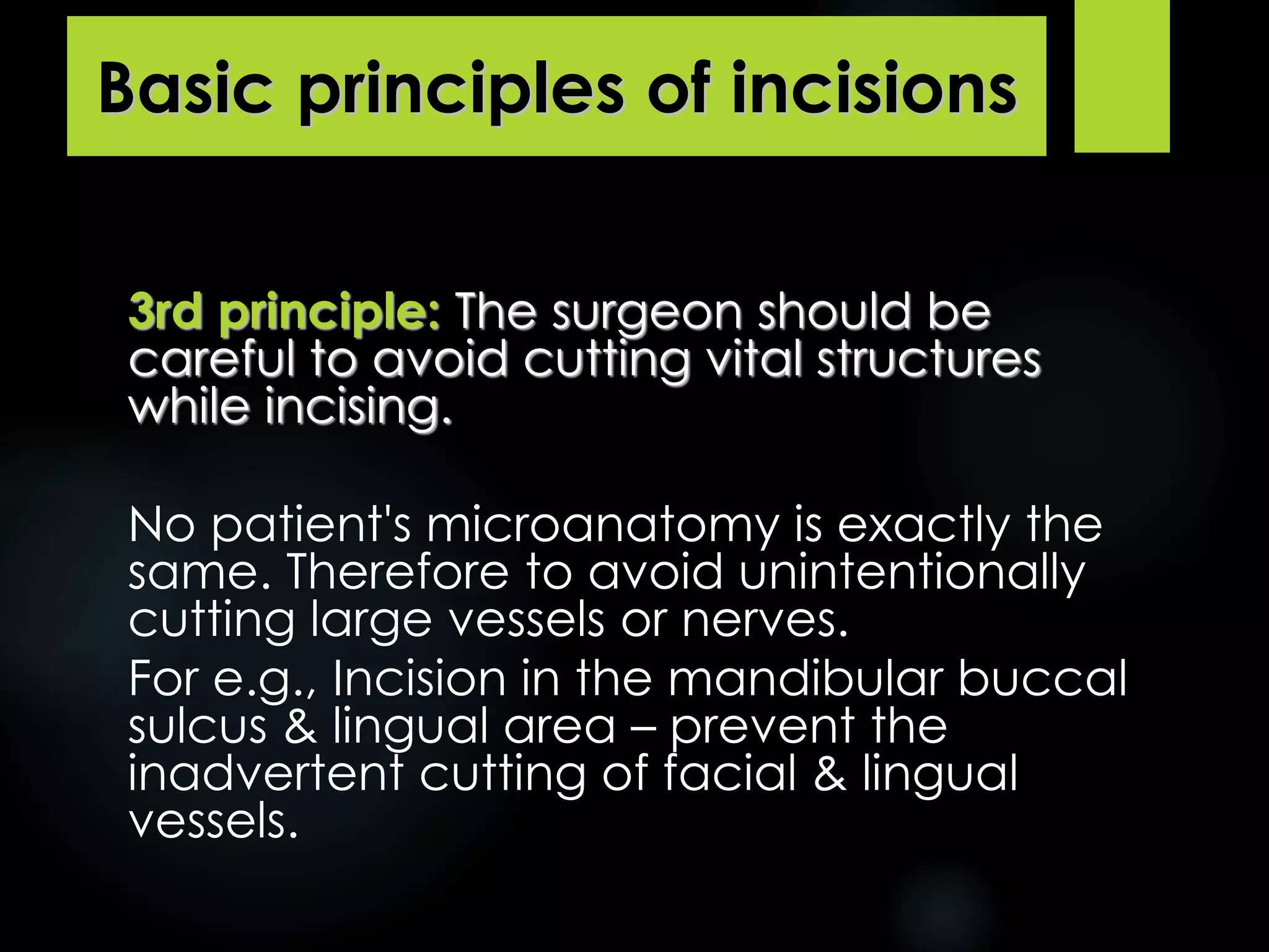
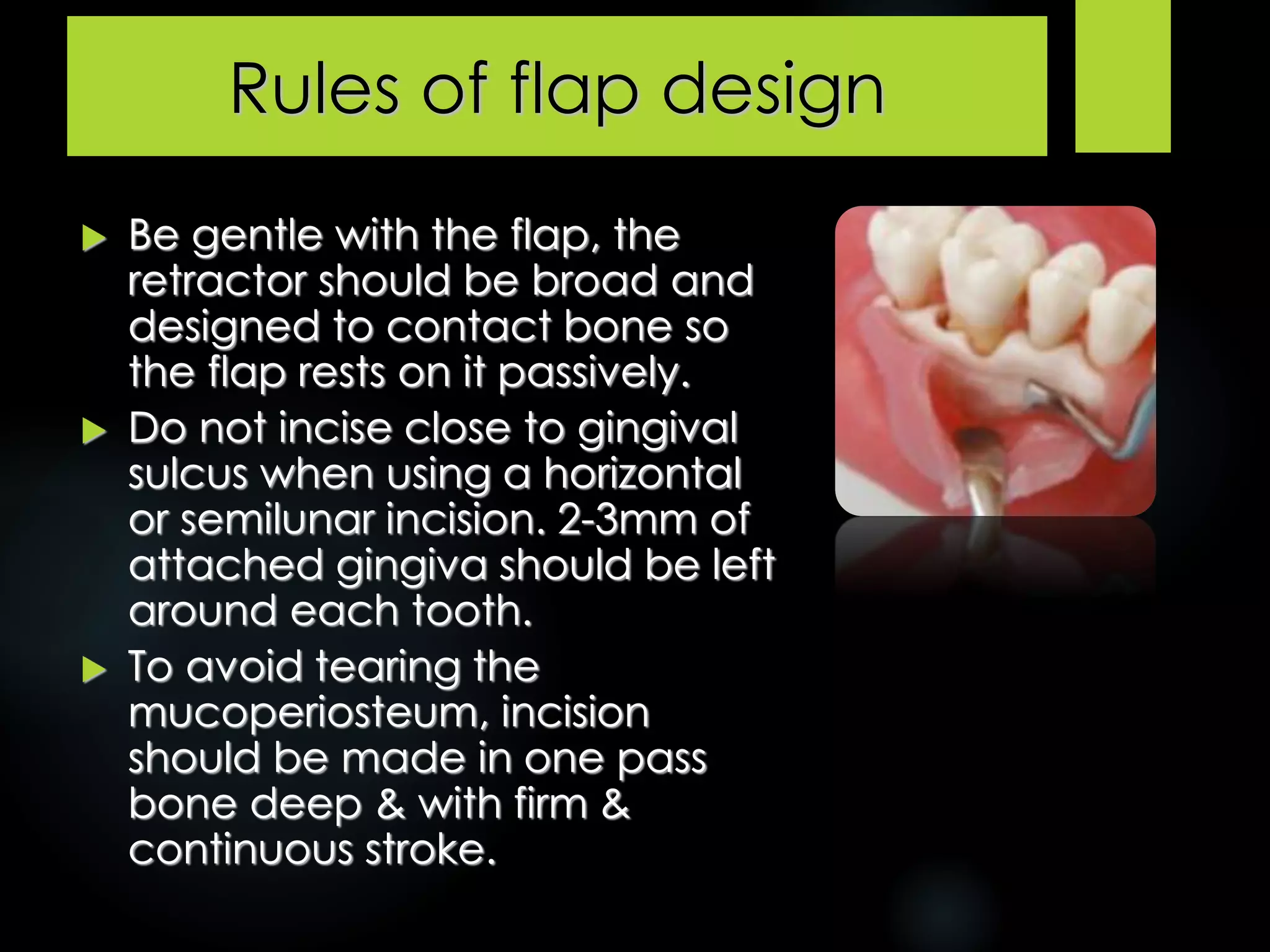
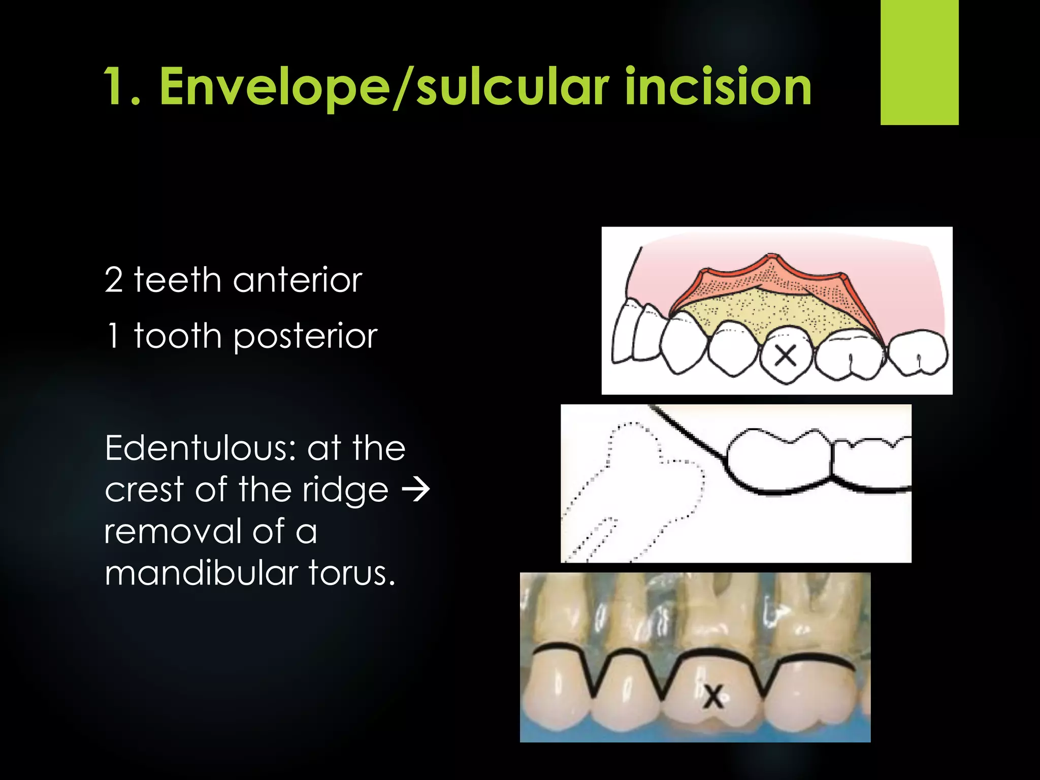
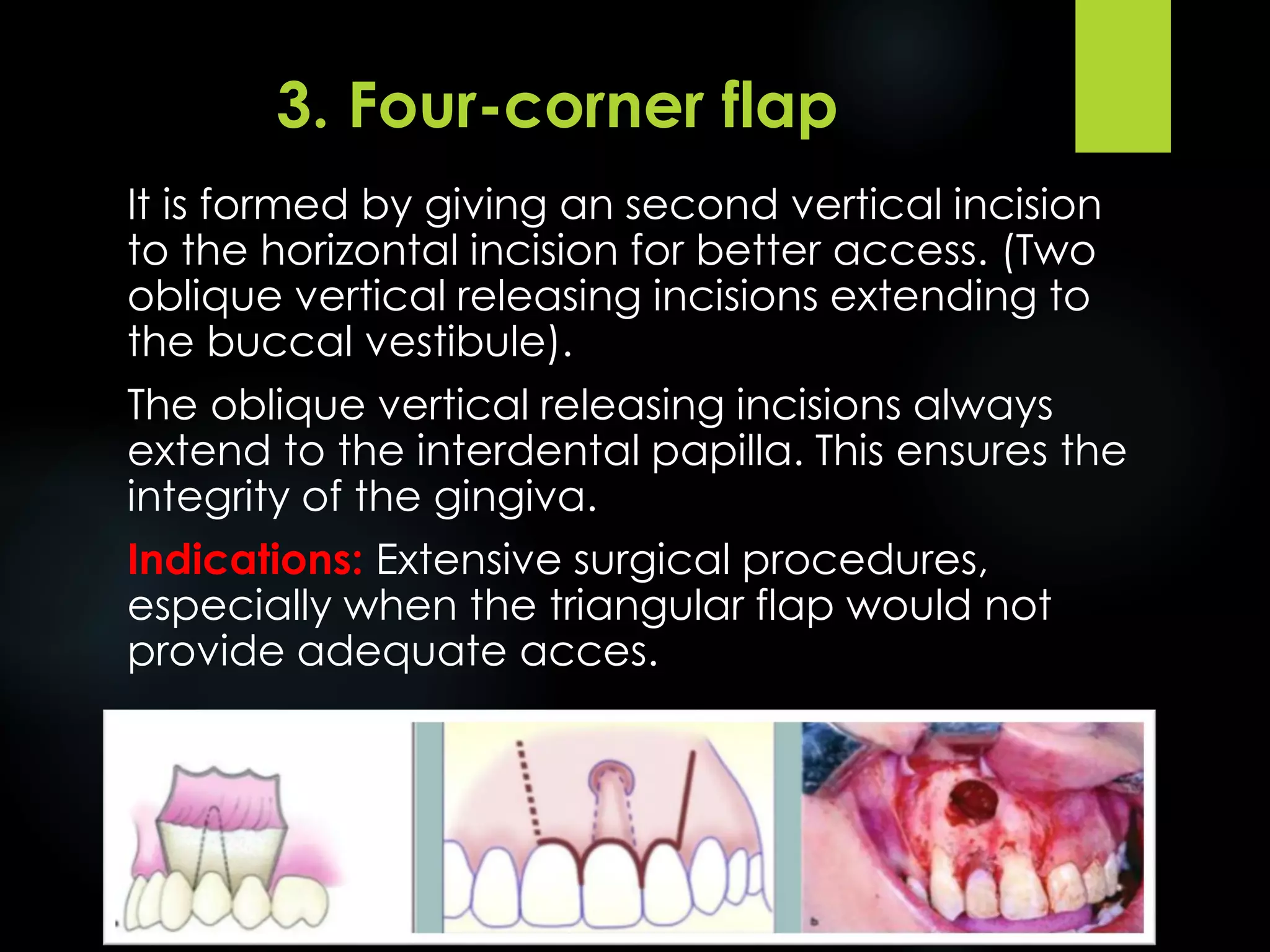
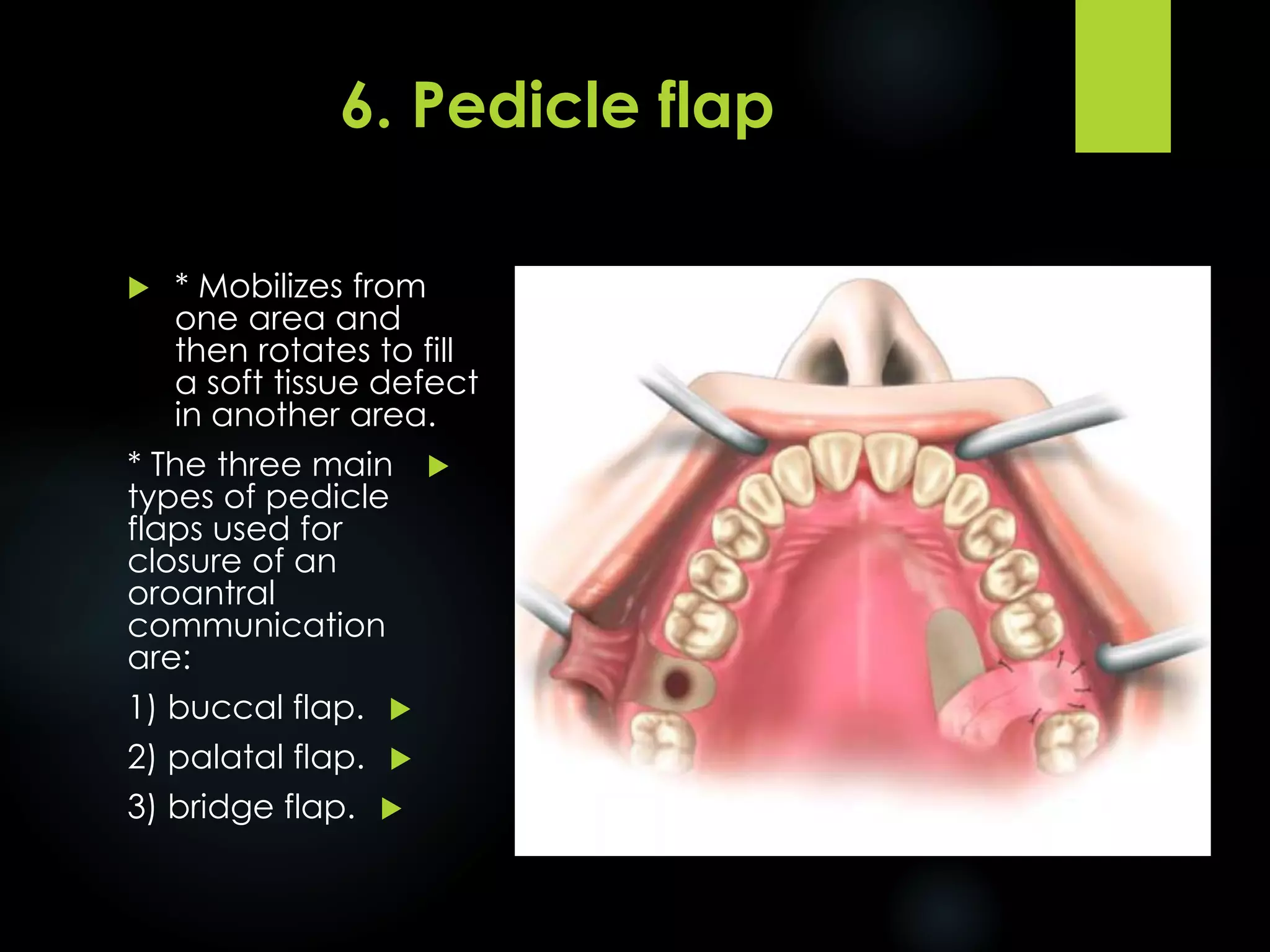
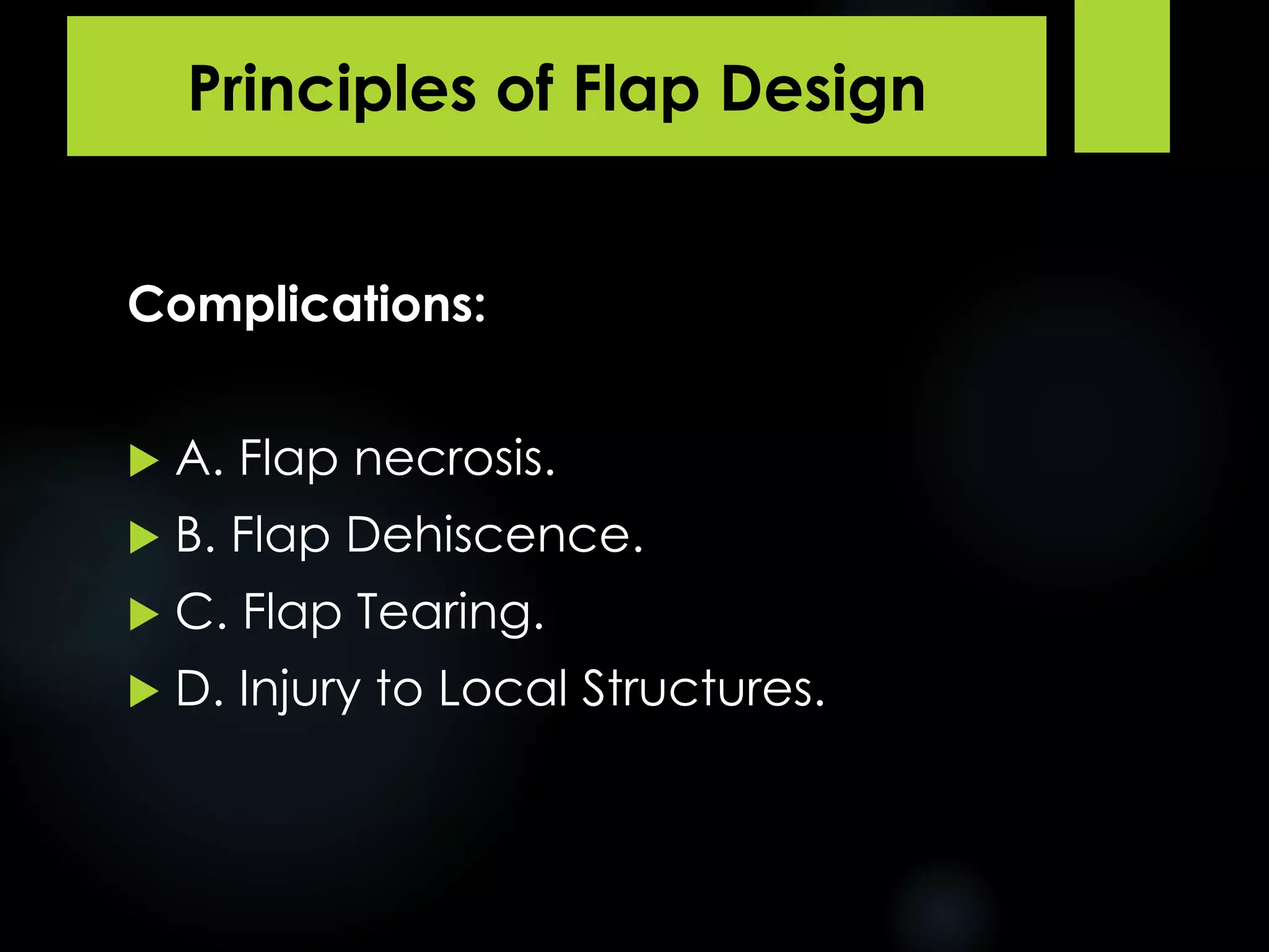
Assist. Prof. Dr. Kareem M Alghanim
BDS , MSc (oral surg.) , PhD (oral med.)
Flap Design for Minor
Oral Surgery
4th Class Oral Surgery Lec.3
INCISIONS
An incision can be described as
a sharp wound produced by a
surgical scalpel.
Basic principles of incisions
1st principle: A sharp blade of
the proper size should be used.
Bone & ligamental tissues dull
blades more rapidly than dose
buccal mucosa.
Basic principles of incisions
2nd principle: is that a firm, continuous
stroke should be used when incising.
Long continuous strokes are
preferable to short interrupted ones.
Mucoperiosteal incision should be firm
that penetrates the mucosa &
periosteium with same stroke.
Basic principles of incisions
3rd principle: The surgeon should be
careful to avoid cutting vital structures
while incising.
No patient's microanatomy is exactly the
same. Therefore to avoid unintentionally
cutting large vessels or nerves.
For e.g., Incision in the mandibular buccal
sulcus & lingual area – prevent the
inadvertent cutting of facial & lingual
vessels.
Basic principles of incisions
4th principle: Incisions through
epithelial surfaces should be
made with blade held
perpendicular to the epithelial
surface.
Basic principles of incisions
5th principle: Incisions in
the oral cavity should be
properly placed.
E.g., Over healthy bone,
wound edges should be at
least 6-8mm away from
the defect.
Incision should lie at the
line angles of the teeth
and not at the facial
surfaces nor in the papilla.
Pen-grip holding of scalpel and incising by moving
hand and wrist not moving the entire forearm
INSTRUMENTR INCISING TISSUE
SCALPEL
1) Handle:-
 No.3, No.7
 Differently shaped.
INSTRUMENTR INCISING TISSUE
SCALPEL
2) Disposable, sterile sharp blade;
 1. No.15 blade:
Is most commonly used for oral surgery.
Relatively small.
Around teeth through mucoperiosteal.
INSTRUMENTR INCISING TISSUE
 2. No.10 blade:
Similar to No.15.
Large skin incisions.
 3. No.11 blade:
Sharp, pointed.
Small stab incisions.
Incising an abscess.
 4. No.12 blade:
Hooked.
Mucogingival procedures.
Posterior aspect of teeth
maxillary tuberosity.
Blade Loaded & Removed
Blade
Loaded
Blade
Removed
Remember..
• Pen Grasp: Allow maximal control
• Hold mobile tissue firmly
• Press down firmlye
• Single- patient use: dulled easily
• Several incisions: single operation- 2nd
blade
• Dull blades: no clean sharp incisons
FLAPS
Definition
Flap is a small incision made in mucosa &
periosteum under local anesthesia to gain
access to the area for raising the
mucoperiosteum to perform dentoalveolar
surgeries.
Rules of flap design
 Avoid severing large vessels
& nerves.
 Place margins far away
from surgical areas to
ensure wound margins over
sound bone, this also gives
room for any adjustments
and avoids collapse of flap
into the bony defect.
 Design the flap for
adequate visibility without
over exposure of bone.
Rules of flap design
 Base of flap should be the
widest portion.
 There should be no sharp
angles on the flap, sharp
corners tend to slough
due to poor circulation.
 Vertical or Oblique
incisions should not be
made over root
eminence, it is best to
incise in through between
adjacent teeth.
Rules of flap design
 Maintain integrity of
interdental papilla, the
papilla at incision line is
allowed to remain
whereas other papilla
should be included in
flap.
Rules of flap design
 Be gentle with the flap, the
retractor should be broad and
designed to contact bone so
the flap rests on it passively.
 Do not incise close to gingival
sulcus when using a horizontal
or semilunar incision. 2-3mm of
attached gingiva should be left
around each tooth.
 To avoid tearing the
mucoperiosteum, incision
should be made in one pass
bone deep & with firm &
continuous stroke.
Rules of flap design
 Elevate the flap away from the line of
vision to provide adequate exposure of
the surgical area.
1. Envelope/sulcular incision.
2. Envelope with one releasing incision (three-
corner flap).
3. Envelope with two releasing incisions (four-
corner flap).
4. Semilunar incision.
5. Y-incision.
6. Pedicle flap.
7- Submarginal Flap. Full-thickness
mucoperiosteal
flap
Types of Mucoperiosteal Flaps
Types
of
Mucoperiosteal
Flaps
1. Envelope/sulcular incision.
2. Envelope with one releasing
incision (three-corner flap).
3. Envelope with two releasing
incisions (four-corner flap).
4. Semilunar incision.
5. Y-incision.
6. Pedicle flap.
7- Submarginal Flap.
Types of Mucoperiosteal Flaps
2 teeth anterior
1 tooth posterior
Edentulous: at the
crest of the ridge 
removal of a
mandibular torus.
1. Envelope/sulcular incision
This type of flap is the result of an extended
horizontal incision in the gingival sulcus along the
cervical lines of the teeth.
Indications:
- Surgical procedure involves the cervical lines of the
teeth labial or buccal and palatal or lingual.
- Cases of removal of impacted teeth.
- Apicoectomies (palatal root of molar).
1. Envelope/sulcular incision
Advantages:
- Avoidance of vertical incision.
- Eeasy reapproximation to original position.
Disadvantages:
- Difficult reflection (mainly palatally).
- Great tension with a risk of the ends tearing.
- Limited visualization in apicoectomies.
- Limited access.
- Possibility of injury of palatal vessels and nerves.
- Defect of attached gingiva.
1 tooth anterior
1 tooth posterior
Greater access in an apical
direction, especially in the
posterior aspect of the
mouth.
2. Three-corner flap
This flap is the result of an L-
shaped incision, with a horizontal
incision made along the gingival
sulcus and a vertical or oblique
incision.
 The vertical incision begins
approximately at the
vestibular fold and extends to
the interdental papilla of the
gingiva.
 The triangular flap is
performed labially or buccally
on both jaws.
 Indications: Surgical removal
of root tips, small cysts, and
apicoectomies.
2. Three-corner flap
Advantages:
 - Ensures an adequate blood supply.
 - Satisfactory visualization.
 - Very good stability and reapproximation.
 - Easily modified with a small releasing incision, or an
additional vertical incision, or even lengthening of the
horizontal incision.
Disadvantages:
 - Limited access to long roots.
 - Tension is created when the flap is held with a retractor.
 - It causes a defect in the attached gingiva.
2. Three-corner flap
3. Four-corner flap
(Envelope with two releasing
incisions / Trapezoidal flap / Three
sided flap).
1 tooth anterior
1 tooth posterior
Rarely indicated
It is formed by giving an second vertical incision
to the horizontal incision for better access. (Two
oblique vertical releasing incisions extending to
the buccal vestibule).
The oblique vertical releasing incisions always
extend to the interdental papilla. This ensures the
integrity of the gingiva.
Indications: Extensive surgical procedures,
especially when the triangular flap would not
provide adequate acces.
3. Four-corner flap
Advantages:
Provides excellent access, allows surgery to be
performed on more than one or two teeth,
produces no tension in the tissues, allows easy
reapproximation of the flap to its original position
and hastens the healing process.
Disadvantages:
Produces a defect in the attached gingiva
(recession of gingiva).
3. Four-corner flap
In case of missing teeth and edentulous arches,
horizontal incision is made over the alveolar crest
whereas vertical incision is carried out in the same
way
3. Four-corner flap
* To approach the root apex.
* Avoids trauma to the papillae and gingival margin.
* Useful for periapical surgery of a limited extent.
* Should not cross major prominences, ex: canine
eminence.
4. Semilunar incision
 This flap is the result of a curved incision, which begins
from the vestibular fold and has a bow-shaped course
with the convex part towards the attached gingiva.
 The lowest point of the incision must be at least 0.5 cm
from the gingival margin, so that the blood supply is not
compromised.
 Each end of the incision must extend at least one tooth
over on each side of the area of bone removal.
 Indications: surgeries requiring periapical exposure,
apicoectomies, removal of small cysts and root tips.
4. Semilunar incision
 Advantages:
 Small incision and easy reflection, no recession of
gingivae, no intervention at the periodontium, easier oral
hygiene compared to other types of flaps.
 Disadvantages:
 Possibility of the incision being performed right over the
bone lesion due to miscalculation, scarring mainly in the
anterior area, difficulty of reapproximation and suturing
due to absence of specific reference points, limited
access and visualization, tendency to tear.
4. Semilunar incision
An incision is made along
the midline of the palate,
as well as two
anterolateral incisions,
which are anterior to the
canines
 Indication: in surgical
procedures involving
the removal of a
maxillary palatal torus.
5. Y-incision
 * Mobilizes from
one area and
then rotates to fill
a soft tissue defect
in another area.

* The three main
types of pedicle
flaps used for
closure of an
oroantral
communication
are:

1) buccal flap.

2) palatal flap.

3) bridge flap.
6. Pedicle flap
 This type of flap is
based on and along
a particular blood
vessel like Greater
Palatine Artery in the
palate.
 Indication: Mainly
used for closure
of an oroantral
communication,
Reconstruction
for malignant
defects.
6. Pedicle flap
Pedicle bridge flap, used
for closure of oroantral
communication
 It is a combination of both vertical & semilunar incisions.
The flap is scalloped to follow gingival architecture.
 Indications: For those cases where there is a
fear that elevation of attached gingiva will
lead to shrinkage & exposure of margins of
restoration.
7- Submarginal Flap
Examples
Examples
Complications:
 A. Flap necrosis.
 B. Flap Dehiscence.
 C. Flap Tearing.
 D. Injury to Local Structures.
Principles of Flap Design
A. Flap necrosis
 Base > Free margin
* to preserve an adequate blood supply
* unless a major artery is present in the base.
 Width of Base > Length of Flap
* less critical in oral cavity, but length < width.
* a long, straight incision with adequate flap
reflection heals more rapidly than a short, torn
incision.
 An axial blood supply in the base.
 Hold the flap with a retractor resting on
intact bone to prevent tension.
A. Flap necrosis
B. Flap Dehiscence
 * The incisions must be made over
intact bone.
 * If the pathologic condition has
eroded the buccocortical plate,
the incision must be at least 6 or 8
mm away from it.
 * The incision is 6 to 8 mm away
from the bony defect created by
surgery.
 * Gently handle the flap's edges.
 * Do not place the flap under
tension.
 * Do not cross bony prominences,
ex: canine eminence.
B. Flap Dehiscence
C. Flap Tearing
Envelope flaps: an incision around the necks of several
teeth extends 2 teeth anterior and 1 tooth posterior
 If not provide sufficient access.
Vertical (oblique) releasing incisions:
 Extends 1 tooth anterior and 1 tooth posterior
 Started at the line angle of a tooth
 Carried obliquely apically into the unattached gingiva
 If cross the papilla  localized periodontal problems
C. Flap Tearing
D. Injury to Local Structures
 Mandible: lingual n. & mental n.
D. Injury to Local Structures
 Maxilla: greater palatine a. & nasopalatine
n./a.
Table: Flap Design Considerations
Summary
 Reflection is performed to separate the mucoperiosteal
flap from the underlying bone.
 The elevator is in direct contact with bone and reflection
starts at the incision, usually at an angle, and is
completed with gentle, steady strokes towards the labial
or buccal vestibule, without damaging the tissues. When
the attachment between bone and periosteumis strong
or if symphysis occurs, then scissors or surgical blades
may be used.
Reflection of the
Mucoperiosteum
THANK
YOU

Flap Design for Minor Oral Surgery

  • 1.
    Assist. Prof. Dr.Kareem M Alghanim BDS , MSc (oral surg.) , PhD (oral med.) Flap Design for Minor Oral Surgery 4th Class Oral Surgery Lec.3
  • 3.
    INCISIONS An incision canbe described as a sharp wound produced by a surgical scalpel.
  • 4.
    Basic principles ofincisions 1st principle: A sharp blade of the proper size should be used. Bone & ligamental tissues dull blades more rapidly than dose buccal mucosa.
  • 5.
    Basic principles ofincisions 2nd principle: is that a firm, continuous stroke should be used when incising. Long continuous strokes are preferable to short interrupted ones. Mucoperiosteal incision should be firm that penetrates the mucosa & periosteium with same stroke.
  • 6.
    Basic principles ofincisions 3rd principle: The surgeon should be careful to avoid cutting vital structures while incising. No patient's microanatomy is exactly the same. Therefore to avoid unintentionally cutting large vessels or nerves. For e.g., Incision in the mandibular buccal sulcus & lingual area – prevent the inadvertent cutting of facial & lingual vessels.
  • 7.
    Basic principles ofincisions 4th principle: Incisions through epithelial surfaces should be made with blade held perpendicular to the epithelial surface.
  • 8.
    Basic principles ofincisions 5th principle: Incisions in the oral cavity should be properly placed. E.g., Over healthy bone, wound edges should be at least 6-8mm away from the defect. Incision should lie at the line angles of the teeth and not at the facial surfaces nor in the papilla.
  • 9.
    Pen-grip holding ofscalpel and incising by moving hand and wrist not moving the entire forearm
  • 10.
    INSTRUMENTR INCISING TISSUE SCALPEL 1)Handle:-  No.3, No.7  Differently shaped.
  • 11.
    INSTRUMENTR INCISING TISSUE SCALPEL 2)Disposable, sterile sharp blade;  1. No.15 blade: Is most commonly used for oral surgery. Relatively small. Around teeth through mucoperiosteal.
  • 12.
    INSTRUMENTR INCISING TISSUE 2. No.10 blade: Similar to No.15. Large skin incisions.  3. No.11 blade: Sharp, pointed. Small stab incisions. Incising an abscess.  4. No.12 blade: Hooked. Mucogingival procedures. Posterior aspect of teeth maxillary tuberosity.
  • 13.
    Blade Loaded &Removed Blade Loaded Blade Removed
  • 14.
    Remember.. • Pen Grasp:Allow maximal control • Hold mobile tissue firmly • Press down firmlye • Single- patient use: dulled easily • Several incisions: single operation- 2nd blade • Dull blades: no clean sharp incisons
  • 15.
    FLAPS Definition Flap is asmall incision made in mucosa & periosteum under local anesthesia to gain access to the area for raising the mucoperiosteum to perform dentoalveolar surgeries.
  • 16.
    Rules of flapdesign  Avoid severing large vessels & nerves.  Place margins far away from surgical areas to ensure wound margins over sound bone, this also gives room for any adjustments and avoids collapse of flap into the bony defect.  Design the flap for adequate visibility without over exposure of bone.
  • 17.
    Rules of flapdesign  Base of flap should be the widest portion.  There should be no sharp angles on the flap, sharp corners tend to slough due to poor circulation.  Vertical or Oblique incisions should not be made over root eminence, it is best to incise in through between adjacent teeth.
  • 18.
    Rules of flapdesign  Maintain integrity of interdental papilla, the papilla at incision line is allowed to remain whereas other papilla should be included in flap.
  • 19.
    Rules of flapdesign  Be gentle with the flap, the retractor should be broad and designed to contact bone so the flap rests on it passively.  Do not incise close to gingival sulcus when using a horizontal or semilunar incision. 2-3mm of attached gingiva should be left around each tooth.  To avoid tearing the mucoperiosteum, incision should be made in one pass bone deep & with firm & continuous stroke.
  • 20.
    Rules of flapdesign  Elevate the flap away from the line of vision to provide adequate exposure of the surgical area.
  • 21.
    1. Envelope/sulcular incision. 2.Envelope with one releasing incision (three- corner flap). 3. Envelope with two releasing incisions (four- corner flap). 4. Semilunar incision. 5. Y-incision. 6. Pedicle flap. 7- Submarginal Flap. Full-thickness mucoperiosteal flap Types of Mucoperiosteal Flaps
  • 22.
    Types of Mucoperiosteal Flaps 1. Envelope/sulcular incision. 2.Envelope with one releasing incision (three-corner flap). 3. Envelope with two releasing incisions (four-corner flap). 4. Semilunar incision. 5. Y-incision. 6. Pedicle flap. 7- Submarginal Flap. Types of Mucoperiosteal Flaps
  • 23.
    2 teeth anterior 1tooth posterior Edentulous: at the crest of the ridge  removal of a mandibular torus. 1. Envelope/sulcular incision
  • 24.
    This type offlap is the result of an extended horizontal incision in the gingival sulcus along the cervical lines of the teeth. Indications: - Surgical procedure involves the cervical lines of the teeth labial or buccal and palatal or lingual. - Cases of removal of impacted teeth. - Apicoectomies (palatal root of molar). 1. Envelope/sulcular incision
  • 25.
  • 26.
    1. Envelope/sulcular incision Advantages: -Avoidance of vertical incision. - Eeasy reapproximation to original position. Disadvantages: - Difficult reflection (mainly palatally). - Great tension with a risk of the ends tearing. - Limited visualization in apicoectomies. - Limited access. - Possibility of injury of palatal vessels and nerves. - Defect of attached gingiva.
  • 27.
    1 tooth anterior 1tooth posterior Greater access in an apical direction, especially in the posterior aspect of the mouth. 2. Three-corner flap
  • 28.
    This flap isthe result of an L- shaped incision, with a horizontal incision made along the gingival sulcus and a vertical or oblique incision.  The vertical incision begins approximately at the vestibular fold and extends to the interdental papilla of the gingiva.  The triangular flap is performed labially or buccally on both jaws.  Indications: Surgical removal of root tips, small cysts, and apicoectomies. 2. Three-corner flap
  • 29.
    Advantages:  - Ensuresan adequate blood supply.  - Satisfactory visualization.  - Very good stability and reapproximation.  - Easily modified with a small releasing incision, or an additional vertical incision, or even lengthening of the horizontal incision. Disadvantages:  - Limited access to long roots.  - Tension is created when the flap is held with a retractor.  - It causes a defect in the attached gingiva. 2. Three-corner flap
  • 30.
    3. Four-corner flap (Envelopewith two releasing incisions / Trapezoidal flap / Three sided flap). 1 tooth anterior 1 tooth posterior Rarely indicated
  • 31.
    It is formedby giving an second vertical incision to the horizontal incision for better access. (Two oblique vertical releasing incisions extending to the buccal vestibule). The oblique vertical releasing incisions always extend to the interdental papilla. This ensures the integrity of the gingiva. Indications: Extensive surgical procedures, especially when the triangular flap would not provide adequate acces. 3. Four-corner flap
  • 32.
    Advantages: Provides excellent access,allows surgery to be performed on more than one or two teeth, produces no tension in the tissues, allows easy reapproximation of the flap to its original position and hastens the healing process. Disadvantages: Produces a defect in the attached gingiva (recession of gingiva). 3. Four-corner flap
  • 33.
    In case ofmissing teeth and edentulous arches, horizontal incision is made over the alveolar crest whereas vertical incision is carried out in the same way 3. Four-corner flap
  • 34.
    * To approachthe root apex. * Avoids trauma to the papillae and gingival margin. * Useful for periapical surgery of a limited extent. * Should not cross major prominences, ex: canine eminence. 4. Semilunar incision
  • 35.
     This flapis the result of a curved incision, which begins from the vestibular fold and has a bow-shaped course with the convex part towards the attached gingiva.  The lowest point of the incision must be at least 0.5 cm from the gingival margin, so that the blood supply is not compromised.  Each end of the incision must extend at least one tooth over on each side of the area of bone removal.  Indications: surgeries requiring periapical exposure, apicoectomies, removal of small cysts and root tips. 4. Semilunar incision
  • 36.
     Advantages:  Smallincision and easy reflection, no recession of gingivae, no intervention at the periodontium, easier oral hygiene compared to other types of flaps.  Disadvantages:  Possibility of the incision being performed right over the bone lesion due to miscalculation, scarring mainly in the anterior area, difficulty of reapproximation and suturing due to absence of specific reference points, limited access and visualization, tendency to tear. 4. Semilunar incision
  • 37.
    An incision ismade along the midline of the palate, as well as two anterolateral incisions, which are anterior to the canines  Indication: in surgical procedures involving the removal of a maxillary palatal torus. 5. Y-incision
  • 38.
     * Mobilizesfrom one area and then rotates to fill a soft tissue defect in another area.  * The three main types of pedicle flaps used for closure of an oroantral communication are:  1) buccal flap.  2) palatal flap.  3) bridge flap. 6. Pedicle flap
  • 39.
     This typeof flap is based on and along a particular blood vessel like Greater Palatine Artery in the palate.  Indication: Mainly used for closure of an oroantral communication, Reconstruction for malignant defects. 6. Pedicle flap
  • 40.
    Pedicle bridge flap,used for closure of oroantral communication
  • 41.
     It isa combination of both vertical & semilunar incisions. The flap is scalloped to follow gingival architecture.  Indications: For those cases where there is a fear that elevation of attached gingiva will lead to shrinkage & exposure of margins of restoration. 7- Submarginal Flap
  • 42.
  • 43.
  • 44.
    Complications:  A. Flapnecrosis.  B. Flap Dehiscence.  C. Flap Tearing.  D. Injury to Local Structures. Principles of Flap Design
  • 45.
    A. Flap necrosis Base > Free margin * to preserve an adequate blood supply * unless a major artery is present in the base.  Width of Base > Length of Flap * less critical in oral cavity, but length < width. * a long, straight incision with adequate flap reflection heals more rapidly than a short, torn incision.  An axial blood supply in the base.  Hold the flap with a retractor resting on intact bone to prevent tension.
  • 46.
  • 47.
    B. Flap Dehiscence * The incisions must be made over intact bone.  * If the pathologic condition has eroded the buccocortical plate, the incision must be at least 6 or 8 mm away from it.  * The incision is 6 to 8 mm away from the bony defect created by surgery.  * Gently handle the flap's edges.  * Do not place the flap under tension.  * Do not cross bony prominences, ex: canine eminence.
  • 48.
  • 49.
    C. Flap Tearing Envelopeflaps: an incision around the necks of several teeth extends 2 teeth anterior and 1 tooth posterior  If not provide sufficient access. Vertical (oblique) releasing incisions:  Extends 1 tooth anterior and 1 tooth posterior  Started at the line angle of a tooth  Carried obliquely apically into the unattached gingiva  If cross the papilla  localized periodontal problems
  • 50.
  • 51.
    D. Injury toLocal Structures  Mandible: lingual n. & mental n.
  • 52.
    D. Injury toLocal Structures  Maxilla: greater palatine a. & nasopalatine n./a.
  • 53.
    Table: Flap DesignConsiderations Summary
  • 54.
     Reflection isperformed to separate the mucoperiosteal flap from the underlying bone.  The elevator is in direct contact with bone and reflection starts at the incision, usually at an angle, and is completed with gentle, steady strokes towards the labial or buccal vestibule, without damaging the tissues. When the attachment between bone and periosteumis strong or if symphysis occurs, then scissors or surgical blades may be used. Reflection of the Mucoperiosteum
  • 55.