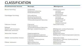
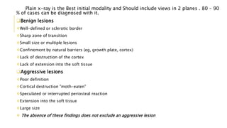
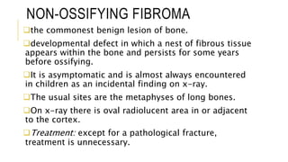
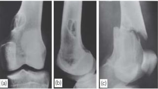
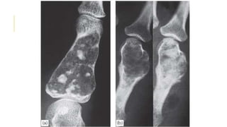
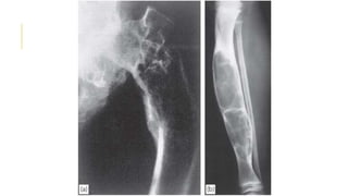
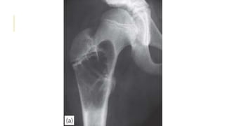
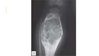
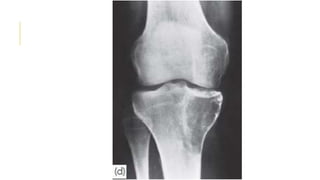
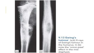
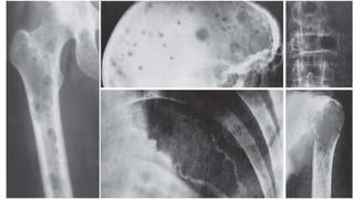
COMMON BONE TUMORS By Ahmed Ayad Lafta
Bone tumors develop when cells
in the bone divide without control,
forming a mass of tissue. Most bone
tumors are benign. However, they
may still weaken bone and lead to
fracture or cause other problems.
Bone cancer destroys normal bone
tissue and may spread to other parts
of the body (called metastasis). Its
may be asymptomatic asincidental
finding or symptomatic as pain ,
CLASSIFICATION
Plain x-ray is the Best initial modality and Should include views in 2 planes . 80 – 90
% of cases can be diagnosed with it.
Benign lesions
Well-defined or sclerotic border
Sharp zone of transition
Small size or multiple lesions
Confinement by natural barriers (eg, growth plate, cortex)
Lack of destruction of the cortex
Lack of extension into the soft tissue
Aggressive lesions
Poor definition
Cortical destruction "moth-eaten"
Speculated or interrupted periosteal reaction
Extension into the soft tissue
Large size
 The absence of these findings does not exclude an aggressive lesion
BENIGN BONE
LESIONS
NON-OSSIFYING FIBROMA
the commonest benign lesion of bone.
developmental defect in which a nest of fibrous tissue
appears within the bone and persists for some years
before ossifying.
It is asymptomatic and is almost always encountered
in children as an incidental finding on x-ray.
The usual sites are the metaphyses of long bones.
On x-ray there is oval radiolucent area in or adjacent
to the cortex.
Treatment: except for a pathological fracture,
treatment is unnecessary.
OSTEOID OSTEOMA
Presents during the second decade.
Proximal femur and tebia is the most common site.
Progressively increasing pain that is worse at night and unrelated to
activity .Pain sometime relieved by aspirin or other NSAIDs, usually
within 20 to 25 minutes
25 percent of osteoid osteomas are not obvious on plain radiographs
 X-ray show Small, round lucency (nidus) with a sclerotic margin
DDX: Stress fracture , Bone infections
TX:
Asymptomatic
Observed with serial examinations and radiographs every four to six
months
 Symptomatic
Radiofrequency ablation or Surgical resection Treatment
CHONDROMA (ENCHONDROMA)
Islands of cartilage may persist in the metaphyses of bones formed by
endochondral ossification;
The lesional tissue is indistinguishable from normal hyaline cartilage,
but there is often a central area of degeneration and calcification.
Chondromas are usually asymptomatic and discovered incidentally on
x-ray or after a pathological fracture.
They are seen at any age (mostly in young people) and in any bone
preformed in cartilage.
On x-ray central, sometimes expanded, radiolucent area near the bone
end with tiny flecks of calcification within the lucent area.
There is a small but significant risk of malignant change
Treatment by curettage if symptomatic
OSTEOCHONDROMA
(CARTILAGECAPPED EXOSTOSIS)
This, one of the commonest ‘tumours’ of bone.
The commonest sites are the fast-growing ends of long bones and
the crest of the ilium.
Multiple lesions may develop as part of a heritable disorder –
hereditary multiple exostosis.
The patient is usually a teenager or young adult when the lump is
first discovered.
The exostosis may go on enlarging up to the end of the normal
growth period for that bone; any further enlargement after that is
suggestive of malignant change.
on x-ray is a well-defined bony protuberance (exostosis) emerging
from the metaphysis. large lesions undergo cartilage degeneration and
calcification and then the x-ray shows the bony exostosis surrounded
by blotches of calcified material.
CHONDROMYXOID FIBROMA
Rare, benign, cartilage-forming tumor of the tubular long bones
mostly lower limbs .
One-quarter of cases occur in the proximal tibia .
Usually presents in the teens.
Patients seldom complain and the lesion is usually discovered by
accident.
On x-ray: Eccentric, lobulated or bubbly lesion in the metaphysis
with Sclerotic border.
Differential diagnosis :Nonossifying fibroma  Aneurysmal bone
cyst  Chondroblastoma  Osteomyelitis  Fibrous dysplasia.
Treatment :Curettage and bone grafting but there is 20% risk of
reccurance.
FIBROUS DYSPLASIA
Fibrous dysplasia is a developmental disorder in which areas of
trabecular bone are replaced by fibrous tissue, osteoid and woven
bone.
Malignant transformation to fibrosarcoma occurs in 5–10% of
patients with polyostotic lesions, but only rarely in monostotic
lesions.
Small, single lesions are asymptomatic. Large lesions may cause
pain and bone deformity.
X-ray show Cyst-like areas in the metaphysis or shaft have a hazy
(so-called ground-glass) appearance. The weight-bearing bones
may be bent, and one of the classic features is the ‘shepherd’s
crook’ deformity of the proximal femur.
Differential diagnosis:  Nonossifying fibroma  Unicameral bone
cyst  Aneurysmal bone cyst  Chondromyxoid fibroma.
Treatment: Small lesions need no treatment. Those that are large
and painful or threatening to fracture (or have fractured) can be
SIMPLE BONE CYST
It appears during childhood.
most commonly appear in the proximal humerus or femur.
The condition is usually discovered after a pathological
fracture or as an incidental finding on x-ray.
x-ray show a large bubble inside the bone. It may occupy the
entire metaphysis but it does not extend beyond the physeal
plate.
Differential diagnosis: - Aneurysmal bone cyst - Fibrous
dysplasia.
Treatment :
Asymptomatic lesions in older children can be left alone .
Active’ cysts (those in young children, usually abutting against
the physeal plate and obviously enlarging in sequential x-
ANEURYSMAL BONE CYST
Occurs chiefly in the spine and the metaphyses of
long bones, It may expand the bone and cause marked
thinning of the cortex.
usually affecting young adults.
There is no risk of malignant transformation.
X-ray: In a growing tubular bone the cyst is always
situated in the metaphysis and may resemble a simple
cyst.
In adults it can be mistaken for a giant-cell tumour
but, unlike the latter, its boundary stops well short of
the articular margin .
 Treatment: cyst should be thoroughly curetted and
GIANT-CELL TUMOR
Giant-cell tumour is a lesion of uncertain origin that appears
after the end of bone growth.
Most commonly in the distal femur, proximal tibia, proximal
humerus and distal radius.
About one-third of these tumors remain truly benign; one-third
become locally invasive and one third metastasize.
Present as pain at the end of a long bone; sometimes there is
slight swelling.
Pathological fracture occurs in 10–15% of cases.
x-ray show a ‘cystic’ (i.e. radiolucent) area situated eccentrically
at the end of a long bone. Unlike any of the other ‘cystic’ lesions,
it always extends right up to the subchondral bone plate.
Treatment : well defined lesion with benign histology could be
treated by curettage and bone grafting While aggressive lesions
PRIMARY MALIGNANT BONE
TUMOURS
OSTEOSARCOMA
In its classic form, osteosarcoma is a highly malignant tumour
arising within the bone and spreading rapidly outwards to the
periosteum and surrounding soft tissues.
Osteosarcoma occurs predominantly in children and adolescents.
It may affect any bone but most commonly the long-bone
metaphyses, especially around the knee and at the proximal end of
the humerus.
Pain is usually the first symptom; it is constant, worse at night and
gradually increases in severity. Sometimes the patient presents with a
lump.
On examination there may be some swelling and local tenderness.
In late cases there is a palpable mass and the overlying tissues may
look inflamed
On x-ray
 Some tumours are entirely osteolytic, others show alternating
areas of lysis and increased bone density.
The tumour margins are poorly defined. Often the cortex is
breached and the tumour extends into the adjacent tissues.
Streaks of new bone appear, radiating outwards from the
cortex – the so-called ‘sunburst’ effect.
Where the tumour emerges from the cortex, reactive new bone
forms in the angle between periosteum and cortex (Codman’s
triangle).
Other imaging studies:
-Radio-isotope scans may reveal skip lesions, but a negative scan does
not exclude them. -CT and MRI reliably show the extent of the tumour.
-Chest x-rays or pulmonary CT to detect lung metastases.
DDx: Chronic bone infection, large gouty tophi and stress
TREATMENT
A biopsy should always be performed before commencing treatment;
it must be planned to allow for complete removal of the track when the
tumour is excised.
The pathological specimen is examined to assess the response to
preoperative chemotherapy.
Multi-agent chemotherapy is given for 8–12 weeks and then, provided
the tumour is resectable and there are no skip lesions, a wide resection
is carried out.
If tumour necrosis is marked (more than 90%), chemotherapy is
continued for another 6–12 months; if the response is poor, a different
chemotherapeutic agent is substituted.
Pulmonary metastases, especially if they are small and peripherally
situated, may be completely resected with a wedge of lung tissue.
Long-term survival after wide resection and chemotherapy is higher
CHONDROSARCOMA
Chondrosarcoma occurs either as a primary
tumour or as a secondary change in a pre-
existing benign chondroma or
osteochondroma.
Chondrosarcomas have their highest incidence
in the fourth and fifth decades, and men are
affected more.
Patients may complain of a dull ache or a
gradually enlarging lump. or as a pathological
fracture.
X-ray examination shows a radiolucent area
TREATMENT
wide excision and prosthetic
replacement.
amputation.
The tumour does not respond
to either radiotherapy or
chemotherapy.
EWING’S SARCOMA
Ewing’s sarcoma is believed to arise from endothelial cells
in the bone marrow.
It occurs most commonly between the ages of 10 and 20
years.
Occure usually in a tubular bone and especially in the
tibia, fibula or clavicle.
The patient presents with pain – often throbbing in
character – and swelling. generalized illness and pyrexia.
X-ray usually shows an area of bone destruction which is
predominantly in the mid diaphysis. New bone formation
may extend along the shaft and sometimes it appears as
fusiform layers of bone around the lesion – the so-called
TREATMENT
The condition which should be excluded as
rapidly as possible is bone infection.
Biopsy is the essential step to recognize this as a
malignant round-cell tumour, distinct from
osteosarcoma.
a course of preoperative neoadjuvant
chemotherapy; then wide excision if the tumour is
in a favourable site.
If it is less accessible radiotherapy followed by
local excision and then a further course of
chemotherapy for 1 year.
METASTATIC BONE DISEASE
After 50 years, bone metastases are seen more frequently
than all primary malignant bone tumours together.
The commonest source is carcinoma of the breast; next in
frequency are carcinomas of the prostate, kidney, lung,
thyroid, bladder and gastrointestinal tract.
In about 10% of cases no primary tumour is found.
The commonest sites for bone metastases are the
vertebrae, pelvis, the proximal half of the femur and the
humerus.
Metastases are usually osteolytic, and pathological
fractures are common while osteoblastic lesions are
uncommon; they usually occur in prostatic carcinoma.
Pain is the commonest – and often the only – clinical feature.
Some deposits remain clinically silent and are discovered
incidentally on x-ray, or after a pathological fracture.
Symptoms of hypercalcaemia may occur (and are often
missed) in patients with skeletal metastases. These include
anorexia, nausea, thirst, polyuria, abdominal pain, general
weakness and depression.
The ESR may be increased and the haemoglobin
concentration is usually low. The serum alkaline phosphatase
concentration is often increased, and in prostatic carcinoma
the acid phosphatase also is elevated.
 On x-ray examination skeletal deposits usually appear as
rarefied areas in the medulla or patches of bone destruction
in the cortex. Vertebral collapse is also common.
TREATMENT
By the time a patient has developed
secondary deposits, the prognosis for survival
is almost hopeless.
Occasionally, radical treatment (by combined
surgery and radiotherapy) of a solitary
secondary deposit and of its parent primary
may be rewarding and even apparently
curative.
In the great majority of cases, and certainly
in those with multiple secondaries,treatment
MULTIPLE MYELOMA
Multiple myeloma is a malignant B-cell
lymphoproliferative disorder of the marrow, with
plasma cells predominating.
myeloma is one of the commonest causes of
osteoporosis and vertebral compression fracture in
men over the age of 45 years.
The patient, typically aged 45–65 years.
presents with weakness, backache, bone pain or a
pathological fracture and Hypercalcaemia symptoms.
Associated features of the marrow cell disorder are
plasma protein abnormalities, increased blood
viscosity and anaemia.
X-rays often show nothing more than
generalized osteoporosis . The ‘classical’
lesions are multiple punched-out defects in the
skull, pelvis and proximal femur, a crushed
vertebra, or a solitary lytic tumour in a large-
bone metaphysis.
Treatment include:
Pain control.
General supportive measures include
correction offluid balance.
Treatment of pathological fractures.
Common benign and malignant bone tumors

Common benign and malignant bone tumors

  • 1.
    COMMON BONE TUMORSBy Ahmed Ayad Lafta
  • 2.
    Bone tumors developwhen cells in the bone divide without control, forming a mass of tissue. Most bone tumors are benign. However, they may still weaken bone and lead to fracture or cause other problems. Bone cancer destroys normal bone tissue and may spread to other parts of the body (called metastasis). Its may be asymptomatic asincidental finding or symptomatic as pain ,
  • 3.
  • 4.
    Plain x-ray isthe Best initial modality and Should include views in 2 planes . 80 – 90 % of cases can be diagnosed with it. Benign lesions Well-defined or sclerotic border Sharp zone of transition Small size or multiple lesions Confinement by natural barriers (eg, growth plate, cortex) Lack of destruction of the cortex Lack of extension into the soft tissue Aggressive lesions Poor definition Cortical destruction "moth-eaten" Speculated or interrupted periosteal reaction Extension into the soft tissue Large size  The absence of these findings does not exclude an aggressive lesion
  • 5.
  • 6.
    NON-OSSIFYING FIBROMA the commonestbenign lesion of bone. developmental defect in which a nest of fibrous tissue appears within the bone and persists for some years before ossifying. It is asymptomatic and is almost always encountered in children as an incidental finding on x-ray. The usual sites are the metaphyses of long bones. On x-ray there is oval radiolucent area in or adjacent to the cortex. Treatment: except for a pathological fracture, treatment is unnecessary.
  • 8.
    OSTEOID OSTEOMA Presents duringthe second decade. Proximal femur and tebia is the most common site. Progressively increasing pain that is worse at night and unrelated to activity .Pain sometime relieved by aspirin or other NSAIDs, usually within 20 to 25 minutes 25 percent of osteoid osteomas are not obvious on plain radiographs  X-ray show Small, round lucency (nidus) with a sclerotic margin DDX: Stress fracture , Bone infections TX: Asymptomatic Observed with serial examinations and radiographs every four to six months  Symptomatic Radiofrequency ablation or Surgical resection Treatment
  • 10.
    CHONDROMA (ENCHONDROMA) Islands ofcartilage may persist in the metaphyses of bones formed by endochondral ossification; The lesional tissue is indistinguishable from normal hyaline cartilage, but there is often a central area of degeneration and calcification. Chondromas are usually asymptomatic and discovered incidentally on x-ray or after a pathological fracture. They are seen at any age (mostly in young people) and in any bone preformed in cartilage. On x-ray central, sometimes expanded, radiolucent area near the bone end with tiny flecks of calcification within the lucent area. There is a small but significant risk of malignant change Treatment by curettage if symptomatic
  • 12.
    OSTEOCHONDROMA (CARTILAGECAPPED EXOSTOSIS) This, oneof the commonest ‘tumours’ of bone. The commonest sites are the fast-growing ends of long bones and the crest of the ilium. Multiple lesions may develop as part of a heritable disorder – hereditary multiple exostosis. The patient is usually a teenager or young adult when the lump is first discovered. The exostosis may go on enlarging up to the end of the normal growth period for that bone; any further enlargement after that is suggestive of malignant change. on x-ray is a well-defined bony protuberance (exostosis) emerging from the metaphysis. large lesions undergo cartilage degeneration and calcification and then the x-ray shows the bony exostosis surrounded by blotches of calcified material.
  • 14.
    CHONDROMYXOID FIBROMA Rare, benign,cartilage-forming tumor of the tubular long bones mostly lower limbs . One-quarter of cases occur in the proximal tibia . Usually presents in the teens. Patients seldom complain and the lesion is usually discovered by accident. On x-ray: Eccentric, lobulated or bubbly lesion in the metaphysis with Sclerotic border. Differential diagnosis :Nonossifying fibroma  Aneurysmal bone cyst  Chondroblastoma  Osteomyelitis  Fibrous dysplasia. Treatment :Curettage and bone grafting but there is 20% risk of reccurance.
  • 16.
    FIBROUS DYSPLASIA Fibrous dysplasiais a developmental disorder in which areas of trabecular bone are replaced by fibrous tissue, osteoid and woven bone. Malignant transformation to fibrosarcoma occurs in 5–10% of patients with polyostotic lesions, but only rarely in monostotic lesions. Small, single lesions are asymptomatic. Large lesions may cause pain and bone deformity. X-ray show Cyst-like areas in the metaphysis or shaft have a hazy (so-called ground-glass) appearance. The weight-bearing bones may be bent, and one of the classic features is the ‘shepherd’s crook’ deformity of the proximal femur. Differential diagnosis:  Nonossifying fibroma  Unicameral bone cyst  Aneurysmal bone cyst  Chondromyxoid fibroma. Treatment: Small lesions need no treatment. Those that are large and painful or threatening to fracture (or have fractured) can be
  • 18.
    SIMPLE BONE CYST Itappears during childhood. most commonly appear in the proximal humerus or femur. The condition is usually discovered after a pathological fracture or as an incidental finding on x-ray. x-ray show a large bubble inside the bone. It may occupy the entire metaphysis but it does not extend beyond the physeal plate. Differential diagnosis: - Aneurysmal bone cyst - Fibrous dysplasia. Treatment : Asymptomatic lesions in older children can be left alone . Active’ cysts (those in young children, usually abutting against the physeal plate and obviously enlarging in sequential x-
  • 20.
    ANEURYSMAL BONE CYST Occurschiefly in the spine and the metaphyses of long bones, It may expand the bone and cause marked thinning of the cortex. usually affecting young adults. There is no risk of malignant transformation. X-ray: In a growing tubular bone the cyst is always situated in the metaphysis and may resemble a simple cyst. In adults it can be mistaken for a giant-cell tumour but, unlike the latter, its boundary stops well short of the articular margin .  Treatment: cyst should be thoroughly curetted and
  • 22.
    GIANT-CELL TUMOR Giant-cell tumouris a lesion of uncertain origin that appears after the end of bone growth. Most commonly in the distal femur, proximal tibia, proximal humerus and distal radius. About one-third of these tumors remain truly benign; one-third become locally invasive and one third metastasize. Present as pain at the end of a long bone; sometimes there is slight swelling. Pathological fracture occurs in 10–15% of cases. x-ray show a ‘cystic’ (i.e. radiolucent) area situated eccentrically at the end of a long bone. Unlike any of the other ‘cystic’ lesions, it always extends right up to the subchondral bone plate. Treatment : well defined lesion with benign histology could be treated by curettage and bone grafting While aggressive lesions
  • 24.
  • 25.
    OSTEOSARCOMA In its classicform, osteosarcoma is a highly malignant tumour arising within the bone and spreading rapidly outwards to the periosteum and surrounding soft tissues. Osteosarcoma occurs predominantly in children and adolescents. It may affect any bone but most commonly the long-bone metaphyses, especially around the knee and at the proximal end of the humerus. Pain is usually the first symptom; it is constant, worse at night and gradually increases in severity. Sometimes the patient presents with a lump. On examination there may be some swelling and local tenderness. In late cases there is a palpable mass and the overlying tissues may look inflamed
  • 26.
    On x-ray  Sometumours are entirely osteolytic, others show alternating areas of lysis and increased bone density. The tumour margins are poorly defined. Often the cortex is breached and the tumour extends into the adjacent tissues. Streaks of new bone appear, radiating outwards from the cortex – the so-called ‘sunburst’ effect. Where the tumour emerges from the cortex, reactive new bone forms in the angle between periosteum and cortex (Codman’s triangle). Other imaging studies: -Radio-isotope scans may reveal skip lesions, but a negative scan does not exclude them. -CT and MRI reliably show the extent of the tumour. -Chest x-rays or pulmonary CT to detect lung metastases. DDx: Chronic bone infection, large gouty tophi and stress
  • 28.
    TREATMENT A biopsy shouldalways be performed before commencing treatment; it must be planned to allow for complete removal of the track when the tumour is excised. The pathological specimen is examined to assess the response to preoperative chemotherapy. Multi-agent chemotherapy is given for 8–12 weeks and then, provided the tumour is resectable and there are no skip lesions, a wide resection is carried out. If tumour necrosis is marked (more than 90%), chemotherapy is continued for another 6–12 months; if the response is poor, a different chemotherapeutic agent is substituted. Pulmonary metastases, especially if they are small and peripherally situated, may be completely resected with a wedge of lung tissue. Long-term survival after wide resection and chemotherapy is higher
  • 29.
    CHONDROSARCOMA Chondrosarcoma occurs eitheras a primary tumour or as a secondary change in a pre- existing benign chondroma or osteochondroma. Chondrosarcomas have their highest incidence in the fourth and fifth decades, and men are affected more. Patients may complain of a dull ache or a gradually enlarging lump. or as a pathological fracture. X-ray examination shows a radiolucent area
  • 31.
    TREATMENT wide excision andprosthetic replacement. amputation. The tumour does not respond to either radiotherapy or chemotherapy.
  • 32.
    EWING’S SARCOMA Ewing’s sarcomais believed to arise from endothelial cells in the bone marrow. It occurs most commonly between the ages of 10 and 20 years. Occure usually in a tubular bone and especially in the tibia, fibula or clavicle. The patient presents with pain – often throbbing in character – and swelling. generalized illness and pyrexia. X-ray usually shows an area of bone destruction which is predominantly in the mid diaphysis. New bone formation may extend along the shaft and sometimes it appears as fusiform layers of bone around the lesion – the so-called
  • 34.
    TREATMENT The condition whichshould be excluded as rapidly as possible is bone infection. Biopsy is the essential step to recognize this as a malignant round-cell tumour, distinct from osteosarcoma. a course of preoperative neoadjuvant chemotherapy; then wide excision if the tumour is in a favourable site. If it is less accessible radiotherapy followed by local excision and then a further course of chemotherapy for 1 year.
  • 35.
    METASTATIC BONE DISEASE After50 years, bone metastases are seen more frequently than all primary malignant bone tumours together. The commonest source is carcinoma of the breast; next in frequency are carcinomas of the prostate, kidney, lung, thyroid, bladder and gastrointestinal tract. In about 10% of cases no primary tumour is found. The commonest sites for bone metastases are the vertebrae, pelvis, the proximal half of the femur and the humerus. Metastases are usually osteolytic, and pathological fractures are common while osteoblastic lesions are uncommon; they usually occur in prostatic carcinoma.
  • 36.
    Pain is thecommonest – and often the only – clinical feature. Some deposits remain clinically silent and are discovered incidentally on x-ray, or after a pathological fracture. Symptoms of hypercalcaemia may occur (and are often missed) in patients with skeletal metastases. These include anorexia, nausea, thirst, polyuria, abdominal pain, general weakness and depression. The ESR may be increased and the haemoglobin concentration is usually low. The serum alkaline phosphatase concentration is often increased, and in prostatic carcinoma the acid phosphatase also is elevated.  On x-ray examination skeletal deposits usually appear as rarefied areas in the medulla or patches of bone destruction in the cortex. Vertebral collapse is also common.
  • 38.
    TREATMENT By the timea patient has developed secondary deposits, the prognosis for survival is almost hopeless. Occasionally, radical treatment (by combined surgery and radiotherapy) of a solitary secondary deposit and of its parent primary may be rewarding and even apparently curative. In the great majority of cases, and certainly in those with multiple secondaries,treatment
  • 39.
    MULTIPLE MYELOMA Multiple myelomais a malignant B-cell lymphoproliferative disorder of the marrow, with plasma cells predominating. myeloma is one of the commonest causes of osteoporosis and vertebral compression fracture in men over the age of 45 years. The patient, typically aged 45–65 years. presents with weakness, backache, bone pain or a pathological fracture and Hypercalcaemia symptoms. Associated features of the marrow cell disorder are plasma protein abnormalities, increased blood viscosity and anaemia.
  • 40.
    X-rays often shownothing more than generalized osteoporosis . The ‘classical’ lesions are multiple punched-out defects in the skull, pelvis and proximal femur, a crushed vertebra, or a solitary lytic tumour in a large- bone metaphysis. Treatment include: Pain control. General supportive measures include correction offluid balance. Treatment of pathological fractures.