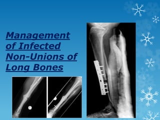
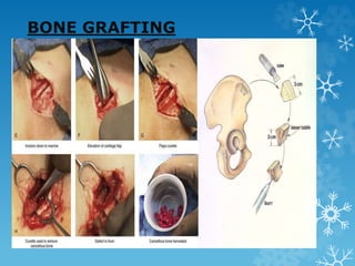
Management
of Infected
Non-Unions of
Long Bones
 Union needs three basic requirements:
1. Stable fixation
2. Biological stimulation, and
3. Restored function.
 Stable fixation can be achieved by any external or
internal fixation devices.
.
 Infected nonunions of the tibia pose significant
challenges for complete resolution and functional
restoration.
 Bones are osteoporotic, deformed, sclerotic, and
nonreactive at the nonunion site.
IMPACT OF NON UNION ON
GENERAL HEALTH
 Hypoproteinemia and anemia result from sero-purulent
wound discharge and chronic negative nitrogen balance.
 Poor appetite from immobility leads to malnourishment.
 Many attempts at union exhaust bone graft donor sites.
 Osteoporosis results from prolonged inability to bear
weight.
 The treatment modalities everywhere is common i.e.,
“ELIMINATE INFECTION FIRST” strategy by process
of “DEBRIDEMENT”.
DEBRIDEMENT
 The infected scarred soft
tissue and necrotic bone
were debrided radically.
 Cortical bone bleeding,
described as the so-called
paprika sign, was accepted
as an indication of vital
tissue.
DEBRIDEMENT
Diagnosis and investigations
Infected nonunions are easy to
diagnose and difficult to treat.
 Long leg films reveal extent of bony
involvement, limb alignment, and length
discrepancy.
 X-rays reveal sequestra, osteoporosis,
deformities, and extent of the bony gap.
 Oblique X-rays can reveal the gap and confirm
the diagnosis of a nonunion.
 Raised acute phase reactant levels confirm
active infection and help monitor
prognosis.
Classification of infected
nonunions
 ASAMI classification is commonly used .Infection is
classified as active or dormant.
Diabetics,
anemic
patients
and
smokers
form bone
poorly.
 The nonunion severity score (NUSS) formulated by helps
to understand the difficulties in treating nonunions and
considers many factors.
 Higher NUSS scores correlate with difficulty in achieving
union.
 They recommend amputation over reconstruction for
scores more than 75. NUSS grades infection only as
previously infected or septic. It does not grade severity
of infection.
Infection severity score
 The infection severity score (ISS)13 grades severity of
infection by analyzing six clinical parameters.
 Maximum score is 25, which is converted to 100. Higher
scores point to need for a second debridement and
possibility of incomplete eradication or recurrence of
infection.
 It could suggest using external rather than internal
fixation in the second stage as a definitive modality to
achieve union.
 Lower scores point to ease of eradication of the
infection.
Control of infection by
debridement
 Debridement remains the cornerstone of treatment of
infection.
 Large sinus tracks and infected scars need excision.
 Loose implants, broken screws, and locking bolts need
removal.
 Granulation tissue and pus must be removed thoroughly.
 Debridement of soft tissue may lead to loss of cover
which may best be filled with a muscle flap.
 Combined surgery of debridement and external fixation
may be safe when the ISS score is less than or equal to
40.
 Higher ISS scores warrant staged surgery.
 Several Surgical treatment options have been proposed,
including
 Bone grafting ,
 Induced-membrane technique,
 Antibiotic cement ,
 Ilizarov methods ,
 Outcome of rail fixator system in reconstructing
bone gap.
 Gap nonunion of forearm bones treated by
modified Nicoll’s technique.
 Other methods include free tissue transfer.
BONE GRAFTING
 Iliac crest bone grafting is the gold standard and
provides osteoconductive, osteoinductive, and
osteogenic material to give biological stimulus.
 Grafts may also be harvested from the opposite upper
tibia or trochanter.
 Posterolateraland central bone grafting achieve union by
approaching the nonunion away from the infected site
and achieving fusion between tibia and the fibula over
the interosseous membrane
BONE GRAFTING
BONE GRAFTING
 Open cancellous bone grafting known as Papineau
method was popular especially in metaphyseal defects
with minimal overlying soft tissue loss.
 A modified Papineau who used a vacuum assisted
closure (VAC) after debridement and after bone grafting
till soft tissue cover surgery.
PERCUTANEOUS
AUTOLOGOUS BONE MARROW
INJECTION
 BOYD Said that “Bone grafting is primarily a second
wounding procedure, in which surgeon hopes that the
response of the body will be more favorable than the
response following the original trauma”.
 The most productive source of cells that influence
osteogenesis is considered to Autlogous bone marrow.
PERCUTANEOUS
AUTOLOGOUS BONE MARROW
INJECTION
 Connolly et al also stated “Autologous bone marrow has
been most useful for the preventive treatment of
nonunion by early injection of delayed union”.
 He also said that the ideal time for bone marrow
injection should be after the initial inflammatory and
osteoclastic resorption period.
 Marrow by itself does not serve as an osteoconductive
agent and therefore cannot fill a large gap,But as
composite graft with demineralized bone matrix, porous
ceramic materials, xenograft .
 The standard procedure
of aspiration of bone
marrow. The procedure
was carried out as an
outpatient procedure.
 Under local infiltration
with 2% xylocaine, a 16-
gauge bone marrow
aspiration needle was
used and to inject the
marrow at fracture site
with needle of 16-gauge
in cases of tibia or ulna.
 Image intensifier was
used to locate the area of
bone marrow injection.
PERCUTANEOUS
AUTOLOGOUS BONE MARROW
INJECTION
 The volume of bone marrow injected was between 50-90
ml in cases of tibia and femur. In ulna the amount
injected was 15 ml only.
 The limiting factor for the amount of bone marrow
injection was the volume that could be injected at a
particular site.
 Adding demineralized bone matrix (DBM),beta tricalcium
phosphate, or hydroxyapatite enhances the marrow
aspirate. Platelet concentrates or gel can be used along
with BMP.
Union potential grading
 Nicoll and Ellis investigated the factors leading to
delayed and nonunion of tibial shaft fractures and
developed a scoring. This score is based on the following
five criteria:
 1. Initial displacement. 2. Comminution. 3. Soft tissue
RESULTS injury, 4. Wound infection, 5. Alignment
PERCUTANEOUS
AUTOLOGOUS BONE MARROW
INJECTION
 Autologous marrow grafting is a simple and effective
method of providing cellular reactivation of osteogenesis
without the complications and risks of cancellous bone
grafting.
 The method can be used as an early intervention,
whenever one suspects a delay in the healing of
fracture.
 This procedure can be done on an outpatient basis and
even under local anesthesia
Union First method in
ASEPTIC NON UNION
 Bone marrow aspirate concentrate also provides a high
concentration of granulocytes and macrophages, which
not only provide progenitors that stimulate bone healing
but also cure the infection in the absence of antibiotic
therapy.
 They used this modality only for gaps less than 15 mm.
BONE RECONSTRUCTION BY
INDUCE MEMBRANE
TECHNIQUE(MASQUELET
TECHNIQUE)
 Inclusion criteria were all patients with posttraumatic
gap nonunion of diaphysis of tibia with or without
infection and with or without accompanying soft tissue
defect.
 Exclusion criteria were patients with neurovascular
injury to the limb, patients with bone loss in epiphyseal
or metaphyseal region, patients with gap nonunion of
etiology other than traumatic in nature.
MASQUELET TECHNIQUE
 Induced membrane favors revascularization and
consolidation of the bone graft.
 Original masquelets technique no antibiotic cement was
used and he believed that “good debridement is the key
to control infection”.
 Induced membrane is richly vascularized by numerous
small capillaries and high concentrations of bone
morphogenic protein-2 (BMP-2), vascular endothelial
growth factor, and transforming growth factor-ß1.
 The membrane prevent soft tissue protrusion in the
bone defect site, provides a scaffold for osteoconduction.
OPERATIVE PROCEDURE
 Technique involves 2 surgeries:
 Stage I surgery :irrigation and debridement of the
infected soft tissue and bone, along with fracture
stabilization with external fixators. Bone cement spacer
without antibiotic was then introduced in the bone gap in
a semisolid stage.
 Stage II surgery :Performed after 4–6 weeks of stage
I surgery in the absence of any clinical signs of infection
includes removal of the cement-spacer, with
preservation of the induced membrane formed at the
spacer surface and filling the bony defect space with
morselized iliac crest bone graft.
MASQUELET TECHNIQUE
MASQUELET TECHNIQUE
MASQUELET TECHNIQUE
MASQUELET TECHNIQUE
 Graft material to be placed within the induced
membrane such as cortical
autograftallograft,RIAmethod, or the use of
recombinant human growth factors and the addition of
stem cells within the membrane.
MASQUELET TECHNIQUE
a) Preoperative X-ray anteroposterior and lateral views of leg showing fixation in situ with infected gap nonunion (b) anteroposterior
and lateral views of leg post stage 1 surgery where after debridement spacer is applied (c) Post stage 2 surgery of same patient where
after
spacer removal bone grafting is done (d) Post stage 2 surgery of same patient after 1 month (e) Post stage 2 surgery of same patient
after
12 months showing graft consolidation and union (f) Post stage 2 surgery of same patient after 17 months showing good graft
consolidation and
union (g and h) Clinical photograph of same patient after 17 months followup showing weight bearing on the limb and good range of
movement
MASQUELET TECHNIQUE
a) Preoperative X-ray anteroposterior and lateral views showing gap nonunion (b) Post stage 1 surgery of same patient showing spacer
within the bone gap (c) Immediate post stage 2 surgery with spacer removal and bone grafting within the induced membrane sleeve (d) Post
stage 2 surgery of same patient after 7 months (e) Post stage 2 surgery of same patient after 12 months showing good graft consolidation
and
union (f) Post stage 2 surgery of same patient after 18 months showing good graft consolidation and union (g) Clinical photograph of the
same
patient after 17 months showing single stance weight bearing on the affected limb
RESULTS
 Radiological and clinical evaluation was done for all
patients at every 6 weeks followup for first 6 months
and every quarterly thereafter.
 The radio opacity of grafted tissues in the bony defect
increased continuously with followup duration.
 An average of 0.5 cm (range 0–1.5 cm) limb length
discrepancy was noted at the latest followup.
 The satisfaction rate among the patients was excellent
as they can manage their routine day-to-day activities
without much difficulty during the course of treatment.
DISADVANATGES
 The main disadvantage of this procedure is that it is a
two-stage procedure with the associated risks of
secondary anesthesia and hospitalization.
 Limited bone graft to harvest in very young children and
the inability to correct residual limb length discrepancy
after the surgery.
 Further patient has to remain non weight bearing during
the initial treatment period as the large bone gaps were
weakly immobilised with external fixators. Thus weight
bearing was started only when the graft showed
consolidation on x ray.
MASQUELET TECHNIQUE
HIGHLIGHTS
 This staged grafting technique within the sleeve of
induced bio-membrane has been described as an
alternative potential treatment strategy.
 This study highlighted that it can provide an effective
and practical management of patients with difficult gap
nonunion.
 It does not require any specialized equipment,
investigations and surgery.
RAIL FIXATOR SYSTEM
Due to many complications such as persistent pain,
deformity of joints and discomfort caused by Ilizarov ring
fixator, inspired the development of rail fixator.
RAIL FIXATOR SYSTEM
 The Rail Fixation System is designed primarily for bone
transport for reconstructing bone loss following open
fracture and sequestrectomy following osteomyelitis.
 This system provides correction in these situations
through the techniques of bone transport,
compression-distraction and bifocal lengthening.
RAIL FIXATOR SYSTEM
 The average bone gap in this series was 7.72 cm (range
3-15 cm).
 Corticotomy was done at single level.Transport was
commenced after 5-7 days of corticotomy.
 Rate of transport was 1.00 mm/day in 4 divided
increments.
 At the conclusion of transport, the defect was closed
byremoving soft-tissue at docking site and giving
compression.
RAIL FIXATOR SYSTEM
RAIL FIXATOR SYSTEM
RAIL FIXATOR SYSTEM
 Partial weight bearing was started at conclusion of
transport.
 Consolidation of docking site was monitored by serial
anteroposterior and lateral X-rays.
 Bone grafting was done in five cases when it was found
callus formation was not adequate at docking site.
 Full weight bearing was advised when three distinct and
complete cortices of regenerate were evident on serial
X-ray
CONCLUSION
 Average duration of rail fixator application was 9 months
(range 6-14 months).
 Pin loosening was the only complication.
 Certain complications8such as heavy apparatus,
persistent pain, deformity of joints and discomfort
caused by Ilizarov ring fixator inspired the development
of monolateral frame devices.
 The rail fixator is relatively simple to apply and patient
compliance is very good when compared with Ilizarov
fixator.
 But filling of bone gap and union does not guarantee
good functional result.
 The functional result is affected by condition of the
nerve, muscles, vessels, joints, and lesser degree to
bone.
 Functional results of the limb were assessed at end of
completion of procedure using ASAMI score.
ASAMI SCORE
CONCLUSION
 Loss of range of motion was more in cases where pins
were close to joint surface and bone gap was more than
10 cm.
 ROM returned to normal in most of cases after proper
physiotherapy.
Local antibiotic delivery
systems
 Removing fibrous tissue, necrotic bone, and opening the
marrow canal improve blood supply and allow nutrients
and antibiotics to reach the site.
 An antibiotic impregnated cement block obliterates dead
space. It allows elution of the antibiotic in very high
concentrations at the local site, in many multiples of the
minimum inhibitory concentration (MIC).
Local antibiotic delivery
systems
 Acrylic bone cement is fashioned into intramedullary
(IM) rods (with a core of a K wire or Rush nail/V nail).
 The rod is inserted from proximal portal after reaming.
 Adding the antibiotic powder or liquid toward the end of
polymerization ensures higher elution of antibiotics.
 In smaller gaps, an absorbable carrier may make the
second surgery unnecessary. The use of calcium
sulfate25 cement paste or powder and absorbable
chitosan polymer26 carriers are good alternatives.
Definitive surgery to achieve
union
 Treatment of infected nonunion of tibia and femur by
bone transport using Ilizarov external fixator and
monolateral external fixator, respectively.
 It is minimally invasive and is able to reliably achieve
union, lengthen and correct complex deformities
 It gives sustained gradual compression at nonunion site,
either in a vertical or in a horizontal mode .
 Fibulectomy and sustained compression at nonunion site
are the choice when there is minimal shortening.
Oblique view of leg bones with knee showing (a) upper tibial infected nonunion (b) After
infection control (c) Ilizarov apparatus in situ (d).
If vertical compression is given, it causes vertical displacement of nonunion ends. With the
help of washers, horizontal compression achieved
perpendicular to plane of nonunion (e) Good union. No loss of length
ILIZAROV TECHNIQUE
 Compression at the nonunion and lengthening at
corticotomy site happen simultaneously for smaller
discrepancies .
 If the shortening is larger or the gap is greater, bone
transport technique (sequential distraction-compression
osteosynthesis) helps fill the gap.
 Many features of the Ilizarov fixator are shared by the
Limb Reconstruction System (LRS)
Bone Transport Technique
 It enables filling up large gaps of bone, in many
instances without the need for bone grafting or bone
graft substitutes.
 External devices such as the Ilizarov, LRS, and Taylor
Spatial Frame (TSF) fixators are most commonly used.
 IM nails can guide the bone transport and reduce
external fixation duration.
 The latency period before bone transport was 7–10 days
& rate of distraction was 0.25 mm per 6 h.
 When bone transport was completed, the tibia or femur
docked ends were compressed by 0.25 mm per day in
order to provide full contact until the patient felt pain at
the docking site.
DISTRACTION
OSTEOGENESIS
“DISTRACTION OSTEOGENESIS “ refers to the
production of new bone between vascular bone surfaces
created by an osteotomy and separated by gradual
distraction
DISTRACTION
OSTEOGENESIS
 Bone transport techniques are a robust biological
solution .
 Even if mild infection persists, it will not cause failure of
the entire process as can happen with the induced
membrane technique.
 A locking nail is inserted after debridement and enables
rapid bone transport. Reduction in axial deviation and
external fixation time and improved patient comfort are
the advantages.
X-ray anteroposterior and lateral views with knee and ankle joints showing (a) Infected
nonunion lower tibia. Beads inserted (b) Ilizarov
fixator applied. Acute compression at nonunion site done gradually at low rate hence no angular
deformity at nonunion site despite an irregular
shaped defect. Simultaneous lengthening at upper level to equalize lengths (c) Union achieved
at distal end with good regenerate at proximal
corticotomy site and no limb length discrepancy
 For smaller gaps, acute compression may fill the defect,
and larger gaps may be filled by gradual compression.
 Compression given at a high rate on irregular bony ends
will result in angular deviation. Union without a
deformity is possible by compressing at a much lower
rate: i.e. 1 mm every 12 days.
X-ray leg bones with knee joint anteroposterior and lateral views showing (a) Infected tibial
nonunion. Irregular bony ends. Had we
resected bone till achieving horizontal bony surfaces, it would have created a lot of
shortening (b) we gave compression at rate of one-fourth mm
twice a week to achieve compression but not cause an angular deviation. No bone grafting
done (c) Sound union, after ilizarov construct removal
REGENERATE PROBLEMS
 Instability of fixation causes the regenerate bone to
deform in valgus and procurvatum.
 Adding bone grafts, bone marrow aspirate, or
bisphosphonates improves regenerate quality.
 Smokers and those on nonsteroidal anti-inflammatory
drugs tend to have poorer regenerate formation.
 Hypovitaminosis D and anemia are the common causes
of poor regenerate formation.
REHABILITATION
 Walking and resumption of employment also help in
early healing to walk for several hours a day. This
improves appetite and vascularity to the lower limb and
promotes early healing.
 Significant whole body exercises are a part of
physiotherapy at Prof. Ilizarov’s Kurgan institute.
 The most common complication was pin-track infection,
and the incidence was 63.64%.
Some important aspects of bone transport should be
paid attention.
(1)We should perform radical debridement in the site of
infected nonunion. This is the key step to control bone
infection.
(2) Distraction usually begins between 7 and 10 days after
the operation at a rate of 0.25 mm per 6 h
If regenerate quality is poor, the speed of distraction
will slow down.
ROLE OF THE FIBULA
 The fibula tends to heal quickly and prevent adequate
compression at the nonunion site.
 Watch for early union of fibula and resect it when the
docking site is being opened, freshened, or grafted.
COMPLICATIONS OF BONE
TRANSPORTATION
 Pin tract infection.
 Joint stiffness.
 Muscle contractures.
 Joint subluxation.
 Axial deviation.
 Neurological or vascular insult.
 Premature consolidation.
 Delayed consolidation.
 Refracture and difficulties with psychological adjustment
are the complications .
Free Vascularized Fibular
Graft
 Microvascular fibular grafting to bridge large gaps was very
popular before the advent of the Ilizarov technique.
 The fibula and tibial diameter mismatch necessitated
prolonged protection.
 It took a long time for the fibula to hypertrophy.
 A recipient vessel far away from the injured zone for
successful microvascular anastomosis is chosen.
 The fibula may face difficulties in uniting at one of the ends.
Infection and anastomotic failure could result in significant
morbidity.
 Combining the free vascularized fibula with the Ilizarov
fixator combines benefits of both methods to fill up gaps in
infected nonunions.
 The muscle flap brings much needed vascularity and helps
in early union and complete eradication of infection
Nicoll’s technique.
 Blocks of the corticocancellous bone with a single cortex
from the iliac crest, augmented with rigid plate fixation
under compression.
 Good results using an Iliac crest graft with medullary
nail fixation using a hun (hunec) nail.
 Bones with segmental bone defects treated by
tricorticocancellous bone graft under optimum
compression with intramedullary fixation.
 The mean intercalary defect was 4.7 cm (range, 1.5–7.5
cm) after debridement.
 Authors have modified the Nicoll technique and used
tricorticocancellous bone strut, to fill the bone gap,
instead of wholly cancellous inserts.
Nicoll’s technique.
 A tricorticocancellous iliac crest bone block, 2–3 mm longer
than the measured gap, was harvested. The width of the
graft was 2–2.5 cm.
 A square nail of predetermined size was negotiated in one
of the fragments and was protruded through the fracture
surface.
 The prepared strut (corticocancellous bone block) was
threaded over the nail and rotated, as per local geometry of
the bone and while maintaining sufficient traction, the strut
was reduced between the fragments and the nail was driven
further across the strut into the other fragment.
 Traction was then left which locked the corticocancellous
block under optimum compression over a nail between the
two fragments.
 Osteoperiosteal flaps were then raised on both bone host
graft junctions and cancellous chips were impacted all
around.
Nicoll’s technique.
 All sclerotic bone ends must be removed, even though
this increases the gap.
 These sclerotic ends are devoid of the blood supply and
are quite incapable of providing an outflow of the
osteogenetic tissue into the graft.
 This outflow from the host bone is regarded by Siffert as
the most important single source of osteogenesis.
Nicoll’s technique.
Nicoll’s technique.
Nicoll’s technique.
 The thin cortical bone on the two sides allows for early
invasion of the periosteal circulation.
 Abundance of the cancellous bone decreases the time of
graft incorporation and gaps in early healing of the
fracture.
 Thus, the procedure serves in three ways, i.e.,
Internal fixation
Defect bridging,
Osteogenesis promoter.
 The intramedullary nail gives a stable fixation to the
fracture and the graft.
 Nonvascularized autografts are the most commonly used
grafts and are considered to be the “gold standard.”
 For physical reasons, solid blocks give a more rapid
union and less fibroblastic reactions than chips or
ribbons.
Nicoll’s technique.
 The only specific complication is the risk of herniation of
muscles at the donor site, which is amicable to
treatment.
 Thus, this simple technique of tricorticocancellous bone
grafting under optimal compression, augmented with
intramedullary fixation, provides a promising solution
for problem of atrophic nonunion with a gap.
Other Methods of Treatment
 Electrical stimulation, low-intensity pulsed ultrasound,
extracorporeal shock wave.
 Antibiotic efficiency and penetration improve by
adding enzyme preparations.
 Increasing vascularity at nonunion site is to drill several
fine wire holes across the nonunion site. This creates
new vascular channels and speeds up healing.
CONCLUSION
 The main principles of treatment of infected nonunions
of the tibia are
(1) Radical and thorough debridement.
(2) Compression of nonunion by external fixation with
lengthening or bone transport.
(3) Conversion to internal or hybrid fixation in less severe
infections &
(4) Augmentation of healing by bone grafting, bone marrow
injections, platelet concentrates, and DBM.

Non union

  • 1.
  • 2.
     Union needsthree basic requirements: 1. Stable fixation 2. Biological stimulation, and 3. Restored function.  Stable fixation can be achieved by any external or internal fixation devices. .
  • 3.
     Infected nonunionsof the tibia pose significant challenges for complete resolution and functional restoration.  Bones are osteoporotic, deformed, sclerotic, and nonreactive at the nonunion site.
  • 4.
    IMPACT OF NONUNION ON GENERAL HEALTH  Hypoproteinemia and anemia result from sero-purulent wound discharge and chronic negative nitrogen balance.  Poor appetite from immobility leads to malnourishment.  Many attempts at union exhaust bone graft donor sites.  Osteoporosis results from prolonged inability to bear weight.
  • 5.
     The treatmentmodalities everywhere is common i.e., “ELIMINATE INFECTION FIRST” strategy by process of “DEBRIDEMENT”.
  • 6.
    DEBRIDEMENT  The infectedscarred soft tissue and necrotic bone were debrided radically.  Cortical bone bleeding, described as the so-called paprika sign, was accepted as an indication of vital tissue.
  • 7.
  • 9.
    Diagnosis and investigations Infectednonunions are easy to diagnose and difficult to treat.  Long leg films reveal extent of bony involvement, limb alignment, and length discrepancy.  X-rays reveal sequestra, osteoporosis, deformities, and extent of the bony gap.  Oblique X-rays can reveal the gap and confirm the diagnosis of a nonunion.  Raised acute phase reactant levels confirm active infection and help monitor prognosis.
  • 10.
    Classification of infected nonunions ASAMI classification is commonly used .Infection is classified as active or dormant.
  • 11.
  • 12.
     The nonunionseverity score (NUSS) formulated by helps to understand the difficulties in treating nonunions and considers many factors.  Higher NUSS scores correlate with difficulty in achieving union.  They recommend amputation over reconstruction for scores more than 75. NUSS grades infection only as previously infected or septic. It does not grade severity of infection.
  • 16.
    Infection severity score The infection severity score (ISS)13 grades severity of infection by analyzing six clinical parameters.  Maximum score is 25, which is converted to 100. Higher scores point to need for a second debridement and possibility of incomplete eradication or recurrence of infection.  It could suggest using external rather than internal fixation in the second stage as a definitive modality to achieve union.  Lower scores point to ease of eradication of the infection.
  • 17.
    Control of infectionby debridement  Debridement remains the cornerstone of treatment of infection.  Large sinus tracks and infected scars need excision.  Loose implants, broken screws, and locking bolts need removal.  Granulation tissue and pus must be removed thoroughly.  Debridement of soft tissue may lead to loss of cover which may best be filled with a muscle flap.
  • 18.
     Combined surgeryof debridement and external fixation may be safe when the ISS score is less than or equal to 40.  Higher ISS scores warrant staged surgery.
  • 19.
     Several Surgicaltreatment options have been proposed, including  Bone grafting ,  Induced-membrane technique,  Antibiotic cement ,  Ilizarov methods ,  Outcome of rail fixator system in reconstructing bone gap.  Gap nonunion of forearm bones treated by modified Nicoll’s technique.  Other methods include free tissue transfer.
  • 20.
    BONE GRAFTING  Iliaccrest bone grafting is the gold standard and provides osteoconductive, osteoinductive, and osteogenic material to give biological stimulus.  Grafts may also be harvested from the opposite upper tibia or trochanter.  Posterolateraland central bone grafting achieve union by approaching the nonunion away from the infected site and achieving fusion between tibia and the fibula over the interosseous membrane
  • 21.
  • 22.
    BONE GRAFTING  Opencancellous bone grafting known as Papineau method was popular especially in metaphyseal defects with minimal overlying soft tissue loss.  A modified Papineau who used a vacuum assisted closure (VAC) after debridement and after bone grafting till soft tissue cover surgery.
  • 23.
    PERCUTANEOUS AUTOLOGOUS BONE MARROW INJECTION BOYD Said that “Bone grafting is primarily a second wounding procedure, in which surgeon hopes that the response of the body will be more favorable than the response following the original trauma”.  The most productive source of cells that influence osteogenesis is considered to Autlogous bone marrow.
  • 24.
    PERCUTANEOUS AUTOLOGOUS BONE MARROW INJECTION Connolly et al also stated “Autologous bone marrow has been most useful for the preventive treatment of nonunion by early injection of delayed union”.  He also said that the ideal time for bone marrow injection should be after the initial inflammatory and osteoclastic resorption period.  Marrow by itself does not serve as an osteoconductive agent and therefore cannot fill a large gap,But as composite graft with demineralized bone matrix, porous ceramic materials, xenograft .
  • 25.
     The standardprocedure of aspiration of bone marrow. The procedure was carried out as an outpatient procedure.  Under local infiltration with 2% xylocaine, a 16- gauge bone marrow aspiration needle was used and to inject the marrow at fracture site with needle of 16-gauge in cases of tibia or ulna.  Image intensifier was used to locate the area of bone marrow injection.
  • 26.
    PERCUTANEOUS AUTOLOGOUS BONE MARROW INJECTION The volume of bone marrow injected was between 50-90 ml in cases of tibia and femur. In ulna the amount injected was 15 ml only.  The limiting factor for the amount of bone marrow injection was the volume that could be injected at a particular site.  Adding demineralized bone matrix (DBM),beta tricalcium phosphate, or hydroxyapatite enhances the marrow aspirate. Platelet concentrates or gel can be used along with BMP.
  • 28.
    Union potential grading Nicoll and Ellis investigated the factors leading to delayed and nonunion of tibial shaft fractures and developed a scoring. This score is based on the following five criteria:  1. Initial displacement. 2. Comminution. 3. Soft tissue RESULTS injury, 4. Wound infection, 5. Alignment
  • 29.
    PERCUTANEOUS AUTOLOGOUS BONE MARROW INJECTION Autologous marrow grafting is a simple and effective method of providing cellular reactivation of osteogenesis without the complications and risks of cancellous bone grafting.  The method can be used as an early intervention, whenever one suspects a delay in the healing of fracture.  This procedure can be done on an outpatient basis and even under local anesthesia
  • 30.
    Union First methodin ASEPTIC NON UNION  Bone marrow aspirate concentrate also provides a high concentration of granulocytes and macrophages, which not only provide progenitors that stimulate bone healing but also cure the infection in the absence of antibiotic therapy.  They used this modality only for gaps less than 15 mm.
  • 31.
    BONE RECONSTRUCTION BY INDUCEMEMBRANE TECHNIQUE(MASQUELET TECHNIQUE)  Inclusion criteria were all patients with posttraumatic gap nonunion of diaphysis of tibia with or without infection and with or without accompanying soft tissue defect.  Exclusion criteria were patients with neurovascular injury to the limb, patients with bone loss in epiphyseal or metaphyseal region, patients with gap nonunion of etiology other than traumatic in nature.
  • 32.
    MASQUELET TECHNIQUE  Inducedmembrane favors revascularization and consolidation of the bone graft.  Original masquelets technique no antibiotic cement was used and he believed that “good debridement is the key to control infection”.  Induced membrane is richly vascularized by numerous small capillaries and high concentrations of bone morphogenic protein-2 (BMP-2), vascular endothelial growth factor, and transforming growth factor-ß1.  The membrane prevent soft tissue protrusion in the bone defect site, provides a scaffold for osteoconduction.
  • 33.
    OPERATIVE PROCEDURE  Techniqueinvolves 2 surgeries:  Stage I surgery :irrigation and debridement of the infected soft tissue and bone, along with fracture stabilization with external fixators. Bone cement spacer without antibiotic was then introduced in the bone gap in a semisolid stage.  Stage II surgery :Performed after 4–6 weeks of stage I surgery in the absence of any clinical signs of infection includes removal of the cement-spacer, with preservation of the induced membrane formed at the spacer surface and filling the bony defect space with morselized iliac crest bone graft.
  • 34.
  • 35.
  • 36.
  • 37.
    MASQUELET TECHNIQUE  Graftmaterial to be placed within the induced membrane such as cortical autograftallograft,RIAmethod, or the use of recombinant human growth factors and the addition of stem cells within the membrane.
  • 38.
    MASQUELET TECHNIQUE a) PreoperativeX-ray anteroposterior and lateral views of leg showing fixation in situ with infected gap nonunion (b) anteroposterior and lateral views of leg post stage 1 surgery where after debridement spacer is applied (c) Post stage 2 surgery of same patient where after spacer removal bone grafting is done (d) Post stage 2 surgery of same patient after 1 month (e) Post stage 2 surgery of same patient after 12 months showing graft consolidation and union (f) Post stage 2 surgery of same patient after 17 months showing good graft consolidation and union (g and h) Clinical photograph of same patient after 17 months followup showing weight bearing on the limb and good range of movement
  • 39.
    MASQUELET TECHNIQUE a) PreoperativeX-ray anteroposterior and lateral views showing gap nonunion (b) Post stage 1 surgery of same patient showing spacer within the bone gap (c) Immediate post stage 2 surgery with spacer removal and bone grafting within the induced membrane sleeve (d) Post stage 2 surgery of same patient after 7 months (e) Post stage 2 surgery of same patient after 12 months showing good graft consolidation and union (f) Post stage 2 surgery of same patient after 18 months showing good graft consolidation and union (g) Clinical photograph of the same patient after 17 months showing single stance weight bearing on the affected limb
  • 40.
    RESULTS  Radiological andclinical evaluation was done for all patients at every 6 weeks followup for first 6 months and every quarterly thereafter.  The radio opacity of grafted tissues in the bony defect increased continuously with followup duration.  An average of 0.5 cm (range 0–1.5 cm) limb length discrepancy was noted at the latest followup.  The satisfaction rate among the patients was excellent as they can manage their routine day-to-day activities without much difficulty during the course of treatment.
  • 41.
    DISADVANATGES  The maindisadvantage of this procedure is that it is a two-stage procedure with the associated risks of secondary anesthesia and hospitalization.  Limited bone graft to harvest in very young children and the inability to correct residual limb length discrepancy after the surgery.  Further patient has to remain non weight bearing during the initial treatment period as the large bone gaps were weakly immobilised with external fixators. Thus weight bearing was started only when the graft showed consolidation on x ray.
  • 42.
    MASQUELET TECHNIQUE HIGHLIGHTS  Thisstaged grafting technique within the sleeve of induced bio-membrane has been described as an alternative potential treatment strategy.  This study highlighted that it can provide an effective and practical management of patients with difficult gap nonunion.  It does not require any specialized equipment, investigations and surgery.
  • 43.
    RAIL FIXATOR SYSTEM Dueto many complications such as persistent pain, deformity of joints and discomfort caused by Ilizarov ring fixator, inspired the development of rail fixator.
  • 44.
    RAIL FIXATOR SYSTEM The Rail Fixation System is designed primarily for bone transport for reconstructing bone loss following open fracture and sequestrectomy following osteomyelitis.  This system provides correction in these situations through the techniques of bone transport, compression-distraction and bifocal lengthening.
  • 45.
    RAIL FIXATOR SYSTEM The average bone gap in this series was 7.72 cm (range 3-15 cm).  Corticotomy was done at single level.Transport was commenced after 5-7 days of corticotomy.  Rate of transport was 1.00 mm/day in 4 divided increments.  At the conclusion of transport, the defect was closed byremoving soft-tissue at docking site and giving compression.
  • 46.
  • 47.
  • 48.
    RAIL FIXATOR SYSTEM Partial weight bearing was started at conclusion of transport.  Consolidation of docking site was monitored by serial anteroposterior and lateral X-rays.  Bone grafting was done in five cases when it was found callus formation was not adequate at docking site.  Full weight bearing was advised when three distinct and complete cortices of regenerate were evident on serial X-ray
  • 49.
    CONCLUSION  Average durationof rail fixator application was 9 months (range 6-14 months).  Pin loosening was the only complication.  Certain complications8such as heavy apparatus, persistent pain, deformity of joints and discomfort caused by Ilizarov ring fixator inspired the development of monolateral frame devices.  The rail fixator is relatively simple to apply and patient compliance is very good when compared with Ilizarov fixator.
  • 50.
     But fillingof bone gap and union does not guarantee good functional result.  The functional result is affected by condition of the nerve, muscles, vessels, joints, and lesser degree to bone.  Functional results of the limb were assessed at end of completion of procedure using ASAMI score.
  • 51.
  • 52.
    CONCLUSION  Loss ofrange of motion was more in cases where pins were close to joint surface and bone gap was more than 10 cm.  ROM returned to normal in most of cases after proper physiotherapy.
  • 53.
    Local antibiotic delivery systems Removing fibrous tissue, necrotic bone, and opening the marrow canal improve blood supply and allow nutrients and antibiotics to reach the site.  An antibiotic impregnated cement block obliterates dead space. It allows elution of the antibiotic in very high concentrations at the local site, in many multiples of the minimum inhibitory concentration (MIC).
  • 54.
    Local antibiotic delivery systems Acrylic bone cement is fashioned into intramedullary (IM) rods (with a core of a K wire or Rush nail/V nail).  The rod is inserted from proximal portal after reaming.  Adding the antibiotic powder or liquid toward the end of polymerization ensures higher elution of antibiotics.  In smaller gaps, an absorbable carrier may make the second surgery unnecessary. The use of calcium sulfate25 cement paste or powder and absorbable chitosan polymer26 carriers are good alternatives.
  • 55.
    Definitive surgery toachieve union  Treatment of infected nonunion of tibia and femur by bone transport using Ilizarov external fixator and monolateral external fixator, respectively.  It is minimally invasive and is able to reliably achieve union, lengthen and correct complex deformities  It gives sustained gradual compression at nonunion site, either in a vertical or in a horizontal mode .  Fibulectomy and sustained compression at nonunion site are the choice when there is minimal shortening.
  • 56.
    Oblique view ofleg bones with knee showing (a) upper tibial infected nonunion (b) After infection control (c) Ilizarov apparatus in situ (d). If vertical compression is given, it causes vertical displacement of nonunion ends. With the help of washers, horizontal compression achieved perpendicular to plane of nonunion (e) Good union. No loss of length
  • 57.
    ILIZAROV TECHNIQUE  Compressionat the nonunion and lengthening at corticotomy site happen simultaneously for smaller discrepancies .  If the shortening is larger or the gap is greater, bone transport technique (sequential distraction-compression osteosynthesis) helps fill the gap.  Many features of the Ilizarov fixator are shared by the Limb Reconstruction System (LRS)
  • 58.
    Bone Transport Technique It enables filling up large gaps of bone, in many instances without the need for bone grafting or bone graft substitutes.  External devices such as the Ilizarov, LRS, and Taylor Spatial Frame (TSF) fixators are most commonly used.  IM nails can guide the bone transport and reduce external fixation duration.  The latency period before bone transport was 7–10 days & rate of distraction was 0.25 mm per 6 h.  When bone transport was completed, the tibia or femur docked ends were compressed by 0.25 mm per day in order to provide full contact until the patient felt pain at the docking site.
  • 59.
    DISTRACTION OSTEOGENESIS “DISTRACTION OSTEOGENESIS “refers to the production of new bone between vascular bone surfaces created by an osteotomy and separated by gradual distraction
  • 60.
  • 61.
     Bone transporttechniques are a robust biological solution .  Even if mild infection persists, it will not cause failure of the entire process as can happen with the induced membrane technique.  A locking nail is inserted after debridement and enables rapid bone transport. Reduction in axial deviation and external fixation time and improved patient comfort are the advantages.
  • 62.
    X-ray anteroposterior andlateral views with knee and ankle joints showing (a) Infected nonunion lower tibia. Beads inserted (b) Ilizarov fixator applied. Acute compression at nonunion site done gradually at low rate hence no angular deformity at nonunion site despite an irregular shaped defect. Simultaneous lengthening at upper level to equalize lengths (c) Union achieved at distal end with good regenerate at proximal corticotomy site and no limb length discrepancy
  • 64.
     For smallergaps, acute compression may fill the defect, and larger gaps may be filled by gradual compression.  Compression given at a high rate on irregular bony ends will result in angular deviation. Union without a deformity is possible by compressing at a much lower rate: i.e. 1 mm every 12 days.
  • 65.
    X-ray leg boneswith knee joint anteroposterior and lateral views showing (a) Infected tibial nonunion. Irregular bony ends. Had we resected bone till achieving horizontal bony surfaces, it would have created a lot of shortening (b) we gave compression at rate of one-fourth mm twice a week to achieve compression but not cause an angular deviation. No bone grafting done (c) Sound union, after ilizarov construct removal
  • 68.
    REGENERATE PROBLEMS  Instabilityof fixation causes the regenerate bone to deform in valgus and procurvatum.  Adding bone grafts, bone marrow aspirate, or bisphosphonates improves regenerate quality.  Smokers and those on nonsteroidal anti-inflammatory drugs tend to have poorer regenerate formation.  Hypovitaminosis D and anemia are the common causes of poor regenerate formation.
  • 69.
    REHABILITATION  Walking andresumption of employment also help in early healing to walk for several hours a day. This improves appetite and vascularity to the lower limb and promotes early healing.  Significant whole body exercises are a part of physiotherapy at Prof. Ilizarov’s Kurgan institute.  The most common complication was pin-track infection, and the incidence was 63.64%.
  • 70.
    Some important aspectsof bone transport should be paid attention. (1)We should perform radical debridement in the site of infected nonunion. This is the key step to control bone infection. (2) Distraction usually begins between 7 and 10 days after the operation at a rate of 0.25 mm per 6 h If regenerate quality is poor, the speed of distraction will slow down.
  • 72.
    ROLE OF THEFIBULA  The fibula tends to heal quickly and prevent adequate compression at the nonunion site.  Watch for early union of fibula and resect it when the docking site is being opened, freshened, or grafted.
  • 74.
    COMPLICATIONS OF BONE TRANSPORTATION Pin tract infection.  Joint stiffness.  Muscle contractures.  Joint subluxation.  Axial deviation.  Neurological or vascular insult.  Premature consolidation.  Delayed consolidation.  Refracture and difficulties with psychological adjustment are the complications .
  • 75.
    Free Vascularized Fibular Graft Microvascular fibular grafting to bridge large gaps was very popular before the advent of the Ilizarov technique.  The fibula and tibial diameter mismatch necessitated prolonged protection.  It took a long time for the fibula to hypertrophy.  A recipient vessel far away from the injured zone for successful microvascular anastomosis is chosen.  The fibula may face difficulties in uniting at one of the ends. Infection and anastomotic failure could result in significant morbidity.  Combining the free vascularized fibula with the Ilizarov fixator combines benefits of both methods to fill up gaps in infected nonunions.  The muscle flap brings much needed vascularity and helps in early union and complete eradication of infection
  • 76.
    Nicoll’s technique.  Blocksof the corticocancellous bone with a single cortex from the iliac crest, augmented with rigid plate fixation under compression.  Good results using an Iliac crest graft with medullary nail fixation using a hun (hunec) nail.  Bones with segmental bone defects treated by tricorticocancellous bone graft under optimum compression with intramedullary fixation.  The mean intercalary defect was 4.7 cm (range, 1.5–7.5 cm) after debridement.  Authors have modified the Nicoll technique and used tricorticocancellous bone strut, to fill the bone gap, instead of wholly cancellous inserts.
  • 77.
    Nicoll’s technique.  Atricorticocancellous iliac crest bone block, 2–3 mm longer than the measured gap, was harvested. The width of the graft was 2–2.5 cm.  A square nail of predetermined size was negotiated in one of the fragments and was protruded through the fracture surface.  The prepared strut (corticocancellous bone block) was threaded over the nail and rotated, as per local geometry of the bone and while maintaining sufficient traction, the strut was reduced between the fragments and the nail was driven further across the strut into the other fragment.  Traction was then left which locked the corticocancellous block under optimum compression over a nail between the two fragments.  Osteoperiosteal flaps were then raised on both bone host graft junctions and cancellous chips were impacted all around.
  • 78.
    Nicoll’s technique.  Allsclerotic bone ends must be removed, even though this increases the gap.  These sclerotic ends are devoid of the blood supply and are quite incapable of providing an outflow of the osteogenetic tissue into the graft.  This outflow from the host bone is regarded by Siffert as the most important single source of osteogenesis.
  • 80.
  • 81.
  • 82.
    Nicoll’s technique.  Thethin cortical bone on the two sides allows for early invasion of the periosteal circulation.  Abundance of the cancellous bone decreases the time of graft incorporation and gaps in early healing of the fracture.  Thus, the procedure serves in three ways, i.e., Internal fixation Defect bridging, Osteogenesis promoter.  The intramedullary nail gives a stable fixation to the fracture and the graft.
  • 83.
     Nonvascularized autograftsare the most commonly used grafts and are considered to be the “gold standard.”  For physical reasons, solid blocks give a more rapid union and less fibroblastic reactions than chips or ribbons.
  • 84.
    Nicoll’s technique.  Theonly specific complication is the risk of herniation of muscles at the donor site, which is amicable to treatment.  Thus, this simple technique of tricorticocancellous bone grafting under optimal compression, augmented with intramedullary fixation, provides a promising solution for problem of atrophic nonunion with a gap.
  • 85.
    Other Methods ofTreatment  Electrical stimulation, low-intensity pulsed ultrasound, extracorporeal shock wave.  Antibiotic efficiency and penetration improve by adding enzyme preparations.  Increasing vascularity at nonunion site is to drill several fine wire holes across the nonunion site. This creates new vascular channels and speeds up healing.
  • 86.
    CONCLUSION  The mainprinciples of treatment of infected nonunions of the tibia are (1) Radical and thorough debridement. (2) Compression of nonunion by external fixation with lengthening or bone transport. (3) Conversion to internal or hybrid fixation in less severe infections & (4) Augmentation of healing by bone grafting, bone marrow injections, platelet concentrates, and DBM.