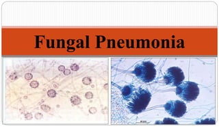
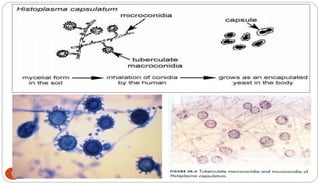
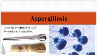
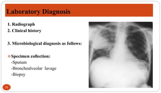
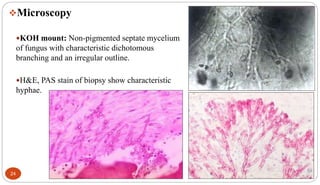
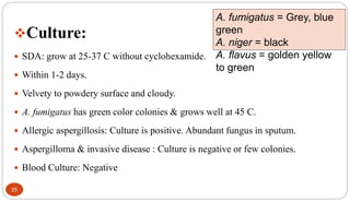
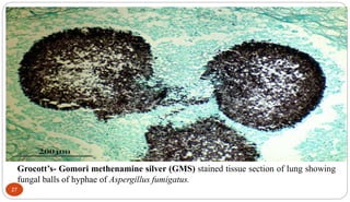
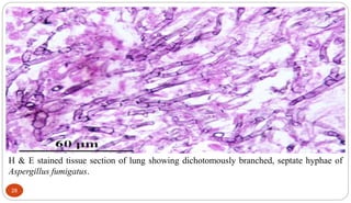
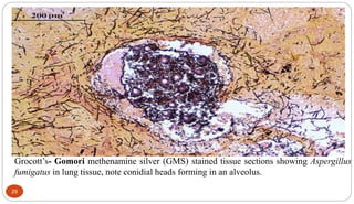
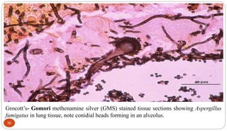
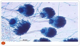
3. Histoplasmosis (Histio – Plasmo – Capsule)
3
Reported first by Samuel Taylor Darling (1905).
Darling’s disease. Cave disease.
Most common fungal infections.
Distributed worldwide but most common in America.
The ecological niche of H. capsulatum is in chicken houses and bat guano.
Typically, patient will have spread chicken manure around his garden and 3 weeks
later will develop pulmonary infection.
4. 4
A systemic disease, mostly of the RE system,
manifesting itself in the lungs, bone marrow,
liver, and the spleen.
Hepatosplenomegaly, primary sign in children
while in adults, more commonly appears as
pulmonary disease.
The majority of patients who develop
histoplasmosis (90-95%) are asymptomatic.
5. Morphology
5
Dimorphic fungi. (yeast in 37 C & hyphal/ mycelial in 22-25 C)
Grows in moist soil high in nitrogen content.
Grow as separate mycelium in soil and artificial culture medium.
Intracellular in animal tissues.
In culture, mycelial phase produces 2 types of unicellular asexual spores.
Macroconidia – large round tuberculate (8-15 um)
Microconidia – small, elliptical (2-4 um)
7. 7
Clinical Manifestation:
1. Acute pulmonary:
Incubation period : 10-16 days.
Mild respiratory illness "flu-like" with general malaise, fever, chills,
headache, myalgia, chest pain and non-productive cough. (Complete recovery
from the acute pulmonary form)
Radiological examination: small, scattered pulmonary infiltrate & hilar
lympadenopathy.
8. 2. Chronic pulmonary:
Similar to Pulmonary tuberculosis.
Remains latent and gradual onset of symptoms.
More pronounced than Acute Pulmonary Histoplasmosis.
Hemoptysis, apical or subapical cavities.
Loss of weight, ulcerative lesions on lips, mouth, nose, larynx & intestine.
Forms Histoplasmoma (2-4mm- later after 10-15 yrs, 3-4 cm)
Chest X- ray also looks like tuberculosis, but can distinguish between these
diseases on the chest film (histoplasmosis usually appears as bilateral
interstitial infiltrates).
8
9. 9
3. Disseminated:
However, if untreated, the disseminated form of disease is usually fatal.
Occurs in any age.
CNS histoplasmosis.
GI histoplasmosis: symptoms similar to ulcerative colitis- Bloody Diarrhea.
Ocular: Presumed Ocular Histoplasomsis Syndrome (POHS)
Fever, anorexia, weight loss, anemia, leukopenia, hepatosplenomegaly,
lymphadenopathy.
HIV infected person at greater risk.
10. 10
I. Specimen
Sputum
Biopsy material from pulmonary disease
Bone marrow aspirate
Biopsy of lymph node
II. Microscopy
Giemsa stain/Wright’s stain: (Sputum/ Pus); Grocott-Gomori methenamine
silver stain & PAS (Biopsies) shows small, oval yeast cells packed within
macrophages or monocytes.
Laboratory Diagnosis
12. 12
III. Culture:
SDA or BHI with cyclohexamide &
chloramphenicol
1-2 weeks; discarded after 4 weeks.
Colony:
White, cottony mycelium, aerial hyphae
or Brown type at 25 C
Yeast forms at 37 C produces whitish
tan colonies
IV. Intradermal skin test: similar to tuberculin test
• Histoplasmin (Culture filtrate antigen of mycelial phase)
• Positive test : indicates past /present infection but do not differentiate active &
past infection.
13. 13
V. Serological test
Complement fixation test
Precipitation test
Latex agglutination test
ELISA
*Takes two weeks for positivity
VI. Histopathology test
Tissue stained by H&E, Giemsa stain, Gomori’s methenamine Silver stain
14. Treatment
14
The drug of choice is amphotericin B, with all its side effects.
Itraconazole and Voriconazole is now also being used.
Ketoconazole : Mild pulmonary histoplasmosis
16. 16
Very common airborne fungus.
Ubiquitous in environment growing in soil, plants and on decomposing organic
matters.
200 species, 20 involved in human disease; A. fumigatus, A. flavus, A. niger,
A. nidulans and A. terreus.
Serious opportunistic threat to AIDS, neutropaenic, leukemic, and transplant
patients.
Infection usually occurs in lungs – spores germinate in lungs and form fungal balls;
can colonize sinuses, ear canals, eyelids, and conjunctiva.
Invasive aspergillosis can produce necrotic pneumonia, and infection of brain, heart,
and other organs.
17. Morphology
17
KOH mount show non-pigmented mycelium with characteristic dichotomous
branching and irregular outline.
18. The Spectrum of Pulmonary Aspergillosis
18
Inhalation of
Aspergillus
conidia or
mycelial
fragments
Normal Host No sequel
Cavitary Dz Aspergilloma
Chronic Lung
Disease
Chronic
necrotising
aspergillosis
Immunocompromise
d Host Invasive PA
Asthma
Allergic Broncho
pulmonary
Aspergillosis
20. 20
Pulmonary aspergillosis
a. Aspergillus asthma:
Hypersentivity state with aspergilla in
atopic individuals.
b. Aspergilloma:
Called fungus ball
Develops in pre-existing lung cavity
forming compact ball of mycelium,
eventually surrounded by a dense fibrous
wall.
Aspergilloma found at post-mortem in the lung
of a child with leukaemia.
21. 21
c. Broncho-pulmonary aspergillosis:
Fungus grows in lumen of bronchioles & produces
mucus plug of fungal mycelium that occlude the
segment of lung tissue.
d. Invasive aspergillosis:
Develops in haematologic malignancy, solid organ
transplantation and HIV.
Establishes first in lung tissues and disseminate to
involve other organs.
Disseminates to brain, kidney, heart and other organs.
Present with pleuritic chest pain and hemoptysis
(pulmonary infarction)
22. 22
Superficial infection:
Aspergillus flavus & Aspergillus fumigatus
colonise in part of nasal sinuses (sinusitis).
External ear (otomycosis): A. niger
Skin, nail and other sites aswell.
23. Laboratory Diagnosis
23
1. Radiograph
2. Clinical history
3. Microbiological diagnosis as follows:
Specimen collection:
-Sputum
-Bronchoalveolar lavage
-Biopsy
24. 24
Microscopy
KOH mount: Non-pigmented septate mycelium
of fungus with characteristic dichotomous
branching and an irregular outline.
H&E, PAS stain of biopsy show characteristic
hyphae.
25. 25
Culture:
SDA: grow at 25-37 C without cyclohexamide.
Within 1-2 days.
Velvety to powdery surface and cloudy.
A. fumigatus has green color colonies & grows well at 45 C.
Allergic aspergillosis: Culture is positive. Abundant fungus in sputum.
Aspergilloma & invasive disease : Culture is negative or few colonies.
Blood Culture: Negative
A. fumigatus = Grey, blue
green
A. niger = black
A. flavus = golden yellow
to green
26. 26
Skin test:
Intradermal skin test to aspergillus antigen extract is used for patients with
suspected allergic bronchopulmonary aspergillosis (ABPA), atopic dermatitis.
Serological test:
Immunodiffusion, CIE & ELISA are widely used for the detection of all forms
of aspergillosis particularly aspergilloma & ABPA.
Serum antibodies to A. fumigatus can be determined by precipitation or
immunodiffusion test.
27. Grocott’s- Gomori methenamine silver (GMS) stained tissue section of lung showing
fungal balls of hyphae of Aspergillus fumigatus.
27
28. H & E stained tissue section of lung showing dichotomously branched, septate hyphae of
Aspergillus fumigatus.
28
29. Grocott’s- Gomori methenamine silver (GMS) stained tissue sections showing Aspergillus
fumigatus in lung tissue, note conidial heads forming in an alveolus.
29
30. Grocott’s- Gomori methenamine silver (GMS) stained tissue sections showing Aspergillus
fumigatus in lung tissue, note conidial heads forming in an alveolus.
30
32. Treatment
32
Amphotericin B
Voriconazole is superior to amphotericin B.
Aspergilloma is treated by surgical excision because antifungal therapy
is of less value.
Allergic forms are treated with corticosteroids.