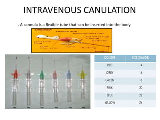
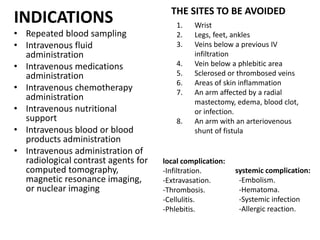
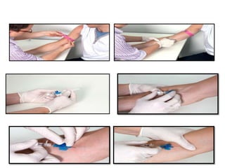
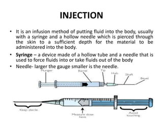
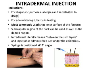
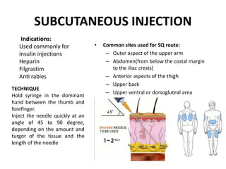
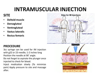
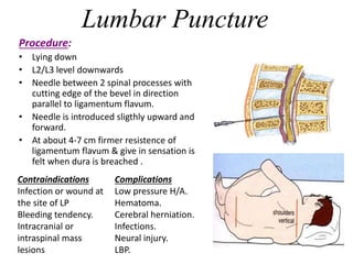
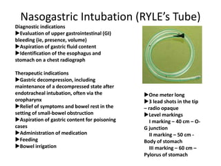
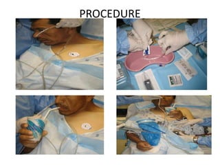
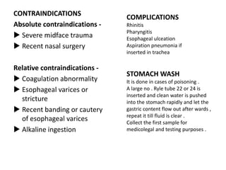
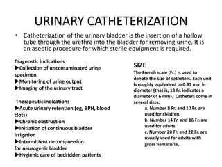
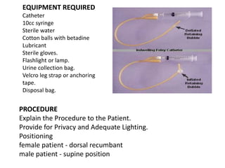
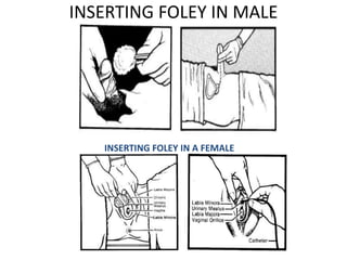
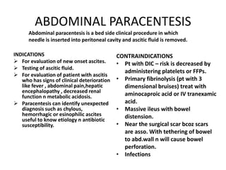
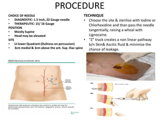
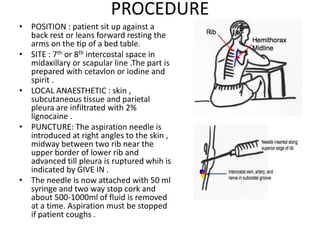
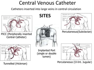
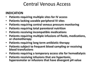
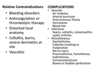
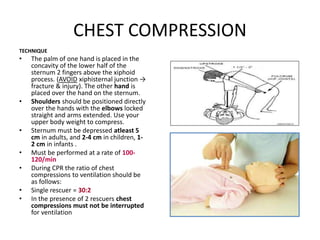
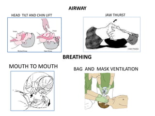
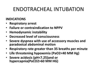
THERAPEUTIC PROCEDURES
PRESENTED BY:
DR DURGESH KUMAR
JUNIOR RESIDENT
DEPARTMENT OF RADIOTHERAPY
VARIOUS THERAPEUTIC PROCEDURE
• I V CANNULATION
• INJECTION TECHNIQUES
• RYLE TUBE INSERTION
• FOLEY CATHETERIZATION
• ABDOMINAL PARACENTESIS
• INTRA PERITONEAL CHEMOTHERAPY
• PLEURAL FLUID ASPIRATION
• LUMBAR PUNCTURE
• CENTRAL VENOUS ACCESS
• CPR
• ENDOTRACHEAL INTUBATION
INTRAVENOUS CANULATION
. A cannula is a flexible tube that can be inserted into the body.
INDICATIONS
• Repeated blood sampling
• Intravenous fluid
administration
• Intravenous medications
administration
• Intravenous chemotherapy
administration
• Intravenous nutritional
support
• Intravenous blood or blood
products administration
• Intravenous administration of
radiological contrast agents for
computed tomography,
magnetic resonance imaging,
or nuclear imaging
THE SITES TO BE AVOIDED
1. Wrist
2. Legs, feet, ankles
3. Veins below a previous IV
infiltration
4. Vein below a phlebitic area
5. Sclerosed or thrombosed veins
6. Areas of skin inflammation
7. An arm affected by a radial
mastectomy, edema, blood clot,
or infection.
8. An arm with an arteriovenous
shunt of fistula
local complication:
-Infiltration.
-Extravasation.
-Thrombosis.
-Cellulitis.
-Phlebitis.
systemic complication:
-Embolism.
-Hematoma.
-Systemic infection
-Allergic reaction.
INJECTION
• It is an infusion method of putting fluid into the body, usually
with a syringe and a hollow needle which is pierced through
the skin to a sufficient depth for the material to be
administered into the body.
• Syringe – a device made of a hollow tube and a needle that is
used to force fluids into or take fluids out of the body
• Needle- larger the gauge smaller is the needle.
INTRADERMAL INJECTION
Indications:
• For diagnostic purposes (allergies and sensitivities to
drugs)
• For administering tuberculin testing
• Most commonly used site: Inner surface of the forearm
• Subscapular region of the back can be used as well as the
deltoid region.
• Intradermal literally means “between the skin layers”
and injection is administered just under the epidermis .
• Syringe is positioned at15˚ angle.
SUBCUTANEOUS INJECTION
• Common sites used for SQ route:
– Outer aspect of the upper arm
– Abdomen(from below the costal margin
to the iliac crests)
– Anterior aspects of the thigh
– Upper back
– Upper ventral or dorsogluteal area
Indications:
Used commonly for
insulin injections
Heparin
Filgrastim
Anti rabies
TECHNIQUE
Hold syringe in the dominant
hand between the thumb and
forefinger.
Inject the needle quickly at an
angle of 45 to 90 degree,
depending on the amount and
turgor of the tissue and the
length of the needle
INTRAMUSCULAR INJECTION
SITE
• Deltoid muscle
• Dorsogluteal
• Ventrogluteal
• Vastus lateralis
• Rectus femoris
PROCEDURE
3cc syringe can be used for IM injection
with g22 or 23 needle; 1-2 inches long
Position the needle at 90˚ angle.
Do not forget to aspirate the plunger once
injected to check for blood.
Inject medication slowly (To minimize
pain) Apply pressure to site and massage
after.
INTRATHECAL INJECTION
four agents are
licensed for
intrathecal
chemotherapy. are
methotrexate,
cytarabine (Ara-C),
hydrocortisone,
and, rarely, thiotepa
Lumbar Puncture
Procedure:
• Lying down
• L2/L3 level downwards
• Needle between 2 spinal processes with
cutting edge of the bevel in direction
parallel to ligamentum flavum.
• Needle is introduced sligthly upward and
forward.
• At about 4-7 cm firmer resistence of
ligamentum flavum & give in sensation is
felt when dura is breached .
Contraindications
Infection or wound at
the site of LP
Bleeding tendency.
Intracranial or
intraspinal mass
lesions
Complications
Low pressure H/A.
Hematoma.
Cerebral herniation.
Infections.
Neural injury.
LBP.
Nasogastric Intubation (RYLE’s Tube)
One meter long
3 lead shots in the tip
– radio opaque
Level markings
I marking – 40 cm – O-
G junction
II marking – 50 cm -
Body of stomach
III marking – 60 cm –
Pylorus of stomach
Diagnostic indications
Evaluation of upper gastrointestinal (GI)
bleeding (ie, presence, volume)
Aspiration of gastric fluid content
Identification of the esophagus and
stomach on a chest radiograph
Therapeutic indications
Gastric decompression, including
maintenance of a decompressed state after
endotracheal intubation, often via the
oropharynx
Relief of symptoms and bowel rest in the
setting of small-bowel obstruction
Aspiration of gastric content for poisoning
cases
Administration of medication
Feeding
Bowel irrigation
PROCEDURE
CONTRAINDICATIONS
Absolute contraindications -
 Severe midface trauma
 Recent nasal surgery
Relative contraindications -
 Coagulation abnormality
 Esophageal varices or
stricture
 Recent banding or cautery
of esophageal varices
 Alkaline ingestion
COMPLICATIONS
Rhinitis
Pharyngitis
Esophageal ulceation
Aspiration pneumonia if
inserted in trachea
STOMACH WASH
It is done in cases of poisoning .
A large no . Ryle tube 22 or 24 is
inserted and clean water is pushed
into the stomach rapidly and let the
gastric content flow out after wards ,
repeat it till fluid is clear .
Collect the first sample for
medicolegal and testing purposes .
URINARY CATHETERIZATION
• Catheterization of the urinary bladder is the insertion of a hollow
tube through the urethra into the bladder for removing urine. It is
an aseptic procedure for which sterile equipment is required.
Diagnostic indications
Collection of uncontaminated urine
specimen
Monitoring of urine output
Imaging of the urinary tract
Therapeutic indications
Acute urinary retention (eg, BPH, blood
clots)
Chronic obstruction
Initiation of continuous bladder
irrigation
Intermittent decompression
for neurogenic bladder
Hygienic care of bedridden patients
SIZE
The French scale (Fr.) is used to
denote the size of catheters. Each unit
is roughly equivalent to 0.33 mm in
diameter (that is, 18 Fr. indicates a
diameter of 6 mm). Catheters come in
several sizes:
a. Number 8 Fr. and 10 Fr. are
used for children.
b. Number 14 Fr. and 16 Fr. are
used for adults.
c. Number 20 Fr. and 22 Fr. are
usually used for adults with
gross hematuria.
EQUIPMENT REQUIRED
Catheter
10cc syringe
Sterile water
Cotton balls with betadine
Lubricant
Sterile gloves.
Flashlight or lamp.
Urine collection bag.
Velcro leg strap or anchoring
tape.
Disposal bag.
PROCEDURE
Explain the Procedure to the Patient.
Provide for Privacy and Adequate Lighting.
Positioning
female patient - dorsal recumbant
male patient - supine position
INSERTING FOLEY IN MALE
INSERTING FOLEY IN A FEMALE
ABDOMINAL PARACENTESIS
INDICATIONS
 For evaluation of new onset ascites.
 Testing of ascitic fluid.
 For evaluation of patient with ascitis
who has signs of clinical deterioration
like fever , abdominal pain,hepatic
encephalopathy , decreased renal
function n metabolic acidosis.
 Paracentesis can identify unexpected
diagnosis such as chylous,
hemorrhagic or esinophilic ascites
useful to know etiology n antibiotic
susceptibility.
Abdominal paracentesis is a bed side clinical procedure in which
needle is inserted into peritoneal cavity and ascitic fluid is removed.
CONTRAINDICATIONS
• Pt with DIC – risk is decreased by
administering platelets or FFPs.
• Primary fibrinolysis (pt with 3
dimensional bruises) treat with
aminocaproic acid or IV tranexamic
acid.
• Massive ileus with bowel
distension.
• Near the surgical scar bcoz scars
are asso. With tethering of bowel
to abd.wall n will cause bowel
perforation.
• Infections
PROCEDURE
CHOICE OF NEEDLE
• DIAGNOSTIC: 1.5 Inch, 22 Gauge needle
• THERAPEUTIC: 15/ 16 Gauge
POSITION
• Mostly Supine
• Head may be elevated
SITE
• Lt lower Quadrant (Dullness on percussion)
• 3cm medial & 3cm above the ant. Sup. Iliac spine
TECHNIQUE
• Choose the site & sterilise with Iodine or
Chlorhexidine and than pass the needle
tangentially, raising a wheal with
Lignocaine.
• “Z” track creates a non linear pathway
b/n Skin& Ascitic fluid & minimise the
chance of leakage.
INTRA PERITONEAL CHEMOTHERAPY
• Used in advanced ovarian tumor or where
disese is less than 1 cm .
• Taxane & platinum based chemotherapy
compounds are used.
• It can be deliverd by using portacath ,
grosshong catheter or single use percutaneous
catheter .
• Before IP therapy peritoneal cavity must be
drained as completely as possible .
PLEURAL FLUID ASPIRATION
INDICATIONS
• DIAGNOSTIC
• LARGE PLEURAL EFFUSION
• CARDIO RESPIRATORY
EMBARASSMENT
• INFECTED PLEURAL FLUID
• ACUTE PULMONARY EDEMA
• PERSISTENT PLEURAL EFFUSION
INSPITE OF ANTI TUBERCULOUS
TREATMENT .
CONTRAINDICATIONS
• Local infection over proposed site of
thoracentesis (e.g. cellulitis, herpes zoster).
Select another entry site .
• Uncontrolled bleeding ,Coagulopathy is a
relative contraindication.
• Caution must be exerted when performing
thoracentesis in mechanically ventilated
patients. The positive pressure of the
ventilator may expand the lung to greater
than normal volumes, increasing the
potential risk of pneumothorax. Ultrasound-
guided thoracentesis is recommended in
this situation
• Defer thoracentesis in patients with severe
hemodynamic or respiratory compromise
until the underlying condition is stabilized
PROCEDURE
• POSITION : patient sit up against a
back rest or leans forward resting the
arms on the tip of a bed table.
• SITE : 7th or 8th intercostal space in
midaxillary or scapular line .The part is
prepared with cetavlon or iodine and
spirit .
• LOCAL ANAESTHETIC : skin ,
subcutaneous tissue and parietal
pleura are infiltrated with 2%
lignocaine .
• PUNCTURE: The aspiration needle is
introduced at right angles to the skin ,
midway between two rib near the
upper border of lower rib and
advanced till pleura is ruptured whih is
indicated by GIVE IN .
• The needle is now attached with 50 ml
syringe and two way stop cork and
about 500-1000ml of fluid is removed
at a time. Aspiration must be stopped
if patient coughs .
Central Venous Catheter
PICC (Peripherally inserted
Central Catheter)
Percutaneous(Subclavian)
Percutaneous (IJ-Int. Jugular)
Tunnelled (Hickman)
Implanted Port
(single or double
lumen)
Catheters inserted into large veins in central circulation
SITES
Central Venous Access
INDICATION
• Patients requiring multiple sites for IV access
• Patients lacking useable peripheral IV sites
• Patients requiring central venous pressure monitoring
• Patients requiring total parenteral nutrition
• Patients receiving incompatible medications
• Patients requiring multiple infusions of fluids, medications,
or chemotherapy
• Patients requiring long term antibiotic therapy
• Patients subject to frequent blood sampling or receiving
blood transfusions
• Patients requiring a temporary access site for hemodialysis
• Patients receiving infusions that are hypertonic,
hyperosmolar or infusions that have divergent pH value
Relative Contraindications
• Bleeding disorders
• Anticoagulation or
thrombolytic therapy
• Distorted local
anatomy
• Cellulitis, burns,
severe dermatitis at
site
• Vasculitis
COMPLICATIONS
• Vascular
Air embolus
Arterial puncture
Arteriovenous fistula
Hematoma
Blood clot
• Infectious
Sepsis, cellulitis, osteomyelitis,
septic arthritis
• Miscellaneous
Dysrhythmias
Catheter knotting or
malposition
Nerve injury
Pneumothorax, hemothorax,
hydrothorax,
hemomediastinum
Bowel or bladder perforation
Location Advantage Disadvantage
Internal
Jugular
• Bleeding can be recognized
and controlled
• Malposition is rare
• Less risk of pneumothorax
• Risk of carotid artery puncture
• pneumothorax possible
Femoral • Easy to find vein
• No risk of pneumothorax
• Preferred site for
emergencies and CPR
• Fewer bad complications
• Highest risk of infection
• Risk of DVT
• Not good for ambulatory
patients
Subclavian • Most comfortable for
conscious patients
• Highest risk of pneumothorax,
should not do on intubated pts
• Should not be done if < 2 years
• Vein is non-compressible
PICC Overview
• Peripherally Inserted Central Catheters
• Designed for patients that need moderate intravenous therapy
(5 days - 1 year)
• Bedside placement
• Lower infection rate than IVs or CVCs (3% infection rate)
• less stenosis or phlebitis
VEINS TO BE SELECTED
• Basilic vein
• Cephalic vein
• Median cubital vein
• Brachial vein
• Greater sapheous vein in neonates
Implantable Ports Devices
• Ports are usually placed in the upper chest, just below
the clavicle .
• Similar to tunneled catheter except access obtained
through s/c reservoir (port) which lies in a minor
surgically devised pocket
• Reservoir connected to tunneled catheter and tip
position at the junction of the superior vena cava and
the right atrium
The port reservoir is constructed from either plastic or
metal ( stainless steel or titanium)
Has a centrally positioned silicone diaphragm (septum)
Accessing device involves puncturing the septum with
a non-coring needle
Successful access is confirmed by aspiration of blood
Silicone septum is typically designed to withstand 1K –
2K punctures during lifetime of port
Device selection
Type of VAD Dwell time Tip placement
Conventional
peripheral
catheters
48 - 72 hrs Peripherally
below axillary
vein
Midline
catheters
2 – 4 weeks Basilic or
cephalic
PICC Over 48 hrs to 1
year
SVC tip
placement
Tunnelled
catheter & ports
Over 6 weeks to
over 1 year
SVC tip
placement
CARDIOPULMONARY RESUSCITATION)
Cardio pulmonary resuscitation (CPR) is a technique
of basic life support for the purpose of oxygenation
to the heart, lungs and brain until and unless the
appropriate medical treatment can come and
restore the normal cardiopulmonary function.
BASIC STEPS OF CAB
• C-COMPRESSION
• A-AIRWAY
• B-BREATHING
CHEST COMPRESSION
TECHNIQUE
• The palm of one hand is placed in the
concavity of the lower half of the
sternum 2 fingers above the xiphoid
process. (AVOID xiphisternal junction →
fracture & injury). The other hand is
placed over the hand on the sternum.
• Shoulders should be positioned directly
over the hands with the elbows locked
straight and arms extended. Use your
upper body weight to compress.
• Sternum must be depressed atleast 5
cm in adults, and 2-4 cm in children, 1-
2 cm in infants .
• Must be performed at a rate of 100-
120/min
• During CPR the ratio of chest
compressions to ventilation should be
as follows:
• Single rescuer = 30:2
• In the presence of 2 rescuers chest
compressions must not be interrupted
for ventilation
AIRWAY
HEAD TILT AND CHIN LIFT JAW THURST
BREATHING
BAG AND MASK VENTILATIONMOUTH TO MOUTH
ENDOTRACHEAL INTUBATION
INDICATIONS
• Respiratory arrest
• Failure or contraindication to NPPV
• Hemodynamic instability
• Decreased level of consciousness
• Severe dyspnea with use of accessory muscles and
paradoxical abdominal motion
• Respiratory rate greater than 35 breaths per minute
• Life threatening hypoxemia (PaO2<40 MM Hg)
• Severe acidosis (pH<7.25)and or
hypercapnia(PaCO2>60 MM HG)
ENDOTRACHEAL INTUBATION

Therapeutic procedures

  • 1.
    THERAPEUTIC PROCEDURES PRESENTED BY: DRDURGESH KUMAR JUNIOR RESIDENT DEPARTMENT OF RADIOTHERAPY
  • 2.
    VARIOUS THERAPEUTIC PROCEDURE •I V CANNULATION • INJECTION TECHNIQUES • RYLE TUBE INSERTION • FOLEY CATHETERIZATION • ABDOMINAL PARACENTESIS • INTRA PERITONEAL CHEMOTHERAPY • PLEURAL FLUID ASPIRATION • LUMBAR PUNCTURE • CENTRAL VENOUS ACCESS • CPR • ENDOTRACHEAL INTUBATION
  • 3.
    INTRAVENOUS CANULATION . Acannula is a flexible tube that can be inserted into the body.
  • 4.
    INDICATIONS • Repeated bloodsampling • Intravenous fluid administration • Intravenous medications administration • Intravenous chemotherapy administration • Intravenous nutritional support • Intravenous blood or blood products administration • Intravenous administration of radiological contrast agents for computed tomography, magnetic resonance imaging, or nuclear imaging THE SITES TO BE AVOIDED 1. Wrist 2. Legs, feet, ankles 3. Veins below a previous IV infiltration 4. Vein below a phlebitic area 5. Sclerosed or thrombosed veins 6. Areas of skin inflammation 7. An arm affected by a radial mastectomy, edema, blood clot, or infection. 8. An arm with an arteriovenous shunt of fistula local complication: -Infiltration. -Extravasation. -Thrombosis. -Cellulitis. -Phlebitis. systemic complication: -Embolism. -Hematoma. -Systemic infection -Allergic reaction.
  • 6.
    INJECTION • It isan infusion method of putting fluid into the body, usually with a syringe and a hollow needle which is pierced through the skin to a sufficient depth for the material to be administered into the body. • Syringe – a device made of a hollow tube and a needle that is used to force fluids into or take fluids out of the body • Needle- larger the gauge smaller is the needle.
  • 7.
    INTRADERMAL INJECTION Indications: • Fordiagnostic purposes (allergies and sensitivities to drugs) • For administering tuberculin testing • Most commonly used site: Inner surface of the forearm • Subscapular region of the back can be used as well as the deltoid region. • Intradermal literally means “between the skin layers” and injection is administered just under the epidermis . • Syringe is positioned at15˚ angle.
  • 8.
    SUBCUTANEOUS INJECTION • Commonsites used for SQ route: – Outer aspect of the upper arm – Abdomen(from below the costal margin to the iliac crests) – Anterior aspects of the thigh – Upper back – Upper ventral or dorsogluteal area Indications: Used commonly for insulin injections Heparin Filgrastim Anti rabies TECHNIQUE Hold syringe in the dominant hand between the thumb and forefinger. Inject the needle quickly at an angle of 45 to 90 degree, depending on the amount and turgor of the tissue and the length of the needle
  • 9.
    INTRAMUSCULAR INJECTION SITE • Deltoidmuscle • Dorsogluteal • Ventrogluteal • Vastus lateralis • Rectus femoris PROCEDURE 3cc syringe can be used for IM injection with g22 or 23 needle; 1-2 inches long Position the needle at 90˚ angle. Do not forget to aspirate the plunger once injected to check for blood. Inject medication slowly (To minimize pain) Apply pressure to site and massage after.
  • 10.
    INTRATHECAL INJECTION four agentsare licensed for intrathecal chemotherapy. are methotrexate, cytarabine (Ara-C), hydrocortisone, and, rarely, thiotepa
  • 11.
    Lumbar Puncture Procedure: • Lyingdown • L2/L3 level downwards • Needle between 2 spinal processes with cutting edge of the bevel in direction parallel to ligamentum flavum. • Needle is introduced sligthly upward and forward. • At about 4-7 cm firmer resistence of ligamentum flavum & give in sensation is felt when dura is breached . Contraindications Infection or wound at the site of LP Bleeding tendency. Intracranial or intraspinal mass lesions Complications Low pressure H/A. Hematoma. Cerebral herniation. Infections. Neural injury. LBP.
  • 12.
    Nasogastric Intubation (RYLE’sTube) One meter long 3 lead shots in the tip – radio opaque Level markings I marking – 40 cm – O- G junction II marking – 50 cm - Body of stomach III marking – 60 cm – Pylorus of stomach Diagnostic indications Evaluation of upper gastrointestinal (GI) bleeding (ie, presence, volume) Aspiration of gastric fluid content Identification of the esophagus and stomach on a chest radiograph Therapeutic indications Gastric decompression, including maintenance of a decompressed state after endotracheal intubation, often via the oropharynx Relief of symptoms and bowel rest in the setting of small-bowel obstruction Aspiration of gastric content for poisoning cases Administration of medication Feeding Bowel irrigation
  • 13.
  • 14.
    CONTRAINDICATIONS Absolute contraindications - Severe midface trauma  Recent nasal surgery Relative contraindications -  Coagulation abnormality  Esophageal varices or stricture  Recent banding or cautery of esophageal varices  Alkaline ingestion COMPLICATIONS Rhinitis Pharyngitis Esophageal ulceation Aspiration pneumonia if inserted in trachea STOMACH WASH It is done in cases of poisoning . A large no . Ryle tube 22 or 24 is inserted and clean water is pushed into the stomach rapidly and let the gastric content flow out after wards , repeat it till fluid is clear . Collect the first sample for medicolegal and testing purposes .
  • 15.
    URINARY CATHETERIZATION • Catheterizationof the urinary bladder is the insertion of a hollow tube through the urethra into the bladder for removing urine. It is an aseptic procedure for which sterile equipment is required. Diagnostic indications Collection of uncontaminated urine specimen Monitoring of urine output Imaging of the urinary tract Therapeutic indications Acute urinary retention (eg, BPH, blood clots) Chronic obstruction Initiation of continuous bladder irrigation Intermittent decompression for neurogenic bladder Hygienic care of bedridden patients SIZE The French scale (Fr.) is used to denote the size of catheters. Each unit is roughly equivalent to 0.33 mm in diameter (that is, 18 Fr. indicates a diameter of 6 mm). Catheters come in several sizes: a. Number 8 Fr. and 10 Fr. are used for children. b. Number 14 Fr. and 16 Fr. are used for adults. c. Number 20 Fr. and 22 Fr. are usually used for adults with gross hematuria.
  • 16.
    EQUIPMENT REQUIRED Catheter 10cc syringe Sterilewater Cotton balls with betadine Lubricant Sterile gloves. Flashlight or lamp. Urine collection bag. Velcro leg strap or anchoring tape. Disposal bag. PROCEDURE Explain the Procedure to the Patient. Provide for Privacy and Adequate Lighting. Positioning female patient - dorsal recumbant male patient - supine position
  • 17.
    INSERTING FOLEY INMALE INSERTING FOLEY IN A FEMALE
  • 18.
    ABDOMINAL PARACENTESIS INDICATIONS  Forevaluation of new onset ascites.  Testing of ascitic fluid.  For evaluation of patient with ascitis who has signs of clinical deterioration like fever , abdominal pain,hepatic encephalopathy , decreased renal function n metabolic acidosis.  Paracentesis can identify unexpected diagnosis such as chylous, hemorrhagic or esinophilic ascites useful to know etiology n antibiotic susceptibility. Abdominal paracentesis is a bed side clinical procedure in which needle is inserted into peritoneal cavity and ascitic fluid is removed. CONTRAINDICATIONS • Pt with DIC – risk is decreased by administering platelets or FFPs. • Primary fibrinolysis (pt with 3 dimensional bruises) treat with aminocaproic acid or IV tranexamic acid. • Massive ileus with bowel distension. • Near the surgical scar bcoz scars are asso. With tethering of bowel to abd.wall n will cause bowel perforation. • Infections
  • 19.
    PROCEDURE CHOICE OF NEEDLE •DIAGNOSTIC: 1.5 Inch, 22 Gauge needle • THERAPEUTIC: 15/ 16 Gauge POSITION • Mostly Supine • Head may be elevated SITE • Lt lower Quadrant (Dullness on percussion) • 3cm medial & 3cm above the ant. Sup. Iliac spine TECHNIQUE • Choose the site & sterilise with Iodine or Chlorhexidine and than pass the needle tangentially, raising a wheal with Lignocaine. • “Z” track creates a non linear pathway b/n Skin& Ascitic fluid & minimise the chance of leakage.
  • 20.
    INTRA PERITONEAL CHEMOTHERAPY •Used in advanced ovarian tumor or where disese is less than 1 cm . • Taxane & platinum based chemotherapy compounds are used. • It can be deliverd by using portacath , grosshong catheter or single use percutaneous catheter . • Before IP therapy peritoneal cavity must be drained as completely as possible .
  • 21.
    PLEURAL FLUID ASPIRATION INDICATIONS •DIAGNOSTIC • LARGE PLEURAL EFFUSION • CARDIO RESPIRATORY EMBARASSMENT • INFECTED PLEURAL FLUID • ACUTE PULMONARY EDEMA • PERSISTENT PLEURAL EFFUSION INSPITE OF ANTI TUBERCULOUS TREATMENT . CONTRAINDICATIONS • Local infection over proposed site of thoracentesis (e.g. cellulitis, herpes zoster). Select another entry site . • Uncontrolled bleeding ,Coagulopathy is a relative contraindication. • Caution must be exerted when performing thoracentesis in mechanically ventilated patients. The positive pressure of the ventilator may expand the lung to greater than normal volumes, increasing the potential risk of pneumothorax. Ultrasound- guided thoracentesis is recommended in this situation • Defer thoracentesis in patients with severe hemodynamic or respiratory compromise until the underlying condition is stabilized
  • 22.
    PROCEDURE • POSITION :patient sit up against a back rest or leans forward resting the arms on the tip of a bed table. • SITE : 7th or 8th intercostal space in midaxillary or scapular line .The part is prepared with cetavlon or iodine and spirit . • LOCAL ANAESTHETIC : skin , subcutaneous tissue and parietal pleura are infiltrated with 2% lignocaine . • PUNCTURE: The aspiration needle is introduced at right angles to the skin , midway between two rib near the upper border of lower rib and advanced till pleura is ruptured whih is indicated by GIVE IN . • The needle is now attached with 50 ml syringe and two way stop cork and about 500-1000ml of fluid is removed at a time. Aspiration must be stopped if patient coughs .
  • 23.
    Central Venous Catheter PICC(Peripherally inserted Central Catheter) Percutaneous(Subclavian) Percutaneous (IJ-Int. Jugular) Tunnelled (Hickman) Implanted Port (single or double lumen) Catheters inserted into large veins in central circulation SITES
  • 24.
    Central Venous Access INDICATION •Patients requiring multiple sites for IV access • Patients lacking useable peripheral IV sites • Patients requiring central venous pressure monitoring • Patients requiring total parenteral nutrition • Patients receiving incompatible medications • Patients requiring multiple infusions of fluids, medications, or chemotherapy • Patients requiring long term antibiotic therapy • Patients subject to frequent blood sampling or receiving blood transfusions • Patients requiring a temporary access site for hemodialysis • Patients receiving infusions that are hypertonic, hyperosmolar or infusions that have divergent pH value
  • 25.
    Relative Contraindications • Bleedingdisorders • Anticoagulation or thrombolytic therapy • Distorted local anatomy • Cellulitis, burns, severe dermatitis at site • Vasculitis COMPLICATIONS • Vascular Air embolus Arterial puncture Arteriovenous fistula Hematoma Blood clot • Infectious Sepsis, cellulitis, osteomyelitis, septic arthritis • Miscellaneous Dysrhythmias Catheter knotting or malposition Nerve injury Pneumothorax, hemothorax, hydrothorax, hemomediastinum Bowel or bladder perforation
  • 26.
    Location Advantage Disadvantage Internal Jugular •Bleeding can be recognized and controlled • Malposition is rare • Less risk of pneumothorax • Risk of carotid artery puncture • pneumothorax possible Femoral • Easy to find vein • No risk of pneumothorax • Preferred site for emergencies and CPR • Fewer bad complications • Highest risk of infection • Risk of DVT • Not good for ambulatory patients Subclavian • Most comfortable for conscious patients • Highest risk of pneumothorax, should not do on intubated pts • Should not be done if < 2 years • Vein is non-compressible
  • 27.
    PICC Overview • PeripherallyInserted Central Catheters • Designed for patients that need moderate intravenous therapy (5 days - 1 year) • Bedside placement • Lower infection rate than IVs or CVCs (3% infection rate) • less stenosis or phlebitis VEINS TO BE SELECTED • Basilic vein • Cephalic vein • Median cubital vein • Brachial vein • Greater sapheous vein in neonates
  • 28.
    Implantable Ports Devices •Ports are usually placed in the upper chest, just below the clavicle . • Similar to tunneled catheter except access obtained through s/c reservoir (port) which lies in a minor surgically devised pocket • Reservoir connected to tunneled catheter and tip position at the junction of the superior vena cava and the right atrium The port reservoir is constructed from either plastic or metal ( stainless steel or titanium) Has a centrally positioned silicone diaphragm (septum) Accessing device involves puncturing the septum with a non-coring needle Successful access is confirmed by aspiration of blood Silicone septum is typically designed to withstand 1K – 2K punctures during lifetime of port
  • 29.
    Device selection Type ofVAD Dwell time Tip placement Conventional peripheral catheters 48 - 72 hrs Peripherally below axillary vein Midline catheters 2 – 4 weeks Basilic or cephalic PICC Over 48 hrs to 1 year SVC tip placement Tunnelled catheter & ports Over 6 weeks to over 1 year SVC tip placement
  • 30.
    CARDIOPULMONARY RESUSCITATION) Cardio pulmonaryresuscitation (CPR) is a technique of basic life support for the purpose of oxygenation to the heart, lungs and brain until and unless the appropriate medical treatment can come and restore the normal cardiopulmonary function. BASIC STEPS OF CAB • C-COMPRESSION • A-AIRWAY • B-BREATHING
  • 31.
    CHEST COMPRESSION TECHNIQUE • Thepalm of one hand is placed in the concavity of the lower half of the sternum 2 fingers above the xiphoid process. (AVOID xiphisternal junction → fracture & injury). The other hand is placed over the hand on the sternum. • Shoulders should be positioned directly over the hands with the elbows locked straight and arms extended. Use your upper body weight to compress. • Sternum must be depressed atleast 5 cm in adults, and 2-4 cm in children, 1- 2 cm in infants . • Must be performed at a rate of 100- 120/min • During CPR the ratio of chest compressions to ventilation should be as follows: • Single rescuer = 30:2 • In the presence of 2 rescuers chest compressions must not be interrupted for ventilation
  • 32.
    AIRWAY HEAD TILT ANDCHIN LIFT JAW THURST BREATHING BAG AND MASK VENTILATIONMOUTH TO MOUTH
  • 33.
    ENDOTRACHEAL INTUBATION INDICATIONS • Respiratoryarrest • Failure or contraindication to NPPV • Hemodynamic instability • Decreased level of consciousness • Severe dyspnea with use of accessory muscles and paradoxical abdominal motion • Respiratory rate greater than 35 breaths per minute • Life threatening hypoxemia (PaO2<40 MM Hg) • Severe acidosis (pH<7.25)and or hypercapnia(PaCO2>60 MM HG)
  • 34.