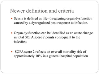
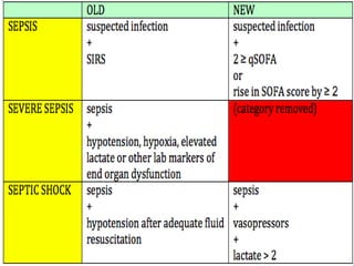
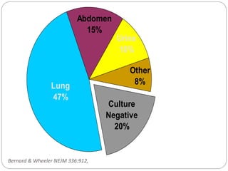
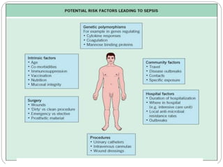
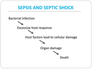
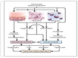
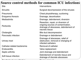
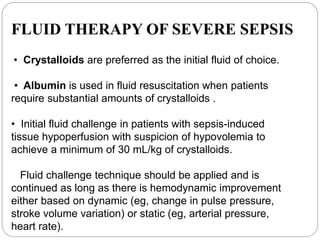
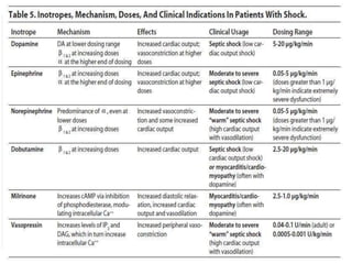
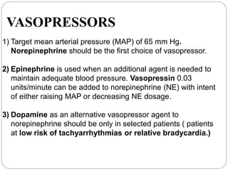
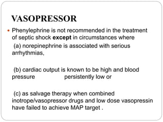
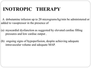
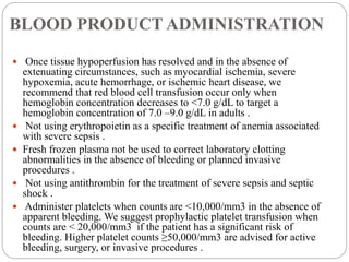
This document discusses sepsis diagnosis and management. It provides historical context on defining sepsis and outlines diagnostic criteria. Sepsis is defined as a life-threatening organ dysfunction caused by a dysregulated response to infection. Common infections that cause sepsis include those of the lung, abdomen, and urine. Management involves initial resuscitation, administering appropriate intravenous antibiotics within 1 hour, and controlling the infection source when possible through procedures like drainage or debridement. Vasopressors, fluid resuscitation, and inotropes may be needed to support blood pressure and organ perfusion.


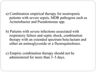
![ORGAN-DYSFUNCTION VARIABLES
• Arterial hypoxemia (ratio of the PaO2 to FIO2, <300)
• Acute oliguria (urine output, <0.5 ml/kg/hr or 45 ml/hr for at least 2 hr)
• Increase in creatinine level of >0.5 mg/dl (>44 μmol/liter)
• Coagulation abnormalities (INR, >1.5; or APTT >60 sec)
• Paralytic ileus (absence of bowel sounds)
• Thrombocytopenia (platelet count, <100,000/mm3)
• Hyperbilirubinemia (plasma total bilirubin, >4 mg/dl [68 μmol/liter])
TISSUE-PERFUSION VARIABLES
• Hyperlactatemia (lactate, >1 mmol/liter)
• Decreased capillary refill or mottling
SEVERE SEPSIS (SEPSIS PLUS ORGAN DYSFUNCTION)
SEPTIC SHOCK (SEPSIS PLUS EITHER HYPOTENSION [REFRACTORY TO
INTRAVENOUS FLUIDS] OR HYPERLACTATEMIA)
INTENSIVE CARE MEDICINE 2003](https://image.slidesharecdn.com/septicshockmanagement1-161215173034/85/Septic-shock-management-1-8-320.jpg)