SlideShare a Scribd company logo
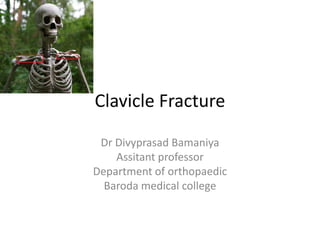
Clavicle Fracture
Dr Divyprasad Bamaniya
Assitant professor
Department of orthopaedic
Baroda medical college
Also known as “collar bone” and
“beauty bone”
Anatomy of clavicle
Mechanism of Injury
• Indirect injury
• Direct injury
Clinical Evaluation
• Clinical Presantation
• Neurovascular examination
• Skin integrity ,open or close
• Rule out chest injury
Associated injuries
• 9 % clavicle fracture associated with rib injury
• Most brachial plexus injury associated with
medial third clavicle fracture
Radiographic evaluation
• Standard AP view
• 30 degree cephalad view
Clavicle fracture AP view
Serendipity view
Allman Classification
Treatment
• Nonoperative / conservative
• Operative
Nonoperative management
• Figure of eight banadage
• Sling
• Minimally displaced fracture treated conservatively
Figure of eight bandage
sling
physiotherapy
Indication for Operative management
• Open fracture
• Fracture with neurovascular injury
• Fracture with skin tent
• Potential for progression to open fracture
Modalities of operative fixation
• Plate fixation
• Intramedullary fixtation
Plate fixation
• Incision
• Open the fracture
• Reduction of fracture
• Fixation with plate and screw
• Advantage: more secure fixation than nail
• Disadvantage: palpable hardware, iatrogenic
neurovscular injury, cosmetic deformity
1.Superior platting
2. anteroinferior platting
Postoperative xray
Intraoperative images
Intramedullary fixation
• 50 % cases operated with nailing associated
with complication
• Hardware migration
• Insertion site skin erosion and infection
Complication
• Neurovascular injury
• Malunion
• Nonunion
• Post-traumatic arthritis
Malunion
• Cosmetic deformity
• Shoulder dysfunction
factors responsible for development of
NONUNION
• Open fracture
• Displaced fracture
• Soft tissue interposition
• Old age
• Poor nutritional status
• Inadequate immobilisation
Shoulder Dislocation
• Also known as glenohumeral dislocation
• Most commonaly dislocated joint of body
• Constitute 45% of all dislocation
Types of shoulder dislocation
• Anterior –most common type
• Poterior – second most common
• Superior
• Inferior / also known as LUXATIO ERECTA
Anatomy of shoulder joint
• Greatest range of motion of any joint in body
• Due to shallow glenoid fossa which 25% of
size of humeral head
• Major contributor for stability is not bone
• But soft tissue envelope composed of capsule
,ligaments,and muscle are major stabiliser of
shoulder joint
Joint capsule
Glenohumeral ligaments
Rotator cuff muscle
• Active stabiliser of shoulder
• Four muscle- subscapularis, pteres minor,
supraspinatus ,infraspinatus
Pteres minor and pteres major
Supraspinatus and infraspinatus
Long head of biceps
Shoulder joint stabiliser
• Passive
• active
Passive shoulder joint stabiliser
1. Joint conformity
2. Vaccum effect of joint
3. Adhesion and cohesion forces due to presence
of synovial fluid
4. Scapular inclination
5. Joint capsule
6. Glenoid labrum
7. Bony prominences like acromion and coracoid
Active shoulder stabiliser
• Long head of biceps
• Rotator cuff muscle
Pathoanatomy of shoulder dislocation
1. Streching and tearing of capsule
2. HAGL lesion- humeral avulsion of
glenohumeral ligament
3. Bankart lesion – bony bankart, soft tissue
bankart
4. HILL-SACHS lesion- posterolateral head
defect cuased by impression fracture on
glenoid rim
Bankart lesion
• Soft tissue bankart • Bony bankart
Hill sachs lesion
Mechanism of injury
1. Indirect
 For anterior shoulder dislocation
upper limb with shoulder abducted ,extension
and external rotation
 For posterior shoulder dislocation
upper limb with shoulder adducted, flexed and
internally rotated
 In inferior shoulder dislocation
shoulder is hyper abducted
2. Direct
impact force directly on shoulder anteriorly
or posteriorly
3.Convulsion and electric shock produce
posterior shoulder dislocation
Clinical evalution
• Injured shoulder held in abduction and external
rotation
• Shoulder is painful with muscular spasm
• On examination sqauaring of shoulder with
relative prominance of acromion and hollow
beneath the acromion posteriorly and palpable
mass anteriorly
• Neurovascular examianation
check integrity of axillary nerve and
musculocutaneus nerve
Clinical image ; left shoulder
dislocation
Test for shoulder dislocation
1. Bryant test: anterior axillary fold at lower level
2. Callway test: vertical circumference of axilla is
increased
3. Dugas test: it is not possible for pt to bring
elbow near to opposite shoulder
4. Hamilton ruler test: because of flattening of
shoulder ruler placed on lateral side of arm and
it touches acromion and lateral condyle of
humerus
Clinical presentation of inferior
shoulder dislocation
• Salute fashion; humerus locked in 110 to 160
degree of abduction and forward elevation
• Humeral head palpable on lateral chest wall
• Almost all cases associated with neurovascular
injury
Salute fashion
Apprehension test
• For recurrent shoulder dislocation
• Passively shoulder placed in abduction
,extension and external rotation this position
reproduce patient sense of instability and pain
Radiographic evalution
• Standard AP view
• Axillary view
Axillary view
Anterior dislocation classification
• Sub corocoid
• Subglenoid
• intrathoracic
Posterior dislocation classification
• Sub acromion
• Subglenoid
• Subspinous
Treatment
nonoperative
1. Traction -counter traction method
2. Hippocratic technique
3. Stimson gravity technique
4. Milch technique
5. Kocher maneuver
Traction- counter traction method
Hippocratic technique
Stimson gravity technique
Kocher maneuver
Indication for operative management
or open reduction
• Shoulder is not reduced by reduction methods
• Shoulder dislocation with greater tubercle
fracture
• Dislocation with Glenoid rim fracture
Operative management of recurrent
shoulder dislocation
1. Bankart repair: detached anterior strcture are
attached to rim of glenoid cavity with suture
2. Puttiplat operation : subscapularis tendon
and capsule overlapped and tightned
3. Latarjet bristow operation: transplantation of
coracoid with its attachment to anterior rim
of glenoid
Bankart repair
Latarjet bristow operation
Complications
1. Recurrent dislocation
2. Osseous lesion
 Hill sachs lesion
 Bankart lesion
 Greater tuberosity facture
 Fracture of acromion and coracoid
3.Soft tissue injury
 Rotator cuff tear
 Capsular injury
4.Vascular injury – involve axillary artery
5.Nerve injury – involve axillary nerve and
musculocutaneus nerve
PROXIMAL HUMERUS FRACTURE
Relevent anatomy
• The humeral head
• The lesser tuberosity
• The greater tuberosity
• The humeral shaft
Deforming muscular forces on the
osseous segment
1. Greater tuberosity displaced superiorly and
posteriorly by supraspinatous and external
rotators
2. The lesser tuberosity displaced medially by
pull of subscapularis
3. The humeral shaft is displaced medially by
pectoralis major
4. The deltoid insertion cause abduction of
proximal fragment
Direction of displacement of osseous
fragment due to muscular forces
Neurovascular supply
a) Major blood supply from anterior and
posterior humeral circumflex arteries
b) Arcuate artery is continuation of ascending
branch of anterior humeral circumflex.
c) Its enter from bicipital groove and supply
most of humeral head
d) Fracture of anatomic neck have poor
prognosis because of precarious vascular
supply of humeral head
Axillary nerve
a) It is particular risk for traction injury because
of its close vicinity
Mechanism of injury
1. Most common is a fall onto outstreched
upper limb from height, commonaly in older
,osteoporotic woman.
2. Proximal humerus fracture in younger patient
associated with road traffic accident
3. Less common mechanism include excessive
shoulder abduction,electric shock, seizure,
benign and malignant involvement of
proximal humerus
Radiographic evalution
• Standard AP view
• Axillary view
Neer’s classification
• Neer divided proximal humerus in four part
• Greater and lesser tuberosity, humeral shaft
and humeral head
• Fracture types:
1. One part fracture: no displaced fragment
regardless of fracture lines
2. Two part fracture: anatomic neck,surgical
neck, greater tuberosity,lesser tuberosity
3. Three part fracture:
 Surgical neck with greater tuberosity
 Surgical neck with lesser tuberosity
4. Four part fracture
5.Fracture dislocation
6. Articular surface fracture
Neer’s classification
Treatment
1. One part fracture / not displaced
• Upto 85 % fracture are nondisplaced
• treated with Sling and immobilization
Two part fracture
A. Anatomic neck fracture
• Associated with high incidence of
osteonecrosis
• They require open reduction internal fixation
(ORIF)
• If fracture is not fixable required shoulder
hemiarthroplasty using neers prosthesis
ORIF
Neer’s prosthesis
Hip prosthesis
B. Surgical neck fracture
Reducible fracture and fracture with good
bone quality fixed percutaneously using k wire
and cannulated screw
Irreducible fracture and fracture with poor
bone quality require ORIF
C. Greater tuberosity fracture
 undisplaced fracture treated non operatively
Displaced fracture around 5 to 10 mm
require ORIF
D. Lesser tuberosity fracture
 its treated when it block internal rotation
Three and four part fracture
management
 displaced fracture require ORIF
 Delto pectoral approach
In Younger patient fracture fixed using plates
older patient benefit from prosthetic
replacement(hemiarthroplasty)
Articular surface fracture
• Hill sach and reverse hill sach lesion
• Patient with more than 40% head involvement
require prosthetic replacement
Complication
1. Vascular injury: axillary artery
2. Neural injury; axillary nerve and brachial
plexus injury
3. Osteonecrosis of head
4. Shoulder stiffness
5. Malunion
6. Nonunion
Acromioclavicular joint injury
• It is synovial plane joint
• It is complex of four ligament :anterior
posterior, superior ,inferior
• Superior is strongest of all
• Horizontal stability conferred by AC ligaments
• Vertical stability maintained by
coracoclavicular ligaments
Classification of acromioclavicular joint
injury
• Classification depending on degree of
direction of displacement of distal clavicle
• Rockwood classification
Clinical image and xray
Management of AC joint injury
• For nonoperative management ice packs and
sling is useful
• For operative management with hook platting,
cancelous screw fixation, fixation with k wire,
reconstrction of ligament are useful
Coracoclavicular ligament
reconstructed using autograft
Scapula fracture
Radiographic evaluation
• AP ,Axillary and scapular Y view required for
fracture diagnosis
Scapular Y view
Anatomic classification
I. Scapular body fracture
II. Apophseal fracture including acromion and
coracoid
III. Fracture of scapular neck and glenoid
Ideberg classification of intra articular
glenoid fracture
Classification of acromial fracture
I. Minimally displaced
II. Displaced but does not reduce subacromial
space
III. Displaced with narrowing of subacromial
space
Classification of coracoid fracture
I. Proximal to coracoclavicular ligament
II. Distal to coracoclavicular ligament
Floating shoulder?
• Double disruption of superior shoulder
suspensory complex(SSSC)
• Superior strut is middle third clavicle and
inferior strut is lateral scapular body
• Traumatic disruption of two or more
component described as floating shoulder
• Historically operative management has been
recommended because of potential instability
• Recent study show nonoperative treatment of
floating shoulder reported good result.
Nonoperative managment
• Non operative treatment required for most
scapular fracture
Indication for operative management
• Displaced intraarticular glenoid fracture involving
greater than 25% of articular surface
• Scapular neck fracture with greater than 40
degree of angulation or 1 cm medial translation
• Scapular neck fracture with associated displaced
clavicle fracture
• fracture of acromion that impinge on subacromial
space
• Fracture of coracoid process result in AC joint
sepration
Xray of operative management
Thank you
Protect your shoulder

More Related Content

What's hot

Proximal humerus fracture Management
Proximal humerus  fracture ManagementProximal humerus  fracture Management
Proximal humerus fracture Managementvaruntandra
 
Acetabular fracture
Acetabular fractureAcetabular fracture
Acetabular fracture
Harjot Gurudatta
 
Septic arthritis sequelae
Septic arthritis sequelaeSeptic arthritis sequelae
Septic arthritis sequelaeorthoprince
 
Perthes disease
Perthes diseasePerthes disease
Perthes disease
vashisth narayan
 
Terrible triad - elbow
Terrible triad - elbow Terrible triad - elbow
Terrible triad - elbow jatinder12345
 
supracondylar fracrture of humerus in children
supracondylar fracrture of humerus in childrensupracondylar fracrture of humerus in children
supracondylar fracrture of humerus in children
Hardik Pawar
 
Surgical approaches to hip joint
Surgical approaches to hip jointSurgical approaches to hip joint
Surgical approaches to hip joint
adityachakri
 
Perilunate dislocations
Perilunate dislocationsPerilunate dislocations
Perilunate dislocations
Rashik Ismail
 
Tension Band Wiring principles and applications
Tension Band Wiring  principles and applicationsTension Band Wiring  principles and applications
Tension Band Wiring principles and applications
Gaurav Singh
 
Primary total knee arthroplasty
Primary total knee arthroplastyPrimary total knee arthroplasty
Primary total knee arthroplastyjatinder12345
 
MULLER AO CLASSIFICATION
MULLER AO CLASSIFICATIONMULLER AO CLASSIFICATION
MULLER AO CLASSIFICATION
inks76
 
Osteotomy around knee dr shankar jangid (1)
Osteotomy around knee dr shankar jangid (1)Osteotomy around knee dr shankar jangid (1)
Osteotomy around knee dr shankar jangid (1)
ShankarJangid5
 
Carpal instability
Carpal instabilityCarpal instability
Carpal instability
azhanrubeesh
 
Proximal femur focal def
Proximal femur focal defProximal femur focal def
Proximal femur focal def
Ponnilavan Ponz
 
Thoracolumbar fracture cme
Thoracolumbar fracture cmeThoracolumbar fracture cme
Thoracolumbar fracture cme
Siti Aishah Mohd Zameri
 
Dr. yt reddy distal radius fractures modified
Dr. yt reddy distal radius fractures modifiedDr. yt reddy distal radius fractures modified
Dr. yt reddy distal radius fractures modifiedvaruntandra
 
supracondylar fracture
supracondylar fracturesupracondylar fracture
supracondylar fracture
MONTHER ALKHAWLANY
 
paediatric injuries around the elbow.
paediatric injuries around the elbow. paediatric injuries around the elbow.
paediatric injuries around the elbow.
yashavardhan yashu
 

What's hot (20)

Proximal humerus fracture Management
Proximal humerus  fracture ManagementProximal humerus  fracture Management
Proximal humerus fracture Management
 
Acetabular fracture
Acetabular fractureAcetabular fracture
Acetabular fracture
 
Septic arthritis sequelae
Septic arthritis sequelaeSeptic arthritis sequelae
Septic arthritis sequelae
 
Perthes disease
Perthes diseasePerthes disease
Perthes disease
 
Terrible triad - elbow
Terrible triad - elbow Terrible triad - elbow
Terrible triad - elbow
 
supracondylar fracrture of humerus in children
supracondylar fracrture of humerus in childrensupracondylar fracrture of humerus in children
supracondylar fracrture of humerus in children
 
Surgical approaches to hip joint
Surgical approaches to hip jointSurgical approaches to hip joint
Surgical approaches to hip joint
 
Perilunate dislocations
Perilunate dislocationsPerilunate dislocations
Perilunate dislocations
 
Tension Band Wiring principles and applications
Tension Band Wiring  principles and applicationsTension Band Wiring  principles and applications
Tension Band Wiring principles and applications
 
Primary total knee arthroplasty
Primary total knee arthroplastyPrimary total knee arthroplasty
Primary total knee arthroplasty
 
MULLER AO CLASSIFICATION
MULLER AO CLASSIFICATIONMULLER AO CLASSIFICATION
MULLER AO CLASSIFICATION
 
Osteotomy around knee dr shankar jangid (1)
Osteotomy around knee dr shankar jangid (1)Osteotomy around knee dr shankar jangid (1)
Osteotomy around knee dr shankar jangid (1)
 
Carpal instability
Carpal instabilityCarpal instability
Carpal instability
 
Proximal femur focal def
Proximal femur focal defProximal femur focal def
Proximal femur focal def
 
Subtrochanteric fracture
Subtrochanteric fractureSubtrochanteric fracture
Subtrochanteric fracture
 
Thoracolumbar fracture cme
Thoracolumbar fracture cmeThoracolumbar fracture cme
Thoracolumbar fracture cme
 
The hip
The hipThe hip
The hip
 
Dr. yt reddy distal radius fractures modified
Dr. yt reddy distal radius fractures modifiedDr. yt reddy distal radius fractures modified
Dr. yt reddy distal radius fractures modified
 
supracondylar fracture
supracondylar fracturesupracondylar fracture
supracondylar fracture
 
paediatric injuries around the elbow.
paediatric injuries around the elbow. paediatric injuries around the elbow.
paediatric injuries around the elbow.
 

Similar to Clavicle fracture & injuries around shoulder

Shoulder fractures around the shoulder
Shoulder fractures around the shoulder Shoulder fractures around the shoulder
Shoulder fractures around the shoulder
bibincmc
 
proximalhumerusfractures-180929171924.pdf
proximalhumerusfractures-180929171924.pdfproximalhumerusfractures-180929171924.pdf
proximalhumerusfractures-180929171924.pdf
Shahzaib404607
 
1PROXIMAL_HUMERUS_FRACTURES_SURGICAL_MANAGEMENT_PHILOS_PLATING_FINAL.pptx
1PROXIMAL_HUMERUS_FRACTURES_SURGICAL_MANAGEMENT_PHILOS_PLATING_FINAL.pptx1PROXIMAL_HUMERUS_FRACTURES_SURGICAL_MANAGEMENT_PHILOS_PLATING_FINAL.pptx
1PROXIMAL_HUMERUS_FRACTURES_SURGICAL_MANAGEMENT_PHILOS_PLATING_FINAL.pptx
VigneshwarArumugam1
 
Surgical approach around hip and knee
Surgical approach around hip and knee Surgical approach around hip and knee
Surgical approach around hip and knee
Kaushal Kafle
 
Share Acromioclavicul-WPS Office.pptx
Share Acromioclavicul-WPS Office.pptxShare Acromioclavicul-WPS Office.pptx
Share Acromioclavicul-WPS Office.pptx
manasil1
 
Fractures around elbow lateral condyle and intercondylar fractures
 Fractures around elbow lateral condyle and intercondylar fractures Fractures around elbow lateral condyle and intercondylar fractures
Fractures around elbow lateral condyle and intercondylar fractures
Siddhartha Sinha
 
Fractures around elbow lateral condyle and intercondylar fractures
Fractures around elbow lateral condyle and intercondylar fracturesFractures around elbow lateral condyle and intercondylar fractures
Fractures around elbow lateral condyle and intercondylar fractures
Siddhartha Sinha
 
Lateral patellar compression syndrome
Lateral patellar compression syndromeLateral patellar compression syndrome
Lateral patellar compression syndrome
Amanj Gardi
 
Supracondylar humeral fracture
Supracondylar humeral fractureSupracondylar humeral fracture
Supracondylar humeral fracture
MinThu62
 
neck of femur fracture
neck of femur fractureneck of femur fracture
neck of femur fracture
mdtawfiqalam
 
Proximal humerus-fractures
Proximal humerus-fracturesProximal humerus-fractures
Proximal humerus-fractures
Prasanthmuddada
 
Hip dislocations and femoral head fractures
Hip dislocations and femoral head fracturesHip dislocations and femoral head fractures
Hip dislocations and femoral head fractures
Ahmed Ashour dr.
 
Pelvic injuries
Pelvic injuriesPelvic injuries
Pelvic injuries
Harsha Nandini
 
Humerus shaft fracture dr anand
Humerus shaft fracture dr anandHumerus shaft fracture dr anand
Humerus shaft fracture dr anand
anandanmm
 
L01 hip dislocation, pipkin
L01 hip dislocation, pipkinL01 hip dislocation, pipkin
L01 hip dislocation, pipkin
Claudiu Cucu
 
Radial head and neck fractures
Radial head and neck fracturesRadial head and neck fractures
Radial head and neck fractures
Johny Wilbert
 
FRACTURES OF LOWER LIMB 1.pdf
FRACTURES OF LOWER LIMB 1.pdfFRACTURES OF LOWER LIMB 1.pdf
FRACTURES OF LOWER LIMB 1.pdf
InsyirahHatta
 
The elbow fracture
The elbow fractureThe elbow fracture
The elbow fracture
MONTHER ALKHAWLANY
 
Humerus fracture
Humerus fractureHumerus fracture
Humerus fracture
Dr. Anshu Sharma
 

Similar to Clavicle fracture & injuries around shoulder (20)

Shoulder fractures around the shoulder
Shoulder fractures around the shoulder Shoulder fractures around the shoulder
Shoulder fractures around the shoulder
 
proximalhumerusfractures-180929171924.pdf
proximalhumerusfractures-180929171924.pdfproximalhumerusfractures-180929171924.pdf
proximalhumerusfractures-180929171924.pdf
 
1PROXIMAL_HUMERUS_FRACTURES_SURGICAL_MANAGEMENT_PHILOS_PLATING_FINAL.pptx
1PROXIMAL_HUMERUS_FRACTURES_SURGICAL_MANAGEMENT_PHILOS_PLATING_FINAL.pptx1PROXIMAL_HUMERUS_FRACTURES_SURGICAL_MANAGEMENT_PHILOS_PLATING_FINAL.pptx
1PROXIMAL_HUMERUS_FRACTURES_SURGICAL_MANAGEMENT_PHILOS_PLATING_FINAL.pptx
 
Surgical approach around hip and knee
Surgical approach around hip and knee Surgical approach around hip and knee
Surgical approach around hip and knee
 
Share Acromioclavicul-WPS Office.pptx
Share Acromioclavicul-WPS Office.pptxShare Acromioclavicul-WPS Office.pptx
Share Acromioclavicul-WPS Office.pptx
 
Fractures around elbow lateral condyle and intercondylar fractures
 Fractures around elbow lateral condyle and intercondylar fractures Fractures around elbow lateral condyle and intercondylar fractures
Fractures around elbow lateral condyle and intercondylar fractures
 
Fractures around elbow lateral condyle and intercondylar fractures
Fractures around elbow lateral condyle and intercondylar fracturesFractures around elbow lateral condyle and intercondylar fractures
Fractures around elbow lateral condyle and intercondylar fractures
 
Lateral patellar compression syndrome
Lateral patellar compression syndromeLateral patellar compression syndrome
Lateral patellar compression syndrome
 
Supracondylar humeral fracture
Supracondylar humeral fractureSupracondylar humeral fracture
Supracondylar humeral fracture
 
Cubitus valgus varus
Cubitus valgus varusCubitus valgus varus
Cubitus valgus varus
 
neck of femur fracture
neck of femur fractureneck of femur fracture
neck of femur fracture
 
Proximal humerus-fractures
Proximal humerus-fracturesProximal humerus-fractures
Proximal humerus-fractures
 
Hip dislocations and femoral head fractures
Hip dislocations and femoral head fracturesHip dislocations and femoral head fractures
Hip dislocations and femoral head fractures
 
Pelvic injuries
Pelvic injuriesPelvic injuries
Pelvic injuries
 
Humerus shaft fracture dr anand
Humerus shaft fracture dr anandHumerus shaft fracture dr anand
Humerus shaft fracture dr anand
 
L01 hip dislocation, pipkin
L01 hip dislocation, pipkinL01 hip dislocation, pipkin
L01 hip dislocation, pipkin
 
Radial head and neck fractures
Radial head and neck fracturesRadial head and neck fractures
Radial head and neck fractures
 
FRACTURES OF LOWER LIMB 1.pdf
FRACTURES OF LOWER LIMB 1.pdfFRACTURES OF LOWER LIMB 1.pdf
FRACTURES OF LOWER LIMB 1.pdf
 
The elbow fracture
The elbow fractureThe elbow fracture
The elbow fracture
 
Humerus fracture
Humerus fractureHumerus fracture
Humerus fracture
 

Recently uploaded

Report Back from SGO 2024: What’s the Latest in Cervical Cancer?
Report Back from SGO 2024: What’s the Latest in Cervical Cancer?Report Back from SGO 2024: What’s the Latest in Cervical Cancer?
Report Back from SGO 2024: What’s the Latest in Cervical Cancer?
bkling
 
KDIGO 2024 guidelines for diabetologists
KDIGO 2024 guidelines for diabetologistsKDIGO 2024 guidelines for diabetologists
KDIGO 2024 guidelines for diabetologists
د.محمود نجيب
 
Alcohol_Dr. Jeenal Mistry MD Pharmacology.pdf
Alcohol_Dr. Jeenal Mistry MD Pharmacology.pdfAlcohol_Dr. Jeenal Mistry MD Pharmacology.pdf
Alcohol_Dr. Jeenal Mistry MD Pharmacology.pdf
Dr Jeenal Mistry
 
POST OPERATIVE OLIGURIA and its management
POST OPERATIVE OLIGURIA and its managementPOST OPERATIVE OLIGURIA and its management
POST OPERATIVE OLIGURIA and its management
touseefaziz1
 
ARTHROLOGY PPT NCISM SYLLABUS AYURVEDA STUDENTS
ARTHROLOGY PPT NCISM SYLLABUS AYURVEDA STUDENTSARTHROLOGY PPT NCISM SYLLABUS AYURVEDA STUDENTS
ARTHROLOGY PPT NCISM SYLLABUS AYURVEDA STUDENTS
Dr. Vinay Pareek
 
Are There Any Natural Remedies To Treat Syphilis.pdf
Are There Any Natural Remedies To Treat Syphilis.pdfAre There Any Natural Remedies To Treat Syphilis.pdf
Are There Any Natural Remedies To Treat Syphilis.pdf
Little Cross Family Clinic
 
micro teaching on communication m.sc nursing.pdf
micro teaching on communication m.sc nursing.pdfmicro teaching on communication m.sc nursing.pdf
micro teaching on communication m.sc nursing.pdf
Anurag Sharma
 
Triangles of Neck and Clinical Correlation by Dr. RIG.pptx
Triangles of Neck and Clinical Correlation by Dr. RIG.pptxTriangles of Neck and Clinical Correlation by Dr. RIG.pptx
Triangles of Neck and Clinical Correlation by Dr. RIG.pptx
Dr. Rabia Inam Gandapore
 
TEST BANK for Operations Management, 14th Edition by William J. Stevenson, Ve...
TEST BANK for Operations Management, 14th Edition by William J. Stevenson, Ve...TEST BANK for Operations Management, 14th Edition by William J. Stevenson, Ve...
TEST BANK for Operations Management, 14th Edition by William J. Stevenson, Ve...
kevinkariuki227
 
Ocular injury ppt Upendra pal optometrist upums saifai etawah
Ocular injury  ppt  Upendra pal  optometrist upums saifai etawahOcular injury  ppt  Upendra pal  optometrist upums saifai etawah
Ocular injury ppt Upendra pal optometrist upums saifai etawah
pal078100
 
THOA 2.ppt Human Organ Transplantation Act
THOA 2.ppt Human Organ Transplantation ActTHOA 2.ppt Human Organ Transplantation Act
THOA 2.ppt Human Organ Transplantation Act
DrSathishMS1
 
Flu Vaccine Alert in Bangalore Karnataka
Flu Vaccine Alert in Bangalore KarnatakaFlu Vaccine Alert in Bangalore Karnataka
Flu Vaccine Alert in Bangalore Karnataka
addon Scans
 
Prix Galien International 2024 Forum Program
Prix Galien International 2024 Forum ProgramPrix Galien International 2024 Forum Program
Prix Galien International 2024 Forum Program
Levi Shapiro
 
24 Upakrama.pptx class ppt useful in all
24 Upakrama.pptx class ppt useful in all24 Upakrama.pptx class ppt useful in all
24 Upakrama.pptx class ppt useful in all
DrSathishMS1
 
Phone Us ❤85270-49040❤ #ℂall #gIRLS In Surat By Surat @ℂall @Girls Hotel With...
Phone Us ❤85270-49040❤ #ℂall #gIRLS In Surat By Surat @ℂall @Girls Hotel With...Phone Us ❤85270-49040❤ #ℂall #gIRLS In Surat By Surat @ℂall @Girls Hotel With...
Phone Us ❤85270-49040❤ #ℂall #gIRLS In Surat By Surat @ℂall @Girls Hotel With...
Savita Shen $i11
 
Charaka Samhita Sutra sthana Chapter 15 Upakalpaniyaadhyaya
Charaka Samhita Sutra sthana Chapter 15 UpakalpaniyaadhyayaCharaka Samhita Sutra sthana Chapter 15 Upakalpaniyaadhyaya
Charaka Samhita Sutra sthana Chapter 15 Upakalpaniyaadhyaya
Dr KHALID B.M
 
The Normal Electrocardiogram - Part I of II
The Normal Electrocardiogram - Part I of IIThe Normal Electrocardiogram - Part I of II
The Normal Electrocardiogram - Part I of II
MedicoseAcademics
 
Ozempic: Preoperative Management of Patients on GLP-1 Receptor Agonists
Ozempic: Preoperative Management of Patients on GLP-1 Receptor Agonists  Ozempic: Preoperative Management of Patients on GLP-1 Receptor Agonists
Ozempic: Preoperative Management of Patients on GLP-1 Receptor Agonists
Saeid Safari
 
Antiulcer drugs Advance Pharmacology .pptx
Antiulcer drugs Advance Pharmacology .pptxAntiulcer drugs Advance Pharmacology .pptx
Antiulcer drugs Advance Pharmacology .pptx
Rohit chaurpagar
 
BRACHYTHERAPY OVERVIEW AND APPLICATORS
BRACHYTHERAPY OVERVIEW  AND  APPLICATORSBRACHYTHERAPY OVERVIEW  AND  APPLICATORS
BRACHYTHERAPY OVERVIEW AND APPLICATORS
Krishan Murari
 

Recently uploaded (20)

Report Back from SGO 2024: What’s the Latest in Cervical Cancer?
Report Back from SGO 2024: What’s the Latest in Cervical Cancer?Report Back from SGO 2024: What’s the Latest in Cervical Cancer?
Report Back from SGO 2024: What’s the Latest in Cervical Cancer?
 
KDIGO 2024 guidelines for diabetologists
KDIGO 2024 guidelines for diabetologistsKDIGO 2024 guidelines for diabetologists
KDIGO 2024 guidelines for diabetologists
 
Alcohol_Dr. Jeenal Mistry MD Pharmacology.pdf
Alcohol_Dr. Jeenal Mistry MD Pharmacology.pdfAlcohol_Dr. Jeenal Mistry MD Pharmacology.pdf
Alcohol_Dr. Jeenal Mistry MD Pharmacology.pdf
 
POST OPERATIVE OLIGURIA and its management
POST OPERATIVE OLIGURIA and its managementPOST OPERATIVE OLIGURIA and its management
POST OPERATIVE OLIGURIA and its management
 
ARTHROLOGY PPT NCISM SYLLABUS AYURVEDA STUDENTS
ARTHROLOGY PPT NCISM SYLLABUS AYURVEDA STUDENTSARTHROLOGY PPT NCISM SYLLABUS AYURVEDA STUDENTS
ARTHROLOGY PPT NCISM SYLLABUS AYURVEDA STUDENTS
 
Are There Any Natural Remedies To Treat Syphilis.pdf
Are There Any Natural Remedies To Treat Syphilis.pdfAre There Any Natural Remedies To Treat Syphilis.pdf
Are There Any Natural Remedies To Treat Syphilis.pdf
 
micro teaching on communication m.sc nursing.pdf
micro teaching on communication m.sc nursing.pdfmicro teaching on communication m.sc nursing.pdf
micro teaching on communication m.sc nursing.pdf
 
Triangles of Neck and Clinical Correlation by Dr. RIG.pptx
Triangles of Neck and Clinical Correlation by Dr. RIG.pptxTriangles of Neck and Clinical Correlation by Dr. RIG.pptx
Triangles of Neck and Clinical Correlation by Dr. RIG.pptx
 
TEST BANK for Operations Management, 14th Edition by William J. Stevenson, Ve...
TEST BANK for Operations Management, 14th Edition by William J. Stevenson, Ve...TEST BANK for Operations Management, 14th Edition by William J. Stevenson, Ve...
TEST BANK for Operations Management, 14th Edition by William J. Stevenson, Ve...
 
Ocular injury ppt Upendra pal optometrist upums saifai etawah
Ocular injury  ppt  Upendra pal  optometrist upums saifai etawahOcular injury  ppt  Upendra pal  optometrist upums saifai etawah
Ocular injury ppt Upendra pal optometrist upums saifai etawah
 
THOA 2.ppt Human Organ Transplantation Act
THOA 2.ppt Human Organ Transplantation ActTHOA 2.ppt Human Organ Transplantation Act
THOA 2.ppt Human Organ Transplantation Act
 
Flu Vaccine Alert in Bangalore Karnataka
Flu Vaccine Alert in Bangalore KarnatakaFlu Vaccine Alert in Bangalore Karnataka
Flu Vaccine Alert in Bangalore Karnataka
 
Prix Galien International 2024 Forum Program
Prix Galien International 2024 Forum ProgramPrix Galien International 2024 Forum Program
Prix Galien International 2024 Forum Program
 
24 Upakrama.pptx class ppt useful in all
24 Upakrama.pptx class ppt useful in all24 Upakrama.pptx class ppt useful in all
24 Upakrama.pptx class ppt useful in all
 
Phone Us ❤85270-49040❤ #ℂall #gIRLS In Surat By Surat @ℂall @Girls Hotel With...
Phone Us ❤85270-49040❤ #ℂall #gIRLS In Surat By Surat @ℂall @Girls Hotel With...Phone Us ❤85270-49040❤ #ℂall #gIRLS In Surat By Surat @ℂall @Girls Hotel With...
Phone Us ❤85270-49040❤ #ℂall #gIRLS In Surat By Surat @ℂall @Girls Hotel With...
 
Charaka Samhita Sutra sthana Chapter 15 Upakalpaniyaadhyaya
Charaka Samhita Sutra sthana Chapter 15 UpakalpaniyaadhyayaCharaka Samhita Sutra sthana Chapter 15 Upakalpaniyaadhyaya
Charaka Samhita Sutra sthana Chapter 15 Upakalpaniyaadhyaya
 
The Normal Electrocardiogram - Part I of II
The Normal Electrocardiogram - Part I of IIThe Normal Electrocardiogram - Part I of II
The Normal Electrocardiogram - Part I of II
 
Ozempic: Preoperative Management of Patients on GLP-1 Receptor Agonists
Ozempic: Preoperative Management of Patients on GLP-1 Receptor Agonists  Ozempic: Preoperative Management of Patients on GLP-1 Receptor Agonists
Ozempic: Preoperative Management of Patients on GLP-1 Receptor Agonists
 
Antiulcer drugs Advance Pharmacology .pptx
Antiulcer drugs Advance Pharmacology .pptxAntiulcer drugs Advance Pharmacology .pptx
Antiulcer drugs Advance Pharmacology .pptx
 
BRACHYTHERAPY OVERVIEW AND APPLICATORS
BRACHYTHERAPY OVERVIEW  AND  APPLICATORSBRACHYTHERAPY OVERVIEW  AND  APPLICATORS
BRACHYTHERAPY OVERVIEW AND APPLICATORS
 

Clavicle fracture & injuries around shoulder

  • 1. Clavicle Fracture Dr Divyprasad Bamaniya Assitant professor Department of orthopaedic Baroda medical college
  • 2. Also known as “collar bone” and “beauty bone”
  • 4.
  • 5.
  • 6.
  • 7. Mechanism of Injury • Indirect injury • Direct injury
  • 8. Clinical Evaluation • Clinical Presantation • Neurovascular examination • Skin integrity ,open or close • Rule out chest injury
  • 9. Associated injuries • 9 % clavicle fracture associated with rib injury • Most brachial plexus injury associated with medial third clavicle fracture
  • 10. Radiographic evaluation • Standard AP view • 30 degree cephalad view
  • 13.
  • 15.
  • 16.
  • 17.
  • 18. Treatment • Nonoperative / conservative • Operative
  • 19. Nonoperative management • Figure of eight banadage • Sling • Minimally displaced fracture treated conservatively
  • 20. Figure of eight bandage
  • 21.
  • 22. sling
  • 24. Indication for Operative management • Open fracture • Fracture with neurovascular injury • Fracture with skin tent • Potential for progression to open fracture
  • 25. Modalities of operative fixation • Plate fixation • Intramedullary fixtation
  • 26. Plate fixation • Incision • Open the fracture • Reduction of fracture • Fixation with plate and screw • Advantage: more secure fixation than nail • Disadvantage: palpable hardware, iatrogenic neurovscular injury, cosmetic deformity
  • 30. Intramedullary fixation • 50 % cases operated with nailing associated with complication • Hardware migration • Insertion site skin erosion and infection
  • 31. Complication • Neurovascular injury • Malunion • Nonunion • Post-traumatic arthritis
  • 32. Malunion • Cosmetic deformity • Shoulder dysfunction
  • 33. factors responsible for development of NONUNION • Open fracture • Displaced fracture • Soft tissue interposition • Old age • Poor nutritional status • Inadequate immobilisation
  • 34.
  • 35. Shoulder Dislocation • Also known as glenohumeral dislocation • Most commonaly dislocated joint of body • Constitute 45% of all dislocation
  • 36. Types of shoulder dislocation • Anterior –most common type • Poterior – second most common • Superior • Inferior / also known as LUXATIO ERECTA
  • 38. • Greatest range of motion of any joint in body • Due to shallow glenoid fossa which 25% of size of humeral head • Major contributor for stability is not bone • But soft tissue envelope composed of capsule ,ligaments,and muscle are major stabiliser of shoulder joint
  • 41. Rotator cuff muscle • Active stabiliser of shoulder • Four muscle- subscapularis, pteres minor, supraspinatus ,infraspinatus
  • 42. Pteres minor and pteres major
  • 44. Long head of biceps
  • 45. Shoulder joint stabiliser • Passive • active
  • 46. Passive shoulder joint stabiliser 1. Joint conformity 2. Vaccum effect of joint 3. Adhesion and cohesion forces due to presence of synovial fluid 4. Scapular inclination 5. Joint capsule 6. Glenoid labrum 7. Bony prominences like acromion and coracoid
  • 47. Active shoulder stabiliser • Long head of biceps • Rotator cuff muscle
  • 48. Pathoanatomy of shoulder dislocation 1. Streching and tearing of capsule 2. HAGL lesion- humeral avulsion of glenohumeral ligament 3. Bankart lesion – bony bankart, soft tissue bankart 4. HILL-SACHS lesion- posterolateral head defect cuased by impression fracture on glenoid rim
  • 49. Bankart lesion • Soft tissue bankart • Bony bankart
  • 51. Mechanism of injury 1. Indirect  For anterior shoulder dislocation upper limb with shoulder abducted ,extension and external rotation  For posterior shoulder dislocation upper limb with shoulder adducted, flexed and internally rotated  In inferior shoulder dislocation shoulder is hyper abducted
  • 52. 2. Direct impact force directly on shoulder anteriorly or posteriorly 3.Convulsion and electric shock produce posterior shoulder dislocation
  • 53. Clinical evalution • Injured shoulder held in abduction and external rotation • Shoulder is painful with muscular spasm • On examination sqauaring of shoulder with relative prominance of acromion and hollow beneath the acromion posteriorly and palpable mass anteriorly • Neurovascular examianation check integrity of axillary nerve and musculocutaneus nerve
  • 54. Clinical image ; left shoulder dislocation
  • 55. Test for shoulder dislocation 1. Bryant test: anterior axillary fold at lower level 2. Callway test: vertical circumference of axilla is increased 3. Dugas test: it is not possible for pt to bring elbow near to opposite shoulder 4. Hamilton ruler test: because of flattening of shoulder ruler placed on lateral side of arm and it touches acromion and lateral condyle of humerus
  • 56.
  • 57. Clinical presentation of inferior shoulder dislocation • Salute fashion; humerus locked in 110 to 160 degree of abduction and forward elevation • Humeral head palpable on lateral chest wall • Almost all cases associated with neurovascular injury
  • 59. Apprehension test • For recurrent shoulder dislocation • Passively shoulder placed in abduction ,extension and external rotation this position reproduce patient sense of instability and pain
  • 60. Radiographic evalution • Standard AP view • Axillary view
  • 62.
  • 63. Anterior dislocation classification • Sub corocoid • Subglenoid • intrathoracic
  • 64. Posterior dislocation classification • Sub acromion • Subglenoid • Subspinous
  • 65. Treatment nonoperative 1. Traction -counter traction method 2. Hippocratic technique 3. Stimson gravity technique 4. Milch technique 5. Kocher maneuver
  • 70. Indication for operative management or open reduction • Shoulder is not reduced by reduction methods • Shoulder dislocation with greater tubercle fracture • Dislocation with Glenoid rim fracture
  • 71. Operative management of recurrent shoulder dislocation 1. Bankart repair: detached anterior strcture are attached to rim of glenoid cavity with suture 2. Puttiplat operation : subscapularis tendon and capsule overlapped and tightned 3. Latarjet bristow operation: transplantation of coracoid with its attachment to anterior rim of glenoid
  • 74. Complications 1. Recurrent dislocation 2. Osseous lesion  Hill sachs lesion  Bankart lesion  Greater tuberosity facture  Fracture of acromion and coracoid 3.Soft tissue injury  Rotator cuff tear  Capsular injury 4.Vascular injury – involve axillary artery 5.Nerve injury – involve axillary nerve and musculocutaneus nerve
  • 75.
  • 76. PROXIMAL HUMERUS FRACTURE Relevent anatomy • The humeral head • The lesser tuberosity • The greater tuberosity • The humeral shaft
  • 77. Deforming muscular forces on the osseous segment 1. Greater tuberosity displaced superiorly and posteriorly by supraspinatous and external rotators 2. The lesser tuberosity displaced medially by pull of subscapularis 3. The humeral shaft is displaced medially by pectoralis major 4. The deltoid insertion cause abduction of proximal fragment
  • 78. Direction of displacement of osseous fragment due to muscular forces
  • 79. Neurovascular supply a) Major blood supply from anterior and posterior humeral circumflex arteries b) Arcuate artery is continuation of ascending branch of anterior humeral circumflex. c) Its enter from bicipital groove and supply most of humeral head d) Fracture of anatomic neck have poor prognosis because of precarious vascular supply of humeral head
  • 80.
  • 81. Axillary nerve a) It is particular risk for traction injury because of its close vicinity
  • 82. Mechanism of injury 1. Most common is a fall onto outstreched upper limb from height, commonaly in older ,osteoporotic woman. 2. Proximal humerus fracture in younger patient associated with road traffic accident 3. Less common mechanism include excessive shoulder abduction,electric shock, seizure, benign and malignant involvement of proximal humerus
  • 83. Radiographic evalution • Standard AP view • Axillary view
  • 84. Neer’s classification • Neer divided proximal humerus in four part • Greater and lesser tuberosity, humeral shaft and humeral head • Fracture types: 1. One part fracture: no displaced fragment regardless of fracture lines 2. Two part fracture: anatomic neck,surgical neck, greater tuberosity,lesser tuberosity
  • 85. 3. Three part fracture:  Surgical neck with greater tuberosity  Surgical neck with lesser tuberosity 4. Four part fracture 5.Fracture dislocation 6. Articular surface fracture
  • 87. Treatment 1. One part fracture / not displaced • Upto 85 % fracture are nondisplaced • treated with Sling and immobilization
  • 88. Two part fracture A. Anatomic neck fracture • Associated with high incidence of osteonecrosis • They require open reduction internal fixation (ORIF) • If fracture is not fixable required shoulder hemiarthroplasty using neers prosthesis
  • 89. ORIF
  • 92. B. Surgical neck fracture Reducible fracture and fracture with good bone quality fixed percutaneously using k wire and cannulated screw Irreducible fracture and fracture with poor bone quality require ORIF
  • 93. C. Greater tuberosity fracture  undisplaced fracture treated non operatively Displaced fracture around 5 to 10 mm require ORIF D. Lesser tuberosity fracture  its treated when it block internal rotation
  • 94. Three and four part fracture management  displaced fracture require ORIF  Delto pectoral approach In Younger patient fracture fixed using plates older patient benefit from prosthetic replacement(hemiarthroplasty)
  • 95. Articular surface fracture • Hill sach and reverse hill sach lesion • Patient with more than 40% head involvement require prosthetic replacement
  • 96. Complication 1. Vascular injury: axillary artery 2. Neural injury; axillary nerve and brachial plexus injury 3. Osteonecrosis of head 4. Shoulder stiffness 5. Malunion 6. Nonunion
  • 97. Acromioclavicular joint injury • It is synovial plane joint • It is complex of four ligament :anterior posterior, superior ,inferior • Superior is strongest of all • Horizontal stability conferred by AC ligaments • Vertical stability maintained by coracoclavicular ligaments
  • 98.
  • 99. Classification of acromioclavicular joint injury • Classification depending on degree of direction of displacement of distal clavicle • Rockwood classification
  • 100.
  • 102. Management of AC joint injury • For nonoperative management ice packs and sling is useful • For operative management with hook platting, cancelous screw fixation, fixation with k wire, reconstrction of ligament are useful
  • 105. Radiographic evaluation • AP ,Axillary and scapular Y view required for fracture diagnosis
  • 107. Anatomic classification I. Scapular body fracture II. Apophseal fracture including acromion and coracoid III. Fracture of scapular neck and glenoid
  • 108. Ideberg classification of intra articular glenoid fracture
  • 109. Classification of acromial fracture I. Minimally displaced II. Displaced but does not reduce subacromial space III. Displaced with narrowing of subacromial space
  • 110. Classification of coracoid fracture I. Proximal to coracoclavicular ligament II. Distal to coracoclavicular ligament
  • 111. Floating shoulder? • Double disruption of superior shoulder suspensory complex(SSSC)
  • 112. • Superior strut is middle third clavicle and inferior strut is lateral scapular body • Traumatic disruption of two or more component described as floating shoulder • Historically operative management has been recommended because of potential instability • Recent study show nonoperative treatment of floating shoulder reported good result.
  • 113. Nonoperative managment • Non operative treatment required for most scapular fracture
  • 114. Indication for operative management • Displaced intraarticular glenoid fracture involving greater than 25% of articular surface • Scapular neck fracture with greater than 40 degree of angulation or 1 cm medial translation • Scapular neck fracture with associated displaced clavicle fracture • fracture of acromion that impinge on subacromial space • Fracture of coracoid process result in AC joint sepration
  • 115. Xray of operative management