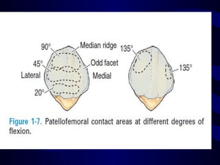
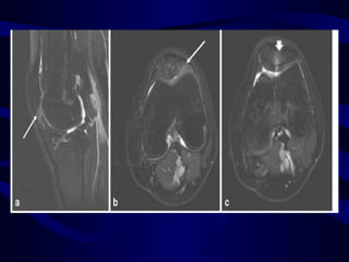
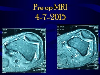
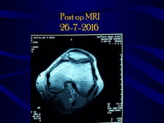
Lateral Patellar
Compression syndrome
Prepared by:
Dr.Amanj Mohsin Mustafa
4th
year candidate at KBMS
Supervised by :
Dr abdulkader alany
Overview
• Anatomy and Biomechanics
• Definition
• Causes of lateral compression
• Sign and symptom
• Examination
• Radiological finding and measurements
• Treatment
• Summmary
• Take home message
• Reference
Anatomy
• largest sesamoid bone in body,sits in
femoral trochlea
• asymmetrical oval with its apex directed
distally
Biomechanics
• Aid knee for extension
• Increase lever arm
• Stress disturbution
• Bears greatets load
• 0.5 B. W with normal walking
• 7 times B. w during squating
• 20 times during jumping
Definition
• Improper tracking of patella in trochlear
groove but with out presence of
instability is often associated with
overload & increased pressure on
lateral facet due to pathologic lateral soft-
tissue restraints
• Chronic anterior knee pain
Causes
1.tight lateral retinaculum leads to excessive
lateral tilt without excessive patellar mobility
2.Miserable Triad
is a term used for anatomic characteristics that
lead to an increased Q angle and an
exacerbation of patellofemoral dysplasia.
•  femoral anteversion
•  genu valgum
•  external tibial torsion / pronated feet
Sign and symptom
• Anterior Knee pain that is out of
proportion with physical examination
• pain with stair climbing & descending
• theatre sign (pain with sitting for long
periods of time)
• Pain & tenderness at lateral side of
patella & some time on medial side
because tension on medial retinaculum
• Patients deny instability or crepitus
 Physical exam• Look:
• Supine:
any muscle waisting, scar , discoloration
Sitting with 90 knee:
any deformity , swelling
Standing :
for any valgus ,external tibial torsion
Walking : again for deformity and rotation
Feel :
tenderness usually at lateral side of patella
present
Movement:
Active movement
Passive movement
Tests :
• Measurement Q-angle by standing
• Apprehension test for patella
• inability to evert the lateral edge of the patella
• Palm of the patella to check for articular
surface
Radiological findings and
measurements
1. X-Ray : Merchant view
Radiological findings and
measurements
1. X-Ray : Sunrise view
measurements for Trochlear dysplasia
usually we have to know about such
condition to exclude trochlear
dysplasia
D angle 145
Sulcus angle
0.8- 2.9 mm normal
Normal range less than 20mm
MRI findings
MRI findings
Treatment
• Nonoperative
• indications
mainstay of treatment & should be done
for extensive period of time
• Technique
• therapy should emphasize vastus
medialis strengthening & closed chain
short arc quadriceps exercises
Operative
• arthroscopic lateral release
• objective evidence of lateral tilting (neutral
or negative tilt)( no subluxation)
• pain refractory to extensive rehabilitation
• inability to evert lateral edge of patella
• ideal candidate has no symptoms of
instability
• medial patellar glide of less than one
quadrant
• lateral patellar glide of less than three
quadrants
Laproscopic Release case
• Pre op
Release
Post release
Pre op MRI
4-7-2015
Post op MRI
26-7-2016
Approach to Distal
Realignment
1. objective anatomic malalignment has
been diagnosed
2. nonoperative treatment has failed.
3. If dislocations have occurred, one must
consider whether stabilization of patella
might be needed by medial
patellofemoral ligament imbrication or
reconstruction, along with correction of
underlying malalignment.
patellar realignment surgery
• Soft tissue procedures:
(Skeletally immature)
1)the Roux-Goldthwait procedure
in which lateral half of patellar tendon is
detached distally, passed behind medial
half of tendon, and sutured to pes
anserinus insertion,
• this procedure is no longer
recommended.
2.Galeazzi semitendinosus tenodesis :
Bony Procedures
• Maquet (tubercle anteriorization)
• indicated only for distal pole lesions
• Elevate about 1.2 cm which has
significant effect on decreasing joint
force but care should be taken for more
elevation because there is risk of skin
necrosis
• Anterior elevation of tibial tuberosity,
enhances efficiency of quadriceps by
increasing lever arm while decreasing
patellofemoral joint reaction force.
• increasing angle between vector of
quadriceps pull & patellar tendon
decreases joint reaction force.
• Elmslie-Trillat (medialization)
• indicated only for instability with lateral
translation (not isolated lateral tilt)
• avoid if medial patellar facet arthrosis
technique classically combines lateral
release, medial capsular reefing, and
medial displacement of bony insertion of
patellar tendon with distal displacement
titrated according to degree of patella
alta measured preoperatively.
• Fulkerson alignment surgery (tubercle
anteriorization & medialization)
• Indications (controversial)
1. lateral and distal pole lesions
2.increased Q angle
• contraindications
superior medial O.A
• Laboratory evaluation of this concept in a
cadaver model with increased lateral
facet overload induced by alteration of
proximal vector of quadriceps showed
excellent reduction of lateral facet
pressure.
• study reported a 30% reduction in lateral facet
pressure with anteriorization of 8.8 mm &
medialization of 8.4 mm,
• 65% relief after additional anteromedialization to
14.8/8.4 mm.
• By 20 to 30 degrees of knee flexion, reduction &
equalization of medial & lateral facet pressure were
noted, with greater reduction in more anteriorized
group
Complications
• 1.skin necrosis
• 2.Fracture
• 3.Compartment syndrome
Take Home Message
1.Patellofemoral joint (PFJ) disorders is
(black hole of orthopaedics).
2.Anterior Knee pain is common complain
and always don’t forget to exclude
patella pathology.
Take Home Message
3.Don’t accept Knee MRI with out axial
view specialy in cases with AKP.
4. With arthroscopy never forget to check
for patellar tracking
5.When ever see patient with chronic knee
pain and stable patella think about LPCS
Reference
1.Insall & Scott Surgery of the Knee
(FIFTH EDITION) 2012
2. CAMPBELL’S OPERATIVE ORTHOPAEDICS
(TWELFTH EDITION) 2013
3.Postgraduate Orthopaedics:
The Candidate’s Guide to the FRCS (Tr & Orth)
Examination (Second edition) 2012
4.Measurements and Classifications in
Musculoskeletal Radiology (2014)
5. Orthobullet 2015
6. A systematised MRI approach to evaluating the
patellofemoral joint
Published in final edited form as:
Skeletal Radiol. 2011 April ; 40(4): 375–387.
doi:10.1007/s00256-010-0909-1
• 7. Diagnosis and Treatment of Lateral Patellar
Compression Syndrome
Michael G. Saper, D.O., A.T.C., C.S.C.S., and David A.
Shneider, M.D.
Lateral patellar compression syndrome

Lateral patellar compression syndrome

  • 1.
    Lateral Patellar Compression syndrome Preparedby: Dr.Amanj Mohsin Mustafa 4th year candidate at KBMS Supervised by : Dr abdulkader alany
  • 2.
    Overview • Anatomy andBiomechanics • Definition • Causes of lateral compression • Sign and symptom • Examination • Radiological finding and measurements • Treatment • Summmary • Take home message • Reference
  • 3.
    Anatomy • largest sesamoidbone in body,sits in femoral trochlea • asymmetrical oval with its apex directed distally
  • 5.
    Biomechanics • Aid kneefor extension • Increase lever arm • Stress disturbution • Bears greatets load • 0.5 B. W with normal walking • 7 times B. w during squating • 20 times during jumping
  • 7.
    Definition • Improper trackingof patella in trochlear groove but with out presence of instability is often associated with overload & increased pressure on lateral facet due to pathologic lateral soft- tissue restraints • Chronic anterior knee pain
  • 8.
    Causes 1.tight lateral retinaculumleads to excessive lateral tilt without excessive patellar mobility 2.Miserable Triad is a term used for anatomic characteristics that lead to an increased Q angle and an exacerbation of patellofemoral dysplasia. •  femoral anteversion •  genu valgum •  external tibial torsion / pronated feet
  • 10.
    Sign and symptom •Anterior Knee pain that is out of proportion with physical examination • pain with stair climbing & descending • theatre sign (pain with sitting for long periods of time)
  • 11.
    • Pain &tenderness at lateral side of patella & some time on medial side because tension on medial retinaculum • Patients deny instability or crepitus
  • 12.
     Physical exam•Look: • Supine: any muscle waisting, scar , discoloration Sitting with 90 knee: any deformity , swelling Standing : for any valgus ,external tibial torsion Walking : again for deformity and rotation
  • 13.
    Feel : tenderness usuallyat lateral side of patella present Movement: Active movement Passive movement
  • 14.
    Tests : • MeasurementQ-angle by standing • Apprehension test for patella • inability to evert the lateral edge of the patella • Palm of the patella to check for articular surface
  • 18.
  • 19.
  • 25.
    measurements for Trochleardysplasia usually we have to know about such condition to exclude trochlear dysplasia
  • 26.
  • 27.
  • 30.
  • 31.
  • 32.
  • 35.
  • 36.
    Treatment • Nonoperative • indications mainstayof treatment & should be done for extensive period of time • Technique • therapy should emphasize vastus medialis strengthening & closed chain short arc quadriceps exercises
  • 39.
    Operative • arthroscopic lateralrelease • objective evidence of lateral tilting (neutral or negative tilt)( no subluxation) • pain refractory to extensive rehabilitation • inability to evert lateral edge of patella
  • 40.
    • ideal candidatehas no symptoms of instability • medial patellar glide of less than one quadrant • lateral patellar glide of less than three quadrants
  • 41.
  • 43.
  • 44.
  • 46.
  • 47.
  • 48.
    Approach to Distal Realignment 1.objective anatomic malalignment has been diagnosed 2. nonoperative treatment has failed. 3. If dislocations have occurred, one must consider whether stabilization of patella might be needed by medial patellofemoral ligament imbrication or reconstruction, along with correction of underlying malalignment.
  • 49.
    patellar realignment surgery •Soft tissue procedures: (Skeletally immature) 1)the Roux-Goldthwait procedure in which lateral half of patellar tendon is detached distally, passed behind medial half of tendon, and sutured to pes anserinus insertion,
  • 50.
    • this procedureis no longer recommended.
  • 51.
  • 52.
    Bony Procedures • Maquet(tubercle anteriorization) • indicated only for distal pole lesions • Elevate about 1.2 cm which has significant effect on decreasing joint force but care should be taken for more elevation because there is risk of skin necrosis
  • 53.
    • Anterior elevationof tibial tuberosity, enhances efficiency of quadriceps by increasing lever arm while decreasing patellofemoral joint reaction force. • increasing angle between vector of quadriceps pull & patellar tendon decreases joint reaction force.
  • 55.
    • Elmslie-Trillat (medialization) •indicated only for instability with lateral translation (not isolated lateral tilt) • avoid if medial patellar facet arthrosis
  • 56.
    technique classically combineslateral release, medial capsular reefing, and medial displacement of bony insertion of patellar tendon with distal displacement titrated according to degree of patella alta measured preoperatively.
  • 58.
    • Fulkerson alignmentsurgery (tubercle anteriorization & medialization) • Indications (controversial) 1. lateral and distal pole lesions 2.increased Q angle • contraindications superior medial O.A
  • 59.
    • Laboratory evaluationof this concept in a cadaver model with increased lateral facet overload induced by alteration of proximal vector of quadriceps showed excellent reduction of lateral facet pressure.
  • 60.
    • study reporteda 30% reduction in lateral facet pressure with anteriorization of 8.8 mm & medialization of 8.4 mm, • 65% relief after additional anteromedialization to 14.8/8.4 mm. • By 20 to 30 degrees of knee flexion, reduction & equalization of medial & lateral facet pressure were noted, with greater reduction in more anteriorized group
  • 64.
    Complications • 1.skin necrosis •2.Fracture • 3.Compartment syndrome
  • 65.
    Take Home Message 1.Patellofemoraljoint (PFJ) disorders is (black hole of orthopaedics). 2.Anterior Knee pain is common complain and always don’t forget to exclude patella pathology.
  • 66.
    Take Home Message 3.Don’taccept Knee MRI with out axial view specialy in cases with AKP. 4. With arthroscopy never forget to check for patellar tracking 5.When ever see patient with chronic knee pain and stable patella think about LPCS
  • 67.
    Reference 1.Insall & ScottSurgery of the Knee (FIFTH EDITION) 2012 2. CAMPBELL’S OPERATIVE ORTHOPAEDICS (TWELFTH EDITION) 2013 3.Postgraduate Orthopaedics: The Candidate’s Guide to the FRCS (Tr & Orth) Examination (Second edition) 2012 4.Measurements and Classifications in Musculoskeletal Radiology (2014) 5. Orthobullet 2015
  • 68.
    6. A systematisedMRI approach to evaluating the patellofemoral joint Published in final edited form as: Skeletal Radiol. 2011 April ; 40(4): 375–387. doi:10.1007/s00256-010-0909-1 • 7. Diagnosis and Treatment of Lateral Patellar Compression Syndrome Michael G. Saper, D.O., A.T.C., C.S.C.S., and David A. Shneider, M.D.