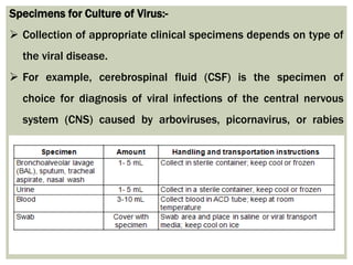
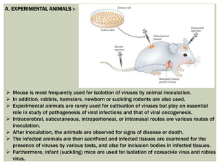
Poxviruses are brick or oval-shaped viruses with large double-stranded DNA genomes. Poxviruses exist throughout the world and cause disease in humans and many other types of animals. Poxvirus infections typically result in the formation of lesions, skin nodules, or disseminated rash.
Largest viruses that infect vertebrates
Can be seen under light microscope
Poxvirus diseases are characterized by skin lesions – localized or generalized
Important diseases caused by poxviruses are-
Smallpox
Monkeypox
Cowpox
Tanapox
Molluscum contagiosum
A picornavirus is a virus belonging to the family Picornaviridae, a family of viruses in the order Picornavirales. Vertebrates, including humans, serve as natural hosts. Picornaviruses are nonenveloped viruses that represent a large family of small, cytoplasmic, plus-strand RNA viruses with a 30-nm icosahedral capsid.
Serological test for virus identificationPlock Ghosh
This presentation consist of detailed study of serological method of virus identification. Basically ELISA is vastly used for virus detection. Western blot method is used for HIV identification.
Viruses are obligate intracellular parasites which means they can only grow or reproduce inside a host cell.
The primary purpose of virus cultivation:
To isolate and identify viruses in clinical samples.
To do research on the viral structure, replication, genetics, and effects on the host cell.
To prepare viruses for vaccine production.
Isolation of the virus is always considered a gold standard for establishing the viral origin of the disease
topics covered
CULTIVATION OF VIRUSES
Animal inoculation
Embryonated eggs
CAM
Allantoic cavity
Amniotic cavity
Yolk sac
Tissue culture
Organ culture
Explant culture
Cell culture
Primary cell culture
diploid cell culture
Continues cell lines
Viruses are obligate intracellular parasites so they depend on host for their survival. They cannot be grown in non-living culture media or on agar plates alone, they must require living cells to support their replication.Cultivation of viruses can be discussed under following headings:
Animal Inoculation
Inoculation into embryonated egg
Cell Culture
Virology is the scientific study of biological viruses. It is a subfield of microbiology that focuses on their detection, structure, classification and evolution, their methods of infection and exploitation of host cells for reproduction, their interaction with host organism physiology and immunity, the diseases they cause, the techniques to isolate and culture them, and their use in research and therapy
Report Back from SGO 2024: What’s the Latest in Cervical Cancer?bkling
Are you curious about what’s new in cervical cancer research or unsure what the findings mean? Join Dr. Emily Ko, a gynecologic oncologist at Penn Medicine, to learn about the latest updates from the Society of Gynecologic Oncology (SGO) 2024 Annual Meeting on Women’s Cancer. Dr. Ko will discuss what the research presented at the conference means for you and answer your questions about the new developments.
Pulmonary Thromboembolism - etilogy, types, medical- Surgical and nursing man...VarunMahajani
Disruption of blood supply to lung alveoli due to blockage of one or more pulmonary blood vessels is called as Pulmonary thromboembolism. In this presentation we will discuss its causes, types and its management in depth.
Anti ulcer drugs and their Advance pharmacology ||
Anti-ulcer drugs are medications used to prevent and treat ulcers in the stomach and upper part of the small intestine (duodenal ulcers). These ulcers are often caused by an imbalance between stomach acid and the mucosal lining, which protects the stomach lining.
||Scope: Overview of various classes of anti-ulcer drugs, their mechanisms of action, indications, side effects, and clinical considerations.
Title: Sense of Taste
Presenter: Dr. Faiza, Assistant Professor of Physiology
Qualifications:
MBBS (Best Graduate, AIMC Lahore)
FCPS Physiology
ICMT, CHPE, DHPE (STMU)
MPH (GC University, Faisalabad)
MBA (Virtual University of Pakistan)
Learning Objectives:
Describe the structure and function of taste buds.
Describe the relationship between the taste threshold and taste index of common substances.
Explain the chemical basis and signal transduction of taste perception for each type of primary taste sensation.
Recognize different abnormalities of taste perception and their causes.
Key Topics:
Significance of Taste Sensation:
Differentiation between pleasant and harmful food
Influence on behavior
Selection of food based on metabolic needs
Receptors of Taste:
Taste buds on the tongue
Influence of sense of smell, texture of food, and pain stimulation (e.g., by pepper)
Primary and Secondary Taste Sensations:
Primary taste sensations: Sweet, Sour, Salty, Bitter, Umami
Chemical basis and signal transduction mechanisms for each taste
Taste Threshold and Index:
Taste threshold values for Sweet (sucrose), Salty (NaCl), Sour (HCl), and Bitter (Quinine)
Taste index relationship: Inversely proportional to taste threshold
Taste Blindness:
Inability to taste certain substances, particularly thiourea compounds
Example: Phenylthiocarbamide
Structure and Function of Taste Buds:
Composition: Epithelial cells, Sustentacular/Supporting cells, Taste cells, Basal cells
Features: Taste pores, Taste hairs/microvilli, and Taste nerve fibers
Location of Taste Buds:
Found in papillae of the tongue (Fungiform, Circumvallate, Foliate)
Also present on the palate, tonsillar pillars, epiglottis, and proximal esophagus
Mechanism of Taste Stimulation:
Interaction of taste substances with receptors on microvilli
Signal transduction pathways for Umami, Sweet, Bitter, Sour, and Salty tastes
Taste Sensitivity and Adaptation:
Decrease in sensitivity with age
Rapid adaptation of taste sensation
Role of Saliva in Taste:
Dissolution of tastants to reach receptors
Washing away the stimulus
Taste Preferences and Aversions:
Mechanisms behind taste preference and aversion
Influence of receptors and neural pathways
Impact of Sensory Nerve Damage:
Degeneration of taste buds if the sensory nerve fiber is cut
Abnormalities of Taste Detection:
Conditions: Ageusia, Hypogeusia, Dysgeusia (parageusia)
Causes: Nerve damage, neurological disorders, infections, poor oral hygiene, adverse drug effects, deficiencies, aging, tobacco use, altered neurotransmitter levels
Neurotransmitters and Taste Threshold:
Effects of serotonin (5-HT) and norepinephrine (NE) on taste sensitivity
Supertasters:
25% of the population with heightened sensitivity to taste, especially bitterness
Increased number of fungiform papillae
Title: Sense of Smell
Presenter: Dr. Faiza, Assistant Professor of Physiology
Qualifications:
MBBS (Best Graduate, AIMC Lahore)
FCPS Physiology
ICMT, CHPE, DHPE (STMU)
MPH (GC University, Faisalabad)
MBA (Virtual University of Pakistan)
Learning Objectives:
Describe the primary categories of smells and the concept of odor blindness.
Explain the structure and location of the olfactory membrane and mucosa, including the types and roles of cells involved in olfaction.
Describe the pathway and mechanisms of olfactory signal transmission from the olfactory receptors to the brain.
Illustrate the biochemical cascade triggered by odorant binding to olfactory receptors, including the role of G-proteins and second messengers in generating an action potential.
Identify different types of olfactory disorders such as anosmia, hyposmia, hyperosmia, and dysosmia, including their potential causes.
Key Topics:
Olfactory Genes:
3% of the human genome accounts for olfactory genes.
400 genes for odorant receptors.
Olfactory Membrane:
Located in the superior part of the nasal cavity.
Medially: Folds downward along the superior septum.
Laterally: Folds over the superior turbinate and upper surface of the middle turbinate.
Total surface area: 5-10 square centimeters.
Olfactory Mucosa:
Olfactory Cells: Bipolar nerve cells derived from the CNS (100 million), with 4-25 olfactory cilia per cell.
Sustentacular Cells: Produce mucus and maintain ionic and molecular environment.
Basal Cells: Replace worn-out olfactory cells with an average lifespan of 1-2 months.
Bowman’s Gland: Secretes mucus.
Stimulation of Olfactory Cells:
Odorant dissolves in mucus and attaches to receptors on olfactory cilia.
Involves a cascade effect through G-proteins and second messengers, leading to depolarization and action potential generation in the olfactory nerve.
Quality of a Good Odorant:
Small (3-20 Carbon atoms), volatile, water-soluble, and lipid-soluble.
Facilitated by odorant-binding proteins in mucus.
Membrane Potential and Action Potential:
Resting membrane potential: -55mV.
Action potential frequency in the olfactory nerve increases with odorant strength.
Adaptation Towards the Sense of Smell:
Rapid adaptation within the first second, with further slow adaptation.
Psychological adaptation greater than receptor adaptation, involving feedback inhibition from the central nervous system.
Primary Sensations of Smell:
Camphoraceous, Musky, Floral, Pepperminty, Ethereal, Pungent, Putrid.
Odor Detection Threshold:
Examples: Hydrogen sulfide (0.0005 ppm), Methyl-mercaptan (0.002 ppm).
Some toxic substances are odorless at lethal concentrations.
Characteristics of Smell:
Odor blindness for single substances due to lack of appropriate receptor protein.
Behavioral and emotional influences of smell.
Transmission of Olfactory Signals:
From olfactory cells to glomeruli in the olfactory bulb, involving lateral inhibition.
Primitive, less old, and new olfactory systems with different path
The prostate is an exocrine gland of the male mammalian reproductive system
It is a walnut-sized gland that forms part of the male reproductive system and is located in front of the rectum and just below the urinary bladder
Function is to store and secrete a clear, slightly alkaline fluid that constitutes 10-30% of the volume of the seminal fluid that along with the spermatozoa, constitutes semen
A healthy human prostate measures (4cm-vertical, by 3cm-horizontal, 2cm ant-post ).
It surrounds the urethra just below the urinary bladder. It has anterior, median, posterior and two lateral lobes
It’s work is regulated by androgens which are responsible for male sex characteristics
Generalised disease of the prostate due to hormonal derangement which leads to non malignant enlargement of the gland (increase in the number of epithelial cells and stromal tissue)to cause compression of the urethra leading to symptoms (LUTS
These simplified slides by Dr. Sidra Arshad present an overview of the non-respiratory functions of the respiratory tract.
Learning objectives:
1. Enlist the non-respiratory functions of the respiratory tract
2. Briefly explain how these functions are carried out
3. Discuss the significance of dead space
4. Differentiate between minute ventilation and alveolar ventilation
5. Describe the cough and sneeze reflexes
Study Resources:
1. Chapter 39, Guyton and Hall Textbook of Medical Physiology, 14th edition
2. Chapter 34, Ganong’s Review of Medical Physiology, 26th edition
3. Chapter 17, Human Physiology by Lauralee Sherwood, 9th edition
4. Non-respiratory functions of the lungs https://academic.oup.com/bjaed/article/13/3/98/278874
Lung Cancer: Artificial Intelligence, Synergetics, Complex System Analysis, S...Oleg Kshivets
RESULTS: Overall life span (LS) was 2252.1±1742.5 days and cumulative 5-year survival (5YS) reached 73.2%, 10 years – 64.8%, 20 years – 42.5%. 513 LCP lived more than 5 years (LS=3124.6±1525.6 days), 148 LCP – more than 10 years (LS=5054.4±1504.1 days).199 LCP died because of LC (LS=562.7±374.5 days). 5YS of LCP after bi/lobectomies was significantly superior in comparison with LCP after pneumonectomies (78.1% vs.63.7%, P=0.00001 by log-rank test). AT significantly improved 5YS (66.3% vs. 34.8%) (P=0.00000 by log-rank test) only for LCP with N1-2. Cox modeling displayed that 5YS of LCP significantly depended on: phase transition (PT) early-invasive LC in terms of synergetics, PT N0—N12, cell ratio factors (ratio between cancer cells- CC and blood cells subpopulations), G1-3, histology, glucose, AT, blood cell circuit, prothrombin index, heparin tolerance, recalcification time (P=0.000-0.038). Neural networks, genetic algorithm selection and bootstrap simulation revealed relationships between 5YS and PT early-invasive LC (rank=1), PT N0—N12 (rank=2), thrombocytes/CC (3), erythrocytes/CC (4), eosinophils/CC (5), healthy cells/CC (6), lymphocytes/CC (7), segmented neutrophils/CC (8), stick neutrophils/CC (9), monocytes/CC (10); leucocytes/CC (11). Correct prediction of 5YS was 100% by neural networks computing (area under ROC curve=1.0; error=0.0).
CONCLUSIONS: 5YS of LCP after radical procedures significantly depended on: 1) PT early-invasive cancer; 2) PT N0--N12; 3) cell ratio factors; 4) blood cell circuit; 5) biochemical factors; 6) hemostasis system; 7) AT; 8) LC characteristics; 9) LC cell dynamics; 10) surgery type: lobectomy/pneumonectomy; 11) anthropometric data. Optimal diagnosis and treatment strategies for LC are: 1) screening and early detection of LC; 2) availability of experienced thoracic surgeons because of complexity of radical procedures; 3) aggressive en block surgery and adequate lymph node dissection for completeness; 4) precise prediction; 5) adjuvant chemoimmunoradiotherapy for LCP with unfavorable prognosis.
2. INTRODUCTION:-
Viruses are obligate intracellular parasites that require living cells in order to
replicate.
The primary purposes of virus cultivation is:
To isolate and identify viruses in clinical samples. Demonstration of virus in
appropriate clinical specimens by culture establishes diagnosis of viral
diseases.
To do research on viral structure, replication, genetics and effects on host cell.
To prepare viruses for vaccine production.
Isolation of virus is always considered as a gold standard for establishing viral
etiology of a disease.
Most of the viruses can be cultivated in
A. Experimental animals
B. Embryonated eggs or
C. Tissue culture.
3. Specimens for Culture of Virus:-
Collection of appropriate clinical specimens depends on type of
the viral disease.
For example, cerebrospinal fluid (CSF) is the specimen of
choice for diagnosis of viral infections of the central nervous
system (CNS) caused by arboviruses, picornavirus, or rabies
virus.
4. A. EXPERIMENTAL ANIMALS :-
Mouse is most frequently used for isolation of viruses by animal inoculation.
In addition, rabbits, hamsters, newborn or suckling rodents are also used.
Experimental animals are rarely used for cultivation of viruses but play an essential
role in study of pathogenesis of viral infections and that of viral oncogenesis.
Intracerebral, subcutaneous, intraperitoneal, or intranasal routes are various routes of
inoculation.
After inoculation, the animals are observed for signs of disease or death.
The infected animals are then sacrificed and infected tissues are examined for the
presence of viruses by various tests, and also for inclusion bodies in infected tissues.
Furthermore, infant (suckling) mice are used for isolation of coxsackie virus and rabies
virus.
6. The viruses are isolated in different sites of the egg, such as
1. Yolk sac,
2. Amniotic cavity,
3. Allantoic cavity, and
4. Chorioallantoic membrane (CAM).
1. Yolk sac: Yolk sac inoculation is used for cultivation of Japanese encephalitis, Saint
Louis encephalitis, and West Nile virus. It is also used for growth of chlamydia and
rickettsia.
2. Amniotic cavity: Inoculation in the amniotic cavity is used mainly for primary isolation
of influenza virus.
3. Allantoic cavity: Inoculation in the allantoic cavity is used for serial passages and for
obtaining large quantities of virus, such as influenza virus, yellow fever (17D strain), and
rabies (Flury strain) viruses for preparation of vaccines.
For production of rabies virus, duck eggs were used due to their bigger size than that of
hen’s egg. This helped in production of large quantities of rabies virus, which are used
for preparation of the inactivated non-neural rabies vaccine.
4. Chorioallantoic membrane: Inoculation of some viruses on CAM produced visible
lesions known as pocks. Each infectious virus particle produces one pock. The pox
viruses, such as variola or vaccinia are identified by demonstration of typical pocks on
the CAM inoculated with the pox virus.
Nowadays, in a virology laboratory, chick embryo inoculation has been replaced by cell
cultures for routine isolation of viruses.
7. C. Tissue Culture
The tissue culture was first applied in diagnostic virology by Steinhardt and
colleagues in 1913.
They maintained the vaccinia virus by culture in tissues of rabbit cornea.
Subsequently, Maitland (1928) used cut tissues in nutrient media for
cultivation of vaccine viruses.
Enders, Weller, and Robins (1949) were the first to culture poliovirus in
tissue cultures of nonneural origin. Since then, most of the virus had been
grown in tissue culture for diagnosis of viral diseases.
Different types of tissue cultures are used to grow viruses. Tissue culture can
be of three different types as follows:
1. Organ Culture
8. 2. Explant Culture
In this method, components of minced tissue are grown as explants
embedded in plasma clots.
Earlier, adenoid tissue explant cultures were used for isolation of
adenoviruses. This method is now seldom used in virology.
3. Cell Culture
Cell culture is now routinely used for growing viruses.
Here tissues are dissociated into component cells by treatment with
proteolytic enzymes (trypsin or collagenase) followed by mechanical
shaking.
The cell cultures are classified into three different types based on their
origin, chromosomal characters, and number of generations for which they
can be maintained.
Primary Cell culture / Diploid Cell culture / Continuous cell lines
9. I. Primary cell culture:
These are a culture of normal cells obtained freshly from the original
tissues that have been cultivated in vitro for the first time and that have not
been subcultured.
These cell cultures can be established from whole animal embryo or from
selected tissues from adult, newborn, or embryos.
Monkey kidney cell culture, human embryonic kidney cell culture, and chick
embryo cell culture are the common examples of primary cell culture.
Primary monkey kidney cell cultures are highly useful for the primary
isolation of myxovirus, paramyxovirus, many enteroviruses, and some
adenoviruses
10. II. Diploid cell strains:
Diploid cell strains are of a single cell type that retains their original diploid
chromosome number and karyotype.
They are usually fibroblasts and can be cultured for maximum 50 serial
passages before they undergo senescence (die off) or undergo a significant
change in their characteristics.
Diploid cells derived from human fibroblasts are useful for isolation of
some fastidious viruses.
They are also used for production of vaccines; for example, WI-38 human
embryonic, lung cell stem is used for the cultivation of fixed rabies virus,
and human fetal diploid cells for isolation of adenovirus, picornaviruses,
HSV, CMV, and VZV
11. III. Continuous cell lines:
Continuous or immortal cell lines are cells of a single type, which are derived from
cancerous tissue and are capable of continuous serial cultivation indefinitely without
senescing.
The cells are usually derived from diploid cell lines or from malignant tissues and
have altered and irregular number of chromosomes.
These cell lines have been used extensively for the growth of a number of viruses.
for example, Hep-2 cell line is excellent for the recovery of respiratory syncytial
viruses, adenoviruses, and HSV.
Most of the viruses can be isolated by using one of these cell lines.
Growth of viruses in cell cultures can be detected by the following methods:
A. Cytopathic effect
B. Hemadsorption
C. Heterologous interference
D. Transformation
E. Light microscopy
F. Immunofluorescence
G. Electron microscopy
12. Growth of viruses in cell cultures can be detected by the following methods:
A. Cytopathic effect:
Many viruses can be detected and initially identified by observation of the
morphological changes in the cultured cells in which they replicate.
For example, adenoviruses produce large granular changes resembling
bunches of grapes,herpes virus produces discrete focal degeneration, and
enteroviruses cause crenation of cells and degeneration of the entire cell sheet
B. Hemadsorption:
Hemadsorption is the process of adsorption of erythrocytes to the surfaces of
infected cells which serves as an indirect measurement of viral protein
synthesis.
Viruses, such as influenza virus, parainfluenza virus, mumps virus, and
togavirus, when infect cell lines code for the expression of red cell agglutinins,
which are expressed on the infected cell membrane during infections
13. C. Heterologous interference:
This property is used to detect viruses that do not produce
classic CPEs in the cell lines.
In this method, the growth of non-CPE-producing virus in cell
culture can be tested by subsequent challenge with a virus
known to produce CPEs.
For example, rubella virus usually does not produce any CPE,
but prevents the replication of picornaviruses, which is
inoculated as a cytopathic challenge virus.
D. Transformation:
Oncogenic viruses that are associated with formation of tumors
induce cell transformation and loss of contact inhibition in the
infected cell lines.
Examples of such oncogenic viruses that produce
transformation in cell lines are some herpes viruses,
adenoviruses, hepadanoviruses, papovavirus, and retroviruses.
14. E. Light microscopy:
Viral antigens in infected cell cultures are demonstrated by
staining virus-infected cells of tissue sections with specific viral
antibody conjugated with horseradish peroxidase.
F. Immunofluorescence:
Direct immunofluorescence using specific antibodies is frequently
used to detect viral antigens in inoculated cell lines for
identification of viruses.
G. Electron microscopy:
The viruses can also be demonstrated in infected cell lines by EM.