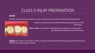
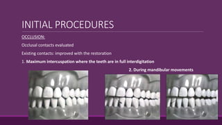
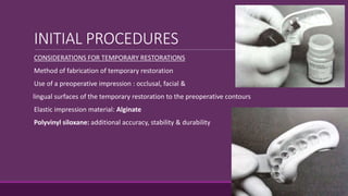
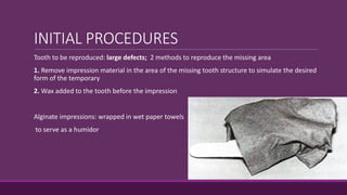
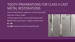
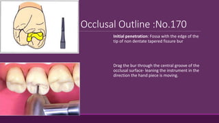
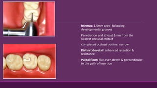
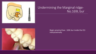
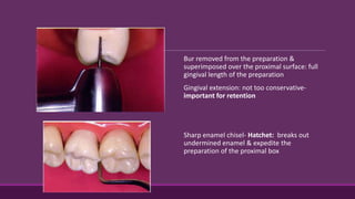
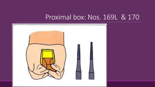
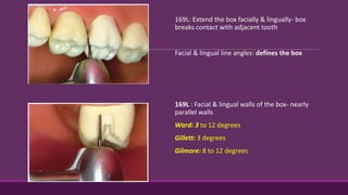
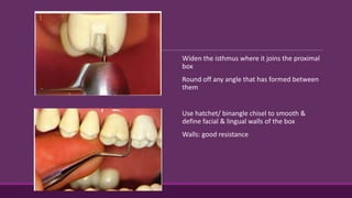
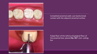
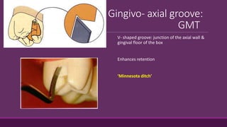
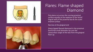
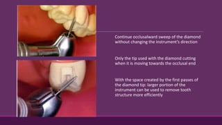
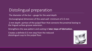
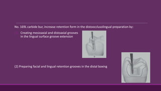
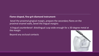
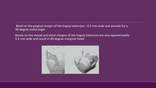
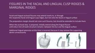
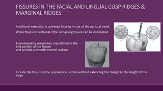
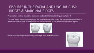
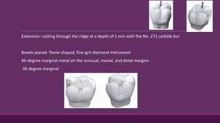
This document provides guidelines for preparing class II inlay restorations. It describes initial procedures like evaluating occlusion and administering anesthesia. It discusses preparing the occlusal outline, proximal box, bevels, and flares. Modifications for specific tooth shapes and situations are covered. Preparation variations like slices and flares are explained. Special considerations for abutment teeth and root surface lesions are also summarized. The document provides a thorough overview of class II inlay preparation techniques.
a detailed account of the principles of tooth preparation with main reference from Shillingburg
The presentation is available on request. Mail me at apurvathampi@gmail.com
An inlay may cap none, or may cap all but one cusp.
Sturdevant’s 4th ed. page579
Inlays may be used as single-tooth restorations for proximo-occlusal or gingival lesions with minimal to moderate extensions
Shillingburg page 1
An inlay may be defined as a restoration which has been constructed out of mouth from gold, porcelain, or other material & then cemented into the prepared cavity of a tooth.
William McGehee pg410
Bevels and flares are very important components of resin restoration procedure. This presentation focuses on bevels and flares in restorative procedure.
This presentation specifically deals with the maxillary and mandibular Major connectors used in a cast partial denture. it also mentions the uses, advantages and disadvantages of each,
A concise and brief presentation on cleaning and shaping of root canals. Colorful and well pictured. Ideal for UG students and PG students to get a good understanding of BMP techniques.
this contains the steps for the class 1 cavity preparation for amalgam in detail. also contains the difference between composite and amalgam cavity preparation.
COMPLETE CROWN PROSTHESIS- A restoration that covers all the coronal tooth surfaces (mesial, distal, facial, lingual and occlusal) (GPT8)
Steps for all metal full veneer crown
1. occlusal reduction
2. axial reduction
3. proximal reduction
4. finishing
5. buccal seating groove
METAL CERAMIC FULL VENEER CROWN - Combines strength of metal and the aesthetics of ceramic.
It is of 2 types:
a. metal with complete ceramic coverage
b. metal with ceramic facing.
The cast metal restoration is versatile and is especially applicable to Class II onlay preparations. The process has many steps, involves many dental materials, and requires meticulous attention to prepration.
Tooth treatment planned to be restored with an intracoronal restoration, but the decay or fracture is so extensive that a direct restoration, such as amalgam or composite, would not be able to sustain or bear forces.
Additionally, when decay or fracture incorporate areas of cusp or remaining tooth structure that undermines perimeter walls of a tooth, an onlay might be indicated.
Diseases of the pulp:Part 1- Development, Physiology, Histology of Dental PulpDeepthi P Ramachandran
The development, physiology, histology of the dental pulp is briefly discussed. The features of the pulp as a connective tissue, its cells,fibers, innervation, vascularity are dealt with
Ozempic: Preoperative Management of Patients on GLP-1 Receptor Agonists Saeid Safari
Preoperative Management of Patients on GLP-1 Receptor Agonists like Ozempic and Semiglutide
ASA GUIDELINE
NYSORA Guideline
2 Case Reports of Gastric Ultrasound
Explore natural remedies for syphilis treatment in Singapore. Discover alternative therapies, herbal remedies, and lifestyle changes that may complement conventional treatments. Learn about holistic approaches to managing syphilis symptoms and supporting overall health.
Title: Sense of Smell
Presenter: Dr. Faiza, Assistant Professor of Physiology
Qualifications:
MBBS (Best Graduate, AIMC Lahore)
FCPS Physiology
ICMT, CHPE, DHPE (STMU)
MPH (GC University, Faisalabad)
MBA (Virtual University of Pakistan)
Learning Objectives:
Describe the primary categories of smells and the concept of odor blindness.
Explain the structure and location of the olfactory membrane and mucosa, including the types and roles of cells involved in olfaction.
Describe the pathway and mechanisms of olfactory signal transmission from the olfactory receptors to the brain.
Illustrate the biochemical cascade triggered by odorant binding to olfactory receptors, including the role of G-proteins and second messengers in generating an action potential.
Identify different types of olfactory disorders such as anosmia, hyposmia, hyperosmia, and dysosmia, including their potential causes.
Key Topics:
Olfactory Genes:
3% of the human genome accounts for olfactory genes.
400 genes for odorant receptors.
Olfactory Membrane:
Located in the superior part of the nasal cavity.
Medially: Folds downward along the superior septum.
Laterally: Folds over the superior turbinate and upper surface of the middle turbinate.
Total surface area: 5-10 square centimeters.
Olfactory Mucosa:
Olfactory Cells: Bipolar nerve cells derived from the CNS (100 million), with 4-25 olfactory cilia per cell.
Sustentacular Cells: Produce mucus and maintain ionic and molecular environment.
Basal Cells: Replace worn-out olfactory cells with an average lifespan of 1-2 months.
Bowman’s Gland: Secretes mucus.
Stimulation of Olfactory Cells:
Odorant dissolves in mucus and attaches to receptors on olfactory cilia.
Involves a cascade effect through G-proteins and second messengers, leading to depolarization and action potential generation in the olfactory nerve.
Quality of a Good Odorant:
Small (3-20 Carbon atoms), volatile, water-soluble, and lipid-soluble.
Facilitated by odorant-binding proteins in mucus.
Membrane Potential and Action Potential:
Resting membrane potential: -55mV.
Action potential frequency in the olfactory nerve increases with odorant strength.
Adaptation Towards the Sense of Smell:
Rapid adaptation within the first second, with further slow adaptation.
Psychological adaptation greater than receptor adaptation, involving feedback inhibition from the central nervous system.
Primary Sensations of Smell:
Camphoraceous, Musky, Floral, Pepperminty, Ethereal, Pungent, Putrid.
Odor Detection Threshold:
Examples: Hydrogen sulfide (0.0005 ppm), Methyl-mercaptan (0.002 ppm).
Some toxic substances are odorless at lethal concentrations.
Characteristics of Smell:
Odor blindness for single substances due to lack of appropriate receptor protein.
Behavioral and emotional influences of smell.
Transmission of Olfactory Signals:
From olfactory cells to glomeruli in the olfactory bulb, involving lateral inhibition.
Primitive, less old, and new olfactory systems with different path
New Directions in Targeted Therapeutic Approaches for Older Adults With Mantl...i3 Health
i3 Health is pleased to make the speaker slides from this activity available for use as a non-accredited self-study or teaching resource.
This slide deck presented by Dr. Kami Maddocks, Professor-Clinical in the Division of Hematology and
Associate Division Director for Ambulatory Operations
The Ohio State University Comprehensive Cancer Center, will provide insight into new directions in targeted therapeutic approaches for older adults with mantle cell lymphoma.
STATEMENT OF NEED
Mantle cell lymphoma (MCL) is a rare, aggressive B-cell non-Hodgkin lymphoma (NHL) accounting for 5% to 7% of all lymphomas. Its prognosis ranges from indolent disease that does not require treatment for years to very aggressive disease, which is associated with poor survival (Silkenstedt et al, 2021). Typically, MCL is diagnosed at advanced stage and in older patients who cannot tolerate intensive therapy (NCCN, 2022). Although recent advances have slightly increased remission rates, recurrence and relapse remain very common, leading to a median overall survival between 3 and 6 years (LLS, 2021). Though there are several effective options, progress is still needed towards establishing an accepted frontline approach for MCL (Castellino et al, 2022). Treatment selection and management of MCL are complicated by the heterogeneity of prognosis, advanced age and comorbidities of patients, and lack of an established standard approach for treatment, making it vital that clinicians be familiar with the latest research and advances in this area. In this activity chaired by Michael Wang, MD, Professor in the Department of Lymphoma & Myeloma at MD Anderson Cancer Center, expert faculty will discuss prognostic factors informing treatment, the promising results of recent trials in new therapeutic approaches, and the implications of treatment resistance in therapeutic selection for MCL.
Target Audience
Hematology/oncology fellows, attending faculty, and other health care professionals involved in the treatment of patients with mantle cell lymphoma (MCL).
Learning Objectives
1.) Identify clinical and biological prognostic factors that can guide treatment decision making for older adults with MCL
2.) Evaluate emerging data on targeted therapeutic approaches for treatment-naive and relapsed/refractory MCL and their applicability to older adults
3.) Assess mechanisms of resistance to targeted therapies for MCL and their implications for treatment selection
Anti ulcer drugs and their Advance pharmacology ||
Anti-ulcer drugs are medications used to prevent and treat ulcers in the stomach and upper part of the small intestine (duodenal ulcers). These ulcers are often caused by an imbalance between stomach acid and the mucosal lining, which protects the stomach lining.
||Scope: Overview of various classes of anti-ulcer drugs, their mechanisms of action, indications, side effects, and clinical considerations.
Title: Sense of Taste
Presenter: Dr. Faiza, Assistant Professor of Physiology
Qualifications:
MBBS (Best Graduate, AIMC Lahore)
FCPS Physiology
ICMT, CHPE, DHPE (STMU)
MPH (GC University, Faisalabad)
MBA (Virtual University of Pakistan)
Learning Objectives:
Describe the structure and function of taste buds.
Describe the relationship between the taste threshold and taste index of common substances.
Explain the chemical basis and signal transduction of taste perception for each type of primary taste sensation.
Recognize different abnormalities of taste perception and their causes.
Key Topics:
Significance of Taste Sensation:
Differentiation between pleasant and harmful food
Influence on behavior
Selection of food based on metabolic needs
Receptors of Taste:
Taste buds on the tongue
Influence of sense of smell, texture of food, and pain stimulation (e.g., by pepper)
Primary and Secondary Taste Sensations:
Primary taste sensations: Sweet, Sour, Salty, Bitter, Umami
Chemical basis and signal transduction mechanisms for each taste
Taste Threshold and Index:
Taste threshold values for Sweet (sucrose), Salty (NaCl), Sour (HCl), and Bitter (Quinine)
Taste index relationship: Inversely proportional to taste threshold
Taste Blindness:
Inability to taste certain substances, particularly thiourea compounds
Example: Phenylthiocarbamide
Structure and Function of Taste Buds:
Composition: Epithelial cells, Sustentacular/Supporting cells, Taste cells, Basal cells
Features: Taste pores, Taste hairs/microvilli, and Taste nerve fibers
Location of Taste Buds:
Found in papillae of the tongue (Fungiform, Circumvallate, Foliate)
Also present on the palate, tonsillar pillars, epiglottis, and proximal esophagus
Mechanism of Taste Stimulation:
Interaction of taste substances with receptors on microvilli
Signal transduction pathways for Umami, Sweet, Bitter, Sour, and Salty tastes
Taste Sensitivity and Adaptation:
Decrease in sensitivity with age
Rapid adaptation of taste sensation
Role of Saliva in Taste:
Dissolution of tastants to reach receptors
Washing away the stimulus
Taste Preferences and Aversions:
Mechanisms behind taste preference and aversion
Influence of receptors and neural pathways
Impact of Sensory Nerve Damage:
Degeneration of taste buds if the sensory nerve fiber is cut
Abnormalities of Taste Detection:
Conditions: Ageusia, Hypogeusia, Dysgeusia (parageusia)
Causes: Nerve damage, neurological disorders, infections, poor oral hygiene, adverse drug effects, deficiencies, aging, tobacco use, altered neurotransmitter levels
Neurotransmitters and Taste Threshold:
Effects of serotonin (5-HT) and norepinephrine (NE) on taste sensitivity
Supertasters:
25% of the population with heightened sensitivity to taste, especially bitterness
Increased number of fungiform papillae
Lung Cancer: Artificial Intelligence, Synergetics, Complex System Analysis, S...Oleg Kshivets
RESULTS: Overall life span (LS) was 2252.1±1742.5 days and cumulative 5-year survival (5YS) reached 73.2%, 10 years – 64.8%, 20 years – 42.5%. 513 LCP lived more than 5 years (LS=3124.6±1525.6 days), 148 LCP – more than 10 years (LS=5054.4±1504.1 days).199 LCP died because of LC (LS=562.7±374.5 days). 5YS of LCP after bi/lobectomies was significantly superior in comparison with LCP after pneumonectomies (78.1% vs.63.7%, P=0.00001 by log-rank test). AT significantly improved 5YS (66.3% vs. 34.8%) (P=0.00000 by log-rank test) only for LCP with N1-2. Cox modeling displayed that 5YS of LCP significantly depended on: phase transition (PT) early-invasive LC in terms of synergetics, PT N0—N12, cell ratio factors (ratio between cancer cells- CC and blood cells subpopulations), G1-3, histology, glucose, AT, blood cell circuit, prothrombin index, heparin tolerance, recalcification time (P=0.000-0.038). Neural networks, genetic algorithm selection and bootstrap simulation revealed relationships between 5YS and PT early-invasive LC (rank=1), PT N0—N12 (rank=2), thrombocytes/CC (3), erythrocytes/CC (4), eosinophils/CC (5), healthy cells/CC (6), lymphocytes/CC (7), segmented neutrophils/CC (8), stick neutrophils/CC (9), monocytes/CC (10); leucocytes/CC (11). Correct prediction of 5YS was 100% by neural networks computing (area under ROC curve=1.0; error=0.0).
CONCLUSIONS: 5YS of LCP after radical procedures significantly depended on: 1) PT early-invasive cancer; 2) PT N0--N12; 3) cell ratio factors; 4) blood cell circuit; 5) biochemical factors; 6) hemostasis system; 7) AT; 8) LC characteristics; 9) LC cell dynamics; 10) surgery type: lobectomy/pneumonectomy; 11) anthropometric data. Optimal diagnosis and treatment strategies for LC are: 1) screening and early detection of LC; 2) availability of experienced thoracic surgeons because of complexity of radical procedures; 3) aggressive en block surgery and adequate lymph node dissection for completeness; 4) precise prediction; 5) adjuvant chemoimmunoradiotherapy for LCP with unfavorable prognosis.
3. CLASS II INLAY PREPARATION
INLAYS:
Entirely intracoronal restorations, most commonly with occlusal and proximal extensions
Indirect intracoronal restoration fabricated using the lost wax
technique technique.
Class II Inlay: An indirect restoration that caps one or more cusps
of a posterior tooth but not all the cusps
ONLAY: An indirect restoration, which is partly intracoronal and partly extracoronal that covers
all the cusps of a posterior tooth.
4. INITIAL PROCEDURES
OCCLUSION:
Occlusal contacts evaluated
Existing contacts: improved with the restoration
1. Maximum intercuspation where the teeth are in full interdigitation
2. During mandibular movements
6. INITIAL PROCEDURES
CONSIDERATIONS FOR TEMPORARY RESTORATIONS
Method of fabrication of temporary restoration
Use of a preoperative impression : occlusal, facial &
lingual surfaces of the temporary restoration to the preoperative contours
Elastic impression material: Alginate
Polyvinyl siloxane: additional accuracy, stability & durability
7. INITIAL PROCEDURES
Tooth to be reproduced: large defects; 2 methods to reproduce the missing area
1. Remove impression material in the area of the missing tooth structure to simulate the desired
form of the temporary
2. Wax added to the tooth before the impression
Alginate impressions: wrapped in wet paper towels
to serve as a humidor
8. TOOTH PREPARATIONS FOR CLASS II CAST
METAL RESTORATIONS
Plane cut tapered fissure carbide burs: vertical internal walls
Side & end surfaces: straight
Uniformly tapered walls; smooth pulpal & gingival walls
No. 271: sides & ends meet in a slightly rounded manner
No. 169L
No. 8862: Slender fine grit flame shaped diamond
9. Occlusal Outline :No.170
Initial penetration: Fossa with the edge of the
tip of non dentate tapered fissure bur
Drag the bur through the central groove of the
occlusal surface- leaning the instrument in the
direction the hand piece is moving.
10. Isthmus: 1.5mm deep- following
developmental grooves
Penetration end at least 1mm from the
nearest occlusal contact
Completed occlusal outline: narrow
Distinct dovetail: enhanced retention &
resistance
Pulpal floor: Flat, even depth & perpendicular
to the path of insertion
11. Undermining the Marginal ridge-
No.169L bur
Begin proximal box : 169L bur inside the CEJ
interproximally
12. Bur removed from the preparation &
superimposed over the proximal surface: full
gingival length of the preparation
Gingival extension: not too conservative-
important for retention
Sharp enamel chisel- Hatchet: breaks out
undermined enamel & expedite the
preparation of the proximal box
14. 169L: Extend the box facially & lingually- box
breaks contact with adjacent tooth
Facial & lingual line angles: defines the box
169L : Facial & lingual walls of the box- nearly
parallel walls
Ward: 3 to 12 degrees
Gillett: 3 degrees
Gilmore: 8 to 12 degrees
15. Widen the isthmus where it joins the proximal
box
Round off any angle that has formed between
them
Use hatchet/ binangle chisel to smooth &
define facial & lingual walls of the box
Walls: good resistance
16. Completed proximal walls: just barely break
contact with the adjacent proximal surface
Pulpal floor of the isthmus & gingival floor of
the proximal box: planed No. 957 end- cutting
bur
17. Gingivo- axial groove:
GMT
V- shaped groove: junction of the axial wall &
gingival floor of the box
Enhances retention
‘Minnesota ditch’
18. Flares: Flame shaped
Diamond
Flat plane cut across the curving proximal
surface equally at the expense of the facial/
lingual wall of the proximal box & the outer
enamel surface
Narrow at the gingival end
Sharp tipped flamed diamond in the proximal
box & the small diameter tip cuts the
cavosurface angle of the box from the gingival
floor up
19. Continue occlusalward sweep of the diamond
without changing the instrument’s direction
Only the tip used with the diamond cutting
when it is moving towards the occlusal end
With the space created by the first passes of
the diamond tip: larger portion of the
instrument can be used to remove tooth
structure more efficiently
20. Sand paper disk: shaping the flares
Extreme caution: Avoid laceration to the soft tissues
Better done only for preparations made under rubber dam isolation
21. Gingival bevel:
Flame shaped Diamond
Lean the flame diamond against the pulpal
axial line angle : proper gingival bevel
Marginal bevel:30 to 45 degrees- optimal
blend of strength & marginal fit
GMT: ragged finish line
Unacceptably rough
23. Occlusal bevel:
Flame diamond
Bevel: along the entire periphery of the
occlusal portion of the preparation
Bevel of 15 to 20 degrees beginning at the
junction of the occlusal 1/3rd & pulpal 2/3rds
of the isthmus wall- Ingraham et al
Convex part of the diamond used- hollow
ground bevel or slightly concave bevel- Tucker
more easily read finish line
25. Bevel & Flare finishing:
Flame bur
Flame shaped carbide bur: the most consistent
bevels & gives smooth finish lines
Smoothness diminishes vulnerability of the
finish line
Refining the occlusal bevel: concave bevel with
distinct finish line- easily identified in the
impression
Inlay easily waxed & finished against it
26.
27. VARIATIONS IN PROXIMAL MARGIN
DESIGN
Principal Determinants of Positions of Proximal Margin:
Extent of hard tissue injury
Convenience required for finishing the preparation
Convenience for finishing the casting
Several basic designs: finish & extend the walls and margins of proximal box resulting from
caries removal
Box
Full tapered Slice
Modified Slice
Modified Flare
Auxiliary Slice
28. BOX PREPARATION
The direct wax technique requires margins which allow easy
manipulation of the wax
Bulk of wax consistent with subsequent finishing procedures: allowed
Buccal & lingual proximal walls finished at nearly 90 degrees to the outer
tooth surface
Cervical bevel: hand instruments providing a lap joint with a bulk of of
wax suitable for carving
Type I gold alloy: suitable for easy manipulation of castings formed from
a well- formed well adapted wax pattern
29. SLICE PREPARATIONS
Involves full proximal disking & was used for indirect technique of wax pattern fabrication
Taking impressions with a non elastic material: compound facilitated by reducing the proximal
contour with a full slice
Accurate elastic impression materials: generalized reduction of the contour not required
30. SLICE PREPARATIONS
Decision whether a full- tapered slice or modified slice preparation :
Careful analysis of all factors related to operation: tooth position, form, extent of hard tissue
injury
Slice indicated: Proximal extension can be gained with limited
bucco- lingual width of the proximal box
Depth of the proximal box is best kept conservative:
danger of pulp encroachment as in young patients
Tooth form: intraoral/ radiographic
31. SLICE PREPARATIONS
Broad proximal contact: buccolingually & cervico incisally
Convenient external outline forms with minimal loss of tooth tissue when flattened contacts are
disked
Narrow occlusally positioned proximal contact: Modified slice
Removal of less tooth tissue from the cervical floor to the occlusal surface
32. SLICE PREPARATIONS
External support of weakened tooth structure & areas subjected to high stress
Auxiliary Slice: wraps partially around the proximal line angles & provide additional tooth
support
Resistance is enhanced with minimal loss of tooth tissue
Excellent definition of finishing line, sound enamel margin
Enables good adaptation & finishing of margins of the casting
33. SLICE PREPARATIONS
External retention form around the lingual proximal line angle when the buccal wall is not sound
for providing retention
Prevents lingual displacement when given around the buccal line angle
Conservatively include the margins of a previous amalgam cavity/ cement base/ areas of
demineralized enamel
34. MODIFIED FLARE PREPARATION
Hybrid between box & slice preparations
Buccal & lingual proximal walls formed initially with minimal extension
Disked in a plane that only slightly reduces the proximal wall dimension
Enhances the obtuseness of the cavosurface angles
Excessive disking reduces retention
35. Selection of box/ slice/ modified flare preparations: mechanical, biologic or esthetic
considerations
Mesial buccal proximal margins of maxillary premolars & molars: slice preparation avoided for
better esthetics
Decision made at the specific time of treatment planning for individual case
36. SPECIAL MODIFICATIONS FOR CLASS II
CAVITY PREPARATIONS
Exceptions : Mandibular bicuspids
Mandibular first premolar: Occlusal preparation needs two thirds of the occlusal width prepared
by removal of structure situated buccally to the central groove & one third from the lingual
aspect
Pulpal floor slanted to the lingual side
Cervical floor may or may not follow the pulpal floor
37. SPECIAL MODIFICATIONS FOR CLASS II
CAVITY PREPARATIONS
Mandibular second Bicuspid:
Central groove forms : H, Y or U patterns
H type: protection of the integrity o the lingual cusp
U and Y types: 2/3rds- 1/3rds relationship & requires operation with great care to avoid undue
weakening of the lingual cusp
Extension to include the lingual groove
38. MODIFICATIONS IN INLAY TOOTH
PREPARATIONS
-Mesio-occlusodistal preparation
-Modifications or esthetics
-Facial or lingual groove extension
-Abutment teeth & extension gingivally to include root- surface lesions
-Maxillary first molar with unaffected, strong oblique ridge
-Fissures in the Facial and Lingual Cusp Ridges & Marginal Ridges
-Capping cusps
-Including Portions of the Facial & Lingual Smooth Surfaces Affected by Caries or Other Injury
40. Whether the remaining marginal ridge would withstand occlusal forces without fracture
Ridge enamel: Gnarled enamel- stronger
Caries on both proximal surfaces: definite indication for MOD
41. ESTHETICS
Mesiofacial proximal wall – maxillary premolars & first molars: minimal flare
Margin barely visible from a facial viewing position
Secondary flare omitted
Wall & margin developed
: chisel or enamel hatchet & final smoothing with a fine- grit paper disk
: narrow diamond / bur when access permits
42. FACIAL & LINGUAL SURFACE GROOVE
EXTENSION
Faulty facial grove on the occlusal surface- continuous with the faulty facial surface groove:
Mandibular molar
faulty distal oblique groove on the occlusal surface continuous with faulty lingual surface groove:
Maxillary molar
Preparation outline extended to include the fissure to its termination
Further gingivally to improve retention form.
Sufficient retention form – though the facial or lingual surface grooves are not fissured.
43. No. 271 carbide bur held parallel to the line of draw, extend through the facial ridge
The depth of the cut: 1.5 mm.
The floor (pulpal wall) should be continuous with the pulpal wall of the occlusal portion
44. With the bur still aligned with the path of draw,
the side of the bur is used to cut the facial surface
portion of this extension
The diameter of the bur serves as a depth gauge for the axial wall, which is in dentin.
The blade portion of the No. 271 bur is 0.8 mm in diameter at its tip end and 1 mm at the neck
The axial wall depth should approximate 1 mm or slightly more
The bur should be tilted lingually as it is drawn occlusally, to develop the uniform depth of the
axial wall
45. Included facial or lingual groove- beveled
With the flame-shaped, fine-grit diamond- provide for 30-degree marginal metal Light bevel on
the mesial and distal margins-
continuous with the occlusal and gingival bevels
40-degree metal at these margins
The bevel width- approximately 0.5 mm
46. ABUTMENT TEETH
Facial, lingual, and gingival margins- the proximal surfaces of abutments for RPDs
Increase the surface area for development of guiding planes
Occlusal outline form wide faciolingually- accommodate any contemplated rest preparation(s)
without involving the margins of the restoration
Accomplished by simply increasing the width of the bevels
47. EXTENSION
GINGIVALLY TO INCLUDE ROOT-SURFACE LESIONS
Primarily by lengthening the gingival bevel- a longer clinical crown due to gingival recession
Slightly extend (gingivally) the gingival floor, and although
Minimal movement of the axial wall pulpally
Additional extension of the gingival floor if necessary, narrower pulpally than when
the floor level is at a normal position
Extending the preparation gingivally without these modifications- dangerous encroachment of the axial
wall on the pulp
48. MAXILLARY FIRST MOLAR WITH UNAFFECTED, STRONG
OBLIQUE RIDGE
Strong oblique ridge preserved
Distal surface lesion – after the insertion of a MO restoration
Prepared for a distoocclusolingual inlay
Distolingual cusp capping-prevents subsequent fracture
49. MAXILLARY FIRST MOLAR WITH UNAFFECTED, STRONG
OBLIQUE RIDGE
Retention form
(1) creating a maximum of 2-degree occlusal divergence of the vertical walls
(2) accentuating some line angles
(3) extending the lingual surface groove to create an axial wall height in this extension of at least
2.5 mm occlusogingivally
50. MAXILLARY FIRST MOLAR WITH UNAFFECTED, STRONG
OBLIQUE RIDGE
Resistance form
(1) routine capping of the distolingual cusp and
(2) maintaining sound tooth structure between the lingual surface groove extension and the
distolingual wall of the proximal boxing
51. Distolingual preparation
Distolingual cusp reduced with the side of the No. 271 carbide bur- uniform 1.5
mm
Remaining occlusal step prepared with the No. 271 carbide bur., followed by the
proximal box portion
The lingual groove extension only after the position of the
distolingual wall of the proximal boxing is established.
Maintain a minimum of 3 mm of sound tooth structure
between this extension and the distolingual wall.
Use the side of the No. 271 carbide bur
If not possible- then a more extensive type of preparation indicated
52. Distolingual preparation
The diameter of the bur – gauge for the axial depth
Occlusogingival dimension of this axial wall- minimum of 2.5 mm
2-mm depth- portion of the pulpal floor that connects the proximal boxing to
the lingual surface groove extension.
Strengthens the wax pattern and casting in later steps of fabrication.
Creates a definite 0.5-mm step from the reduced
distolingual cusp to the pulpal floor.
53. No. 169L carbide bur, increase retention form in the distoocclusolingual preparation by:
(1) Creating mesioaxial and distoaxial grooves
in the lingual surface groove extension
(2) Preparing facial and lingual retention grooves in the distal boxing
54. Flame-shaped, fine-grit diamond instrument-
bevel the proximal gingival margin, prepare the secondary flares on the
proximal enamel walls, bevel the lingual margins
A lingual counterbevel- distolingual cusp wide enough for a 30-degree metal at
the margin
Beyond any occlusal contacts
55. Bevel on the gingival margin of the lingual extension - 0.5 mm wide and provide for a
30-degree metal angle
Bevels on the mesial and distal margins of the lingual extension are also approximately
0.5 mm wide and result in 40-degree marginal metal
56. FISSURES IN THE FACIAL AND LINGUAL CUSP RIDGES &
MARGINAL RIDGES
Facial and lingual occlusal fissures may extend nearly to, or through
the respective facial and lingual cusp ridges, but not onto the facial or lingual surface
The preparation margin should not cross such fissures, but should be extended to include them
When the occlusal step is prepared, initially extend along the lingual fissure
until only 2 mm of tooth structure remains between the bur and the lingual surface
Additional lingual extension at this time is incorrect because it may remove the supporting
dentin unnecessarily
57. FISSURES IN THE FACIAL AND LINGUAL CUSP RIDGES &
MARGINAL RIDGES
Additional extension is achieved later by virtue of the occlusal bevel
Wider than conventional if the remaining fissure can be eliminated
Enameloplasty sometimes may eliminate the
end portion of the fissure
and provide a smooth enamel surface
Include the fissure in the preparation outline without extending the margin to the height of the
ridge
58. FISSURES IN THE FACIAL AND LINGUAL CUSP
RIDGES & MARGINAL RIDGES
Preparation outline should be extended just onto the facial or lingual surface
Occlusal bevel places the margin on the height of the ridge, then the marginal enamel likely is
weak because of both its sharpness and the inclination of the enamel rods in this region.
If the fissure still remains through the ridge after enameloplasty
59. Extension- cutting through the ridge at a depth of 1 mm with the No. 271 carbide bur
Bevels placed- flame shaped, fine-grit diamond instrument
40-degree marginal metal on the occlusal, mesial, and distal margins
30-degree marginal
60. Fissures that may extend into or through a proximal marginal ridge- similarly managed
Does not extend the preparation outline near the adjacent tooth contact
Particularly applies to a mesial fissure of the maxillary first premolar
61. CAPPING CUSPS
The facial and lingual margins on the occlusal surface
frequently must be extended toward
the cusp tips to the extent of existing restorative materials and to uncover caries
When the occlusal outline is extended up the cusp slopes more than half the distance
from any primary occlusal groove to the cusp tip, capping the cusp should be considered
If the preparation outline is extended two thirds of this distance or more, capping the cusp is
usually necessary to:
(1) protect the weak, underlying cuspal structure from fracture caused by masticatory force
(2) remove the occlusal margin from a region subjected to heavy stress and wear
62. CAPPING CUSPS
At this point in preparation the pulpal floor, depth can be increased from 1.5 mm to 2 mm.
Sufficient reduction in an area that is often underreduced
Result in greater strength and rigidity to the wax pattern and cast restoration.
63. Technique of capping less than all of the
cusps
Reduce the Cusps for Capping as Soon as the Indication for Such Capping Is Determined Because
This Improves Access and Visibility for Subsequent Steps in Preparation.
Cusp in infraocclusion of the desired occlusal plane before reduction- -
The amount of cusp reduction is less
Only to provide the required clearance with the desired occlusal plane.
Depth gauge grooves (depth cuts) prepared with the side of the
No. 271 carbide bur
Prevent thin spots in the restoration
64. With the depth cuts serving as guides, complete the cusp reduction
with the side of the carbide bur
The reduction should provide for a uniform 1.5 mm of metal thickness over the reduced cusp.
On maxillary premolars and first molars, the reduction should be minimal (i.e., 0.75 to 1 mm) on
the facial cusp ridge to decrease the display of metal.
Reduction should increase progressively to 1.5 mm toward the center of the tooth to help
provide rigidity to the capping metal
65. If only one of the two lingual cusps of a molar is reduced for capping, the reduction must extend
to just include the lingual groove between the reduced and unreduced cusps.
This reduction should terminate with a distinct vertical wall that has a height that is the same as
the prescribed cusp reduction.
Applying the bur vertically, as shown, should help establish
a vertical wall of proper depth and direction.
Similar principles apply when only one of the facial cusps is to be reduced
66. A bevel of generous width is prepared on the facial (lingual) margin of a reduced cusp with the
flame shaped, fine-grit diamond instrument
Avoided in esthetically prominent areas
Reverse bevel or counterbevel.
Beyond any occlusal contact
30-degree marginal metal
67. Esthetic requirements- facial margin on maxillary premolars & first molar
Blunting and smoothing of the enamel margin (a stub margin) by the light application of a fine-
grit sandpaper disc or the fine-grit diamond instrument (flame-shaped) held at a
right angle to the facial surface
Slightly round any sharp external corners to strengthen them and
reduce the problems they may generate in future steps
68. Cusp reduction appreciably decreases retention form because of decreasing the height of the
vertical walls
Proximal retention grooves usually are recommended
For additional retention
Extending facial and lingual groove regions of the respective surfaces,
Secondary retention features- collar and skirt
69. Treatment of the distal cusp of the
mandibular first molar -MOD preparation
Satisfactory treatment usually dictates either
(1) extending the distofacial margin (and wall) slightly mesial of the distofacial groove
(2) capping the remaining portion of the distal cusp
70. After cusp reduction, visually verify that the occlusal clearances are sufficient.
A wax interocclusal record- especially in areas difficult to visualize
Central groove/lingual cusp region
First dry the preparation(s) free of visible moisture but not desiccate the dentin.
71. Next lightly press a portion of softened, low-fusing inlay wax over the prepared tooth (teeth);
then immediately request the patient to close into the soft wax and slide the teeth in all
directions
72. During the mandibular movements, observe to verify that
(1) the patient moves in right lateral, left lateral, and protrusive movements
(2) the adjacent unprepared teeth are in contact with the opposing teeth
(3) the wax in the preparation is stable (not loose and rocking)
(4) the wax is not in infraocclusion.
Cool and carefully remove the wax
Hold it up to a light, and note the degree of light transmittedAlternatives
Use wax calipers
Section the wax to verify its thickness
Insufficient thickness calls for more reduction in the indicated area before proceeding.
73. INCLUDING PORTIONS OF THE FACIAL & LINGUAL SMOOTH
SURFACES AFFECTED BY CARIES OR OTHER INJURY
Portion of both a facial/ lingual smooth surface & a proximal surface are affected by caries or
some other factor
A large inlay, an onlay, a three-quarter crown, a full crown, or multiple amalgam or composite
restorations
74. Choice of treatment- degree of tooth circumference involved
Full crown- both the lingual and facial smooth surfaces are defective, especially if the tooth is a
second or third molar.
MODFL inlay or onlay with a lingual groove extension-
When only a portion of the facial smooth surface is carious and the lingual surfaces of the teeth
are conspicuously free of caries
More favorable to the health of the gingival tissues
More conservative in the removal of tooth structure
Treatment choice for the maxillary second molar- caries or decalcification on the distofacial
surface
75. Mesiofacial, distolingual cusps & the distofacial cusp- reduced for capping
If the distofacial cusp defect is primarily shallow decalcification- the flame-shaped diamond
instrument is used to both reduce the involved facial surface and distofacial corner
approximately the depth of the enamel and to establish the gingival margin
76. If an effective distal box or groove & a facial wall is not possible, then the No. 271 carbide bur
should be used to cut a gingival shoulder extending from the distal gingival floor around to
include the affected facial surface
This shoulder partially provides the desired resistance form
Use the No.271 bur to also create a nearly vertical wall
in the remaining facial enamel
77. Width of the shoulder- diameter of the end of the cutting instrument
Vertical walls - appropriate degree of draft to contribute to retention form.
Faciogingival and facial margins are beveled with the flame-shaped, fine-grit diamond
instrument- 30-degree metal at the gingival margin and 40-degree metal along the facial margin
78. Two bevels should blend together, the faciogingival bevel should be continuous with the gingival
bevel on the distal surface.
79. Additional Retention & Resistance features
Arbitrary lingual groove extension
Distolingual skirt extension
Resist forces normally opposed by the missing distofacial wall
Protect the restored tooth from fracture injury