
Recommended
More Related Content
Similar to ENDOCROWNS AN INNOVATIVE APPROACH, INDICATION AND ADVANTAGES, PREPARATION
Similar to ENDOCROWNS AN INNOVATIVE APPROACH, INDICATION AND ADVANTAGES, PREPARATION (20)
More from aishwaryakhare5
More from aishwaryakhare5 (17)
Recently uploaded
Recently uploaded (20)
ENDOCROWNS AN INNOVATIVE APPROACH, INDICATION AND ADVANTAGES, PREPARATION
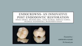
- 1. ENDOCROWNS- AN INNOVATIVE POST ENDODONTIC RESTORATION DISHA ARORA, MUNISH GOEL, VIJAY KUMAR, SHWETA VERMA, PRABHAT MANDHOTRA, MAHENDER SINGH Presented by:- AISHWARYA KHARE PG Second year
- 2. INTRODUCTION • As dentistry is moving towards minimally invasive preparations, with maximal tissue conservation, any method which achieve this will be considered ‘the gold standard’ for restoring ETT. • By following this rationale, endocrowns can be used as a prosthetic option in restoration of endodontically treated teeth excessive tissue loss
- 3. TREATMENT OPTIONS TO RESTORE ENDODONTICALLY TREATED TEETH Amount of tooth structure remaining Treatment options available 1. Minimal loss of coronal structures. Restorative material of choice should be composite resin with suitable bonding system. Contraindication: Patients with para functional habits. Such patients would require crowns to provided strength to avoid fracture of tooth. 2. Up to One-Half of the Coronal Tooth Structure Missing. Complete occlusal coverage with help of endocrown or onlay restoration is sufficient instead of post and core.
- 4. 3. More Than Half of the Coronal Tooth Structure is Missing. When more than half of the coronal tissue is lost, such cases post-core restoration is the only option to ensure tooth-restoration continuum strength and resistance to fracture 4. Most of the Coronal Tooth Structure is Missing Extraction and dental implants might be then acceptable as an alternative to conventional treatment of severely compromised posterior teeth.
- 5. ENDOCROWN RESTORATIONS • The concept of endocrown was given by Pissis. • An Endocrowns can be defined as a monolithic (onepiece) ceramic bonded construction characterized by a supra-cervical butt joint, retaining maximum enamel to improve adhesion. • In 1999, the endocrown was described for the first time by Bindle and Mormann as adhesive endodontic crowns and characterized as total porcelain crowns fixed to endodontically treated posterior teeth
- 6. • Retention of these crowns is achieved by the internal portion of the pulp chamber and on the cavity margins, so macromechanical retention is provided by the pulpal walls, and micromechanical retention is obtained by the use of adhesive cementation.
- 7. INDICATIONS • Cases in which there is excessive loss of tissue of the crown, interproximal space is limited and traditional rehabilitation with post and crown is not possible because of inadequate ceramic thickness. • Molars with clinically low crowns. • Where the root canals are calcified or very slender roots.
- 8. CONTRAINDICATIONS • Less than 3mm pulp chamber depth. • When adhesion cannot be assured. • If only negligible remaining tooth structure is present.
- 9. STEPS IN OCCLUSAL PREPARATION • The first step is to achieve an overall reduction in the height of the occlusal surface of at least 2 mm in the axial direction. This reduction is done by drilling 2-mm-deep grooves as guides, then using a green diamond wheel bur to reduce the occlusal surface.
- 10. • The bur is oriented along the major axis of the tooth and held parallel to the occlusal plane, this allows a flat surface, which determines the position of the cervical margin or “cervical sidewalk.” • The cervical margin should be supragingival; however, if clinical factors or esthetics require, the margin can follow the gingival margin.
- 11. • Differences in level between the various parts of the cervical margin must be linked by a slope of no more than 60° to avoid a staircase effect • Enamel walls less than 2 mm thick should be removed.
- 12. STEPS IN AXIAL PREPARATION • In this step undercuts are eliminated in the access cavity. A total occlusal convergence of 7° is used to make the coronal pulp chamber and endodontic access cavity continuous with help of a cylindricalconical green diamond bur. • The orientation of the bur is along the long axis of the tooth, the preparation is carried out without excessive pressure and without touching the pulpal floor.
- 13. POLISHING THE CERVICAL BAND • The bur used in this step has the same taper as the one used in axial preparation, but a larger diameter and a finer particle size. • It should be guided around the entire surface of the cervical band to remove micro-irregularities and produce a flat, polished surface. The margin line should appear as a regular line with a sharp edge.
- 14. PREPARATION OF THE CAVITY FLOOR • The entrance to the pulpal canal is opened. Gutta percha is removed to a depth not exceeding 2 mm to take advantage of the saddle-like anatomy of the cavity floor. • This should be done with a nonabrasive instrument to maintain the integrity of the canals entrance. No drilling of dentin is carried out. A hot small Burnisher can be used to remove the 2mm of Gutta Percha.
- 15. CEMENTATION • Before bonding, precision fit and marginal adaptation of endocrowns is evaluated. • Cementation surfaces of endocrowns were etched with 5% hydrofluoric acid for 20 seconds, rinsed with water for 30 seconds, ultrasonically cleaned in distilled water for 3 minutes, and dried. • A silane coupling agent (RelyX Ceramic Primer, 3M ESPE) is applied and allowed to dry for 1 minute. Then light cured for 10 seconds
- 19. CASE REPORT 1: ZIRCONIA ENDOCROWN
- 21. CASE REPORT 2: ZIRCONIA ENDOCROWN
- 28. CONCLUSION • Endocrown is the new treatment modality for grossly destructed endodontically treated teeth. • Endocrowns have been used as an alternative to conventional post-core and fixed partial dentures in restoration of endodontically treated teeth with extensive coronal tissue loss. • Compared to traditional methods, better aesthetics and mechanical performance, low cost and short clinical time are the advantages of endocrowns.
- 29. REFERENCES • ENDOCROWN: AN APPROACH FOR RESTORING ENDODONTICALLY TREATED RIGHT MANDIBULAR FIRST MOLARS WITH LARGE CORONAL DESTRUCTION- A CASE REPORT • Endocrown A NEW MODALITY OF TREATMENT: MANAGEMENT OF THREE CASES • Endocrown Restoration on Postendodontic Treatment on Lower First Molar • Clinical efficacy of different marginal forms of endocrowns: study protocol for a randomized controlled trial