
Downloaded 1,519 times

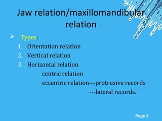


Jaw relations refer to the spatial relationship between the maxilla and mandible. There are several types of jaw relations including orientation, vertical, and horizontal relations. The vertical jaw relation is the distance between two selected points on the maxilla and mandible. It is important to accurately record the vertical jaw relation to establish proper esthetics, phonetics, and function. There are various methods for determining the vertical jaw relation including physiologic methods and using interocclusal records or prior dentures. Facebows are used to transfer the maxillomandibular spatial relationship to articulators.


