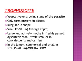
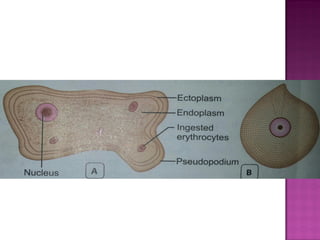
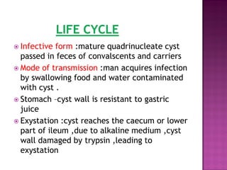
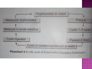
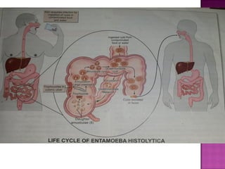
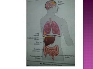
 Amoeba are structurally simple protozoans
which have no fixed shape
 Phylum : Sarcomastigophora
 Subphylum :Sarcodina
 Super class :Rhizopoda
 Order : Amoebida
Amoeba
Free living
Intestinal
 Entamoeba histolytica is an intestinal
amoeba
 All intestinal amoebae are non pathogenic
except Entamoeba histolytica
 All free living amoeba are oppurtunistic
pathogens.
 E. histolytica was discovered by Losch in
1875
 Demonstrated the parasite in the dysenteric
feces of a patient in St.Petersburg in Russia.
 MORPHOLOGY
 LIFE CYCLE
 PATHOGENESIS & CLINICAL FEATURES
 LABORATORY DIAGNOSIS
 TREATMENT
 PREVENTION
 E.histolytica occurs in 3 forms
Trophozoite
Precyst
Cyst
 Vegetative or growing stage of the parasite
 Only form present in tissues
 Irregular in shape
 Size: 12-60 μm( Average 20μm)
 Large and actively motile in freshly passed
dysenteric stool, while smaller in
convalescents and carriers.
 In the lumen, commensal and small in
size(15-20 μm)-MINUTA FORM
Cytoplasm
Outer ectoplasm-clear,transparent,
refractile.
Inner endoplasm-finely granular
(ground glass appearance),with
nucleus
food vacuoles
erythrocytes
leucocytes(occasionally)
tissue debris
 Pseudopodia
Fingerlike projections formed by
sudden jerky movements of ectoplasm in one
direction, followed by the streaming in of
the whole endoplasm
 Typical amoeboid motility is a Crawling or
Gliding
 Pseudopodia formation and motility are
inhibited at low temperature
Nucleus
 It is spherical 4-6μm in size
 contains central karyosome,surrounded by
clear halo and anchored to the nuclear
membrane by fine radiating fibrils called the
Linin network , giving a cartwheel
appearance
 Nuclear membrane is lined by a rim of
chromatin distributed evenly as small
granules
 Trophozoites from acute dysentric stools
often contain phagocytosed erythrocytes-
diagnostic feature ,these are not found in
any other commensal intestinal amoeba
 These divided by binary fission in every 8
hours
 These are killed by drying, heat ,and
chemical sterilization
 Infections are not transmitted by these-
destroyed in stomach and cannot initiate
infection
 Trophozoites undergo encystment in the
lumen
 Before encystment,the trophozoites
extrudesits food vacuoles and become round
or oval,10-20µm in size-precyst
 Contains a large glycogen vacuole and two
chromatid bars
 It then secretes a highly retractile cyst wall
around it and become cyst
 Spherical ,10-20µm
 3 types of cyst
Early cyst
Binucleate cyst
mature quadrinucleate cyst
Early cyst
contains a single nucleus and two other
structures-a mass of glycogen and 1-4
chromatid bodies or chromadial bars
 As the cyst mature, the glycogen mass and
chromidial bars disappear and the nucleus
undergoes 2 succesive mitotic divisions to
form 2 and then 4 nuclei .
 The cyst wall is a highly resistant to gastric
juice and unfavorable environmental
conditions
 Infective form :mature quadrinucleate cyst
passed in feces of convalscents and carriers
 Mode of transmission :man acquires infection
by swallowing food and water contaminated
with cyst .
 Stomach –cyst wall is resistant to gastric
juice
 Exystation :cyst reaches the caecum or lower
part of ileum ,due to alkaline medium ,cyst
wall damaged by trypsin ,leading to
exystation
 E.histolytica causes intestinal and extra
intestinal amoebiasis
Intestinal amoebiasis -PATHOGENESIS
Lumen dwelling amoeba do not cause any
illness .They causes disease only when they
invade the intestinal tissues .
10 % -symptomatic
90% -asymptomatic
 Not all strains of E.histolytica are pathogenic or
invasive .
 Differentiation between pathogenic and non
pathogenic strains can be made by
 susceptibility to complement-mediated lysis
 Phagocytic activity
 by the use of genetic markers
 monoclonal antibodies
 Zymodeme analysis
 The metacystic trophozoites penetrate the columnar
epithelial cells of crypts of Liberkuhn
 Penetration is facilitated by
motility of the trophozoites
tissue lytic enzyme –histolysin
amoebic lectin -mediate adherence
 Mucosal penetration by amoeba produce discrete
ulcers with pinhead center and raised edges .
 Sometimes invasion remains superficial and heal
spontaneously.
 More often, the amoeba penetrates to submucosal
layer and multiplies rapidly- lytic necrosis- abscess-
ulcer
 Ulcer appear initially on the mucosa as raised
nodules with pouting edges .
 They breakdown discharging brownish
necrotic material contains large numbers of
trophozoites.
 The typical amoebic ulcer –flask shaped
 Multiple ulcers may coalesce to form large
necrotic lesions with ragged and undermined
edges,covered with brownish slough
 The ulcers generally do not extend deeper
than submucosal layer.
 Occassionally, a granulamatous
pseudotumoral growth may develop on the
intestinal wall from a chronic ulcer.This
amoebic granuloma or amoeboma may be
mistaken for are malignant tumor
 Small superficial ulcers involving only the mucosa
 Round or oval shaped with ragged and undermined margin
and flask-shaped in cross section
 Marked scarring of intestinal wall with thining ,dilatation
,and sacculation
 Extensive adhesions with neighboring viscera
 Formation of tumor-like masses of granulation tissue
amoeba
 The incubation period is highly variable from
1-4 months
 The clinical course is characterized by
prolonged latency,relapses and intermissions.
 Typical manifestation is amoebic dysentry
 Compared to bacillary desentry ,it is usually
insidious in onset and the abdominal
tenderness is less and localised.
 The stools are large, foul-smelling,and
brownish black, often with bloodstreaked
mucus intermingled with feces.
 The RBCs in stools are clumped and reddish
brown in color.
 Cellular exudate is scanty.
 Charcot-leyden crystals are often present.
 The patient is usually afebrile and non toxic.
 In fulminant colitis, there is confluent
ulceration and necrosis of colon-patient is
febrile and toxic.
 Intestinal amoebiasis not always result in
dysentry.quite often there may be only
diarrhea or vagueabdominal symptoms
popularly called uncomfortable belly or
growling abdomen.
 Chronic involvement of the caecum causes a
condition simulating appendicitis.
 Fulminant amoebic colitis
toxic megacolon
perianal ulceration
Amoeboma
 Extra intestinal Amoebiasis
amoebic hepatitis
amoebic liver abscess
amoebic appendicitis and peritonitis
pulmonary amoebiasis
cerebral amoebiasis
splenic abscess
cutaneous amoebiasis
genitourinary amoebiasis
 Hepatic Amoebiasis
 Pulmonary Amoebiasis
 Metastatic Amoebiasis
 Cutaneous Amoebiasis
 Genitourinary Amoebiasis
 Most common extra intestinal amoebiasis .
 The history of amoebic dysentry is absent in
more than 50% cases
 Several patients with amoebic colitis develop
an enlarged tender liver without detectable
impairment of liver function or fever.
 This acute hepatic involvement (amoebic
hepatitis) may be due to repeated invasion
by amoeba from an active colonic infection
or to toxic substance from the colon reaching
the liver.
 In about 5-10% of person with intestinal
amoebiasis ,liver abscesses may ensue.
 The center of the abscess contains thick
chocolate brown pus(anchovy pus),which is
liqeefied necrotic liver tissue.
 At the periphery, there is almost normal
liver tissue,which contain invading amoeba.
 Liver abscess may
multiple
solitary
 Usually located on right lobe of liver
 Jaundice develops
only when lesions are multiple OR
when they press on the biliary tract
 Incidenca of liver abscess is less common in
women and rare in children under age of 10
 It may occure by direct hematogenous spread
from colon bypassing the liver,but it most
often follows extension of hepatic abscess
through the diaphragm
 Hepatobronchial fistula usually results with
expectoration of chocolate brown sputum
 Patient presents with
severe pleuritic chest pain
dyspnea
non-productive cough
 Involvement of distant organs is by
hematogenous spread and through
lymphatics
 Abscess in
Kidney
Brain
Spleen
Adrenals
Spread to brain leads to severe destruction of
brain tissues and is fatal
 It occurs by direct extension around anus,
colostomy site,or discharging sinuses from
amoebic abscess .
 Extensive gangrenous destruction of the skin
occurs .
 The lesion may be mistaken for condyloma or
epithelioma
 Penile amoebiasis
prepuce and glans are affected
acquired through anal intercourse
In females
 vulva ,vagina, or cervix
 by spread from perineum
 destructive ulcerative lesions resemble
carcinoma
THANK YOU

Entamoeba histolytica

  • 2.
     Amoeba arestructurally simple protozoans which have no fixed shape  Phylum : Sarcomastigophora  Subphylum :Sarcodina  Super class :Rhizopoda  Order : Amoebida
  • 3.
    Amoeba Free living Intestinal  Entamoebahistolytica is an intestinal amoeba  All intestinal amoebae are non pathogenic except Entamoeba histolytica  All free living amoeba are oppurtunistic pathogens.
  • 4.
     E. histolyticawas discovered by Losch in 1875  Demonstrated the parasite in the dysenteric feces of a patient in St.Petersburg in Russia.
  • 5.
     MORPHOLOGY  LIFECYCLE  PATHOGENESIS & CLINICAL FEATURES  LABORATORY DIAGNOSIS  TREATMENT  PREVENTION
  • 6.
     E.histolytica occursin 3 forms Trophozoite Precyst Cyst
  • 7.
     Vegetative orgrowing stage of the parasite  Only form present in tissues  Irregular in shape  Size: 12-60 μm( Average 20μm)  Large and actively motile in freshly passed dysenteric stool, while smaller in convalescents and carriers.  In the lumen, commensal and small in size(15-20 μm)-MINUTA FORM
  • 9.
    Cytoplasm Outer ectoplasm-clear,transparent, refractile. Inner endoplasm-finelygranular (ground glass appearance),with nucleus food vacuoles erythrocytes leucocytes(occasionally) tissue debris
  • 10.
     Pseudopodia Fingerlike projectionsformed by sudden jerky movements of ectoplasm in one direction, followed by the streaming in of the whole endoplasm  Typical amoeboid motility is a Crawling or Gliding  Pseudopodia formation and motility are inhibited at low temperature
  • 11.
    Nucleus  It isspherical 4-6μm in size  contains central karyosome,surrounded by clear halo and anchored to the nuclear membrane by fine radiating fibrils called the Linin network , giving a cartwheel appearance  Nuclear membrane is lined by a rim of chromatin distributed evenly as small granules
  • 12.
     Trophozoites fromacute dysentric stools often contain phagocytosed erythrocytes- diagnostic feature ,these are not found in any other commensal intestinal amoeba  These divided by binary fission in every 8 hours  These are killed by drying, heat ,and chemical sterilization  Infections are not transmitted by these- destroyed in stomach and cannot initiate infection
  • 13.
     Trophozoites undergoencystment in the lumen  Before encystment,the trophozoites extrudesits food vacuoles and become round or oval,10-20µm in size-precyst  Contains a large glycogen vacuole and two chromatid bars  It then secretes a highly retractile cyst wall around it and become cyst
  • 15.
     Spherical ,10-20µm 3 types of cyst Early cyst Binucleate cyst mature quadrinucleate cyst Early cyst contains a single nucleus and two other structures-a mass of glycogen and 1-4 chromatid bodies or chromadial bars
  • 16.
     As thecyst mature, the glycogen mass and chromidial bars disappear and the nucleus undergoes 2 succesive mitotic divisions to form 2 and then 4 nuclei .  The cyst wall is a highly resistant to gastric juice and unfavorable environmental conditions
  • 17.
     Infective form:mature quadrinucleate cyst passed in feces of convalscents and carriers  Mode of transmission :man acquires infection by swallowing food and water contaminated with cyst .  Stomach –cyst wall is resistant to gastric juice  Exystation :cyst reaches the caecum or lower part of ileum ,due to alkaline medium ,cyst wall damaged by trypsin ,leading to exystation
  • 20.
     E.histolytica causesintestinal and extra intestinal amoebiasis Intestinal amoebiasis -PATHOGENESIS Lumen dwelling amoeba do not cause any illness .They causes disease only when they invade the intestinal tissues . 10 % -symptomatic 90% -asymptomatic
  • 21.
     Not allstrains of E.histolytica are pathogenic or invasive .  Differentiation between pathogenic and non pathogenic strains can be made by  susceptibility to complement-mediated lysis  Phagocytic activity  by the use of genetic markers  monoclonal antibodies  Zymodeme analysis
  • 22.
     The metacystictrophozoites penetrate the columnar epithelial cells of crypts of Liberkuhn  Penetration is facilitated by motility of the trophozoites tissue lytic enzyme –histolysin amoebic lectin -mediate adherence  Mucosal penetration by amoeba produce discrete ulcers with pinhead center and raised edges .  Sometimes invasion remains superficial and heal spontaneously.  More often, the amoeba penetrates to submucosal layer and multiplies rapidly- lytic necrosis- abscess- ulcer
  • 23.
     Ulcer appearinitially on the mucosa as raised nodules with pouting edges .  They breakdown discharging brownish necrotic material contains large numbers of trophozoites.  The typical amoebic ulcer –flask shaped  Multiple ulcers may coalesce to form large necrotic lesions with ragged and undermined edges,covered with brownish slough
  • 24.
     The ulcersgenerally do not extend deeper than submucosal layer.  Occassionally, a granulamatous pseudotumoral growth may develop on the intestinal wall from a chronic ulcer.This amoebic granuloma or amoeboma may be mistaken for are malignant tumor
  • 25.
     Small superficialulcers involving only the mucosa  Round or oval shaped with ragged and undermined margin and flask-shaped in cross section  Marked scarring of intestinal wall with thining ,dilatation ,and sacculation  Extensive adhesions with neighboring viscera  Formation of tumor-like masses of granulation tissue amoeba
  • 26.
     The incubationperiod is highly variable from 1-4 months  The clinical course is characterized by prolonged latency,relapses and intermissions.  Typical manifestation is amoebic dysentry  Compared to bacillary desentry ,it is usually insidious in onset and the abdominal tenderness is less and localised.  The stools are large, foul-smelling,and brownish black, often with bloodstreaked mucus intermingled with feces.
  • 28.
     The RBCsin stools are clumped and reddish brown in color.  Cellular exudate is scanty.  Charcot-leyden crystals are often present.  The patient is usually afebrile and non toxic.  In fulminant colitis, there is confluent ulceration and necrosis of colon-patient is febrile and toxic.
  • 29.
     Intestinal amoebiasisnot always result in dysentry.quite often there may be only diarrhea or vagueabdominal symptoms popularly called uncomfortable belly or growling abdomen.  Chronic involvement of the caecum causes a condition simulating appendicitis.
  • 30.
     Fulminant amoebiccolitis toxic megacolon perianal ulceration Amoeboma  Extra intestinal Amoebiasis amoebic hepatitis amoebic liver abscess amoebic appendicitis and peritonitis pulmonary amoebiasis cerebral amoebiasis splenic abscess cutaneous amoebiasis genitourinary amoebiasis
  • 33.
     Hepatic Amoebiasis Pulmonary Amoebiasis  Metastatic Amoebiasis  Cutaneous Amoebiasis  Genitourinary Amoebiasis
  • 34.
     Most commonextra intestinal amoebiasis .  The history of amoebic dysentry is absent in more than 50% cases  Several patients with amoebic colitis develop an enlarged tender liver without detectable impairment of liver function or fever.  This acute hepatic involvement (amoebic hepatitis) may be due to repeated invasion by amoeba from an active colonic infection or to toxic substance from the colon reaching the liver.
  • 35.
     In about5-10% of person with intestinal amoebiasis ,liver abscesses may ensue.  The center of the abscess contains thick chocolate brown pus(anchovy pus),which is liqeefied necrotic liver tissue.  At the periphery, there is almost normal liver tissue,which contain invading amoeba.  Liver abscess may multiple solitary
  • 36.
     Usually locatedon right lobe of liver  Jaundice develops only when lesions are multiple OR when they press on the biliary tract  Incidenca of liver abscess is less common in women and rare in children under age of 10
  • 37.
     It mayoccure by direct hematogenous spread from colon bypassing the liver,but it most often follows extension of hepatic abscess through the diaphragm  Hepatobronchial fistula usually results with expectoration of chocolate brown sputum  Patient presents with severe pleuritic chest pain dyspnea non-productive cough
  • 38.
     Involvement ofdistant organs is by hematogenous spread and through lymphatics  Abscess in Kidney Brain Spleen Adrenals Spread to brain leads to severe destruction of brain tissues and is fatal
  • 39.
     It occursby direct extension around anus, colostomy site,or discharging sinuses from amoebic abscess .  Extensive gangrenous destruction of the skin occurs .  The lesion may be mistaken for condyloma or epithelioma
  • 40.
     Penile amoebiasis prepuceand glans are affected acquired through anal intercourse In females  vulva ,vagina, or cervix  by spread from perineum  destructive ulcerative lesions resemble carcinoma
  • 42.