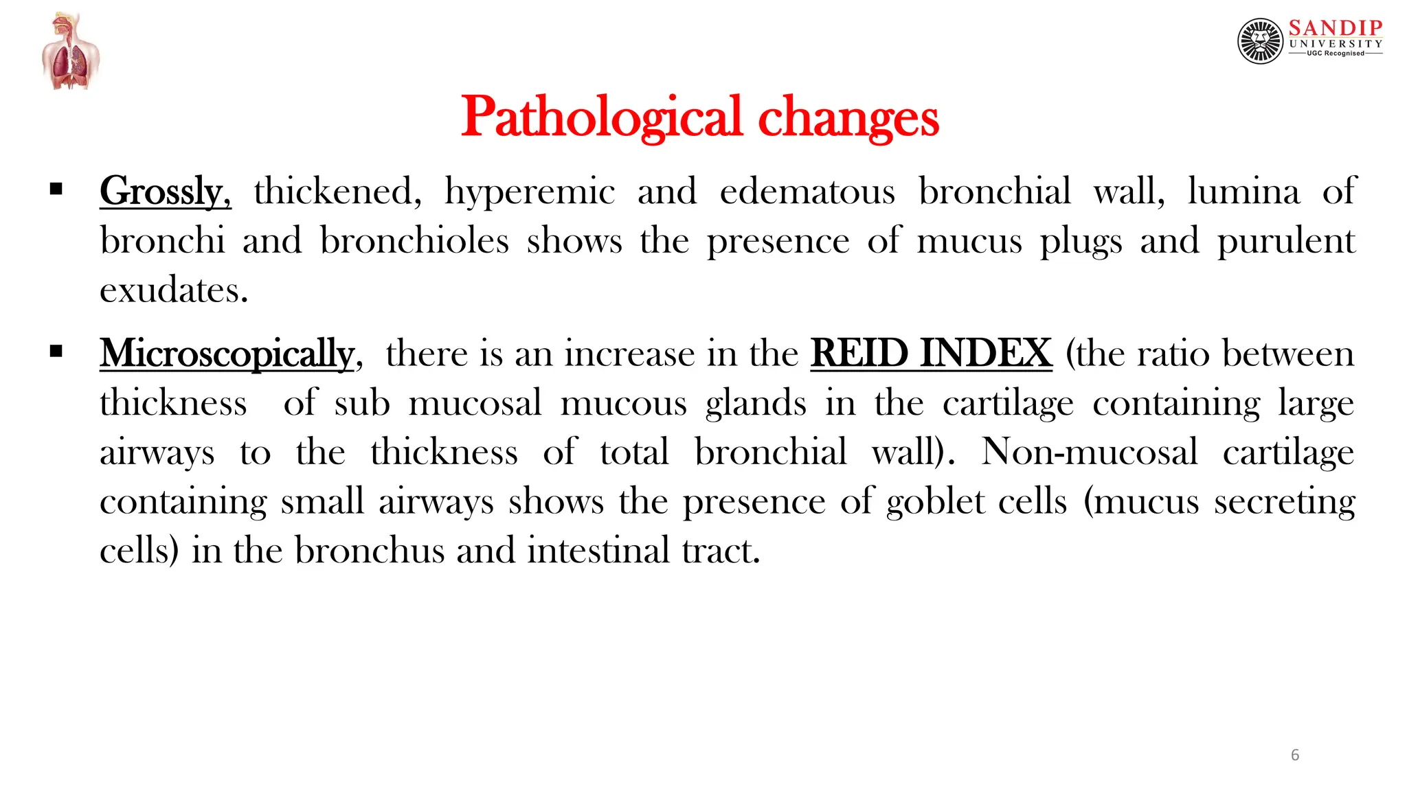
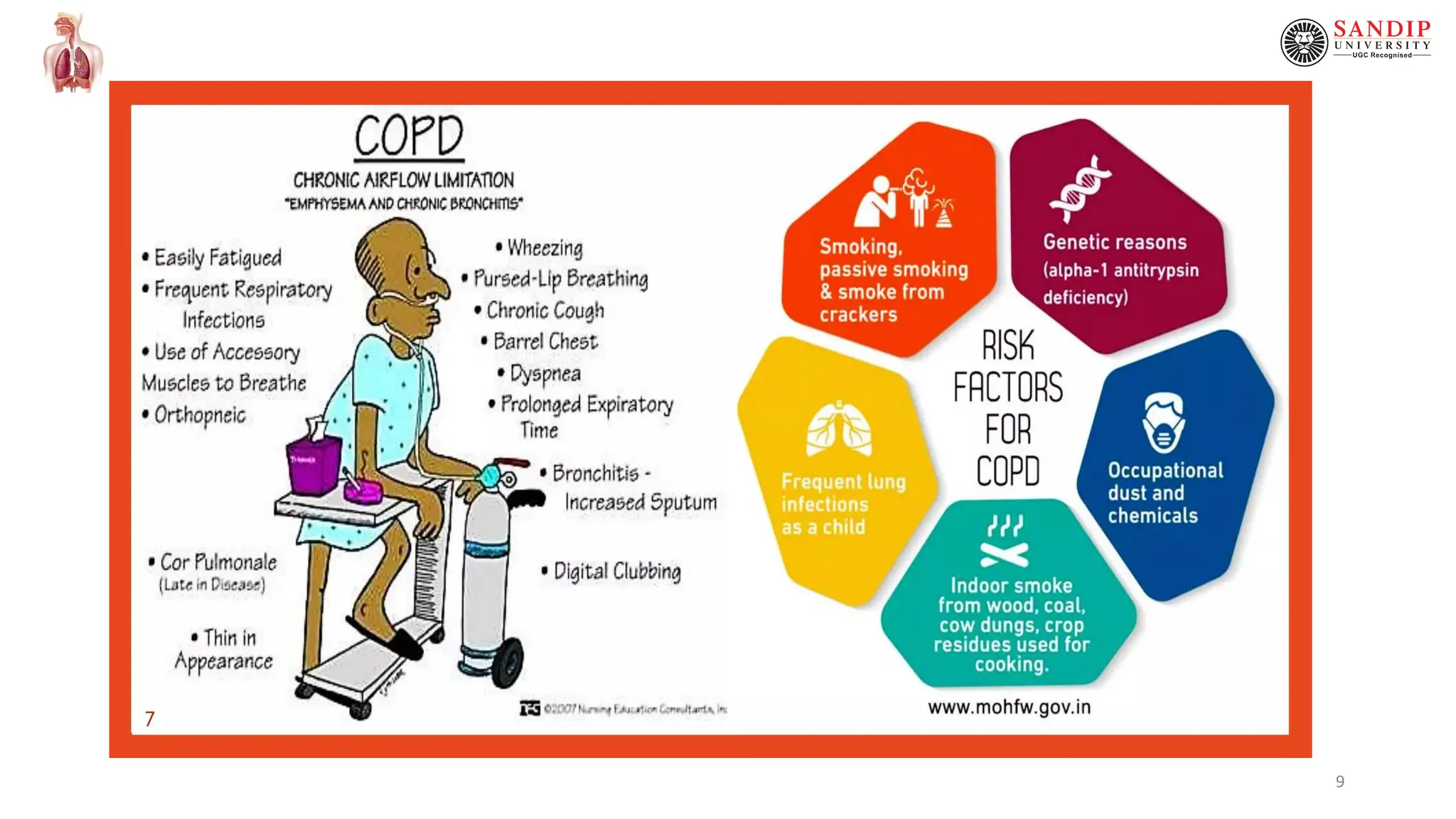
Respiratory Pathophysiology
Chronic Obstructive Pulmonary Disease
(COPD)
Dr. Vishal Balakrushna Jadhav
Assistant Professor (Pharmacology)
School of Pharmaceutical Sciences (SOPS), SUN
1
Content
 Definition
a. Chronic bronchitis
b. Emphysema
 Etiopathogenesis of COPD
 Pathological changes
 Clinical features
 Illustration- COPD and Microscopic changes
2
Definition
 The persistent obstruction of the airways which is either chronic,
partial or complete (partially reversible or completely irreversible)
leads to functional disability of the lungs.
 Causes- Chronic bronchitis and emphysema are the usual causes.
 Chronic bronchitis- The condition characterized by persistent cough
formed due to mucus hypersecretion with expectoration for at least
four months of the year for two consecutive years, more commonly
seen in middle aged males than females, approximately 20% adult
men and 5% adult women are suffered.
 Emphysema- The combination of permanent distention of air spaces
distal to the terminal bronchioles and destruction of the dilated
alveolar wall. 3
Etiopathogenesis of COPD
 Smoking- Cigarette smoke impairs mucocilliary defense mechanism and
leads to mucus hypersecretion by raising the activity of mucocytes.
 Atmospheric air pollutants- includes sulphur dioxide, nitrogen dioxide,
particulate dust and toxic fumes etc.
 Infection- Bacterial, viral, and mycoplasmal infection do not initiate chronic
bronchitis but usually occurs secondarily to chronic bronchitis.
 Familial and genetic make up- Inborn deficiency of Alpha-1 antitrypsin (a
protease inhibitor) leads to an early damage of alveolar mucosa and leads
to the development of emphysema.
 Alpha-1 antitrypsin deficiency is an inherited disorder characterized by low serum
levels of alpha-1 antitrypsin (AAT). Loss of AAT disrupts the protease- antiprotease
balance in the lungs, allowing proteases, specifically neutrophil elastase, to act
uninhibited and destroy lung matrix and alveolar structures. 4
Flowchart indicating Etiopathogenesis of COPD/COAD
Precipitating factors
Alveolar inflammation
Accumulation of WBCs in the inflammed area
Release of enzymes (Neutrophilic Elastases)
Damage of connective tissue wall of alveoli and development of COPD/COAD
5
Pathological changes
 Grossly, thickened, hyperemic and edematous bronchial wall, lumina of
bronchi and bronchioles shows the presence of mucus plugs and purulent
exudates.
 Microscopically, there is an increase in the REID INDEX (the ratio between
thickness of sub mucosal mucous glands in the cartilage containing large
airways to the thickness of total bronchial wall). Non-mucosal cartilage
containing small airways shows the presence of goblet cells (mucus secreting
cells) in the bronchus and intestinal tract.
6
Clinical features
 Persistent cough with copious expectoration of long duration as seen in heavy
smokers
 Recurrent respiratory infection
 Dyspnoea not prominent at rest but is more in exertion or in stress conditions
 Patients become ‘blue bloaters’ due to cyanosis and edema
 Core pulmonale (Pulmonary Heart Disease) characterized by right heart failure
 Wheezing, breathlessness and tightness of the chest in the morning.
7
Complications of COPD
 Pulmonary hypertension,
 Right Ventricular Failure,
 Leakage of air into the pleural spaces,
 Arrhythmias,
 Secondary polycythemia, and
 Respiratory failure
8
9
10
11
Chronic bronchitis
Gross Appearance Histopathological View
13
Emphysema
Gross Appearance Histopathological View
14
Thank You...

Chronic Obstructive Pulmonary Disease...

  • 1.
    Respiratory Pathophysiology Chronic ObstructivePulmonary Disease (COPD) Dr. Vishal Balakrushna Jadhav Assistant Professor (Pharmacology) School of Pharmaceutical Sciences (SOPS), SUN 1
  • 2.
    Content  Definition a. Chronicbronchitis b. Emphysema  Etiopathogenesis of COPD  Pathological changes  Clinical features  Illustration- COPD and Microscopic changes 2
  • 3.
    Definition  The persistentobstruction of the airways which is either chronic, partial or complete (partially reversible or completely irreversible) leads to functional disability of the lungs.  Causes- Chronic bronchitis and emphysema are the usual causes.  Chronic bronchitis- The condition characterized by persistent cough formed due to mucus hypersecretion with expectoration for at least four months of the year for two consecutive years, more commonly seen in middle aged males than females, approximately 20% adult men and 5% adult women are suffered.  Emphysema- The combination of permanent distention of air spaces distal to the terminal bronchioles and destruction of the dilated alveolar wall. 3
  • 4.
    Etiopathogenesis of COPD Smoking- Cigarette smoke impairs mucocilliary defense mechanism and leads to mucus hypersecretion by raising the activity of mucocytes.  Atmospheric air pollutants- includes sulphur dioxide, nitrogen dioxide, particulate dust and toxic fumes etc.  Infection- Bacterial, viral, and mycoplasmal infection do not initiate chronic bronchitis but usually occurs secondarily to chronic bronchitis.  Familial and genetic make up- Inborn deficiency of Alpha-1 antitrypsin (a protease inhibitor) leads to an early damage of alveolar mucosa and leads to the development of emphysema.  Alpha-1 antitrypsin deficiency is an inherited disorder characterized by low serum levels of alpha-1 antitrypsin (AAT). Loss of AAT disrupts the protease- antiprotease balance in the lungs, allowing proteases, specifically neutrophil elastase, to act uninhibited and destroy lung matrix and alveolar structures. 4
  • 5.
    Flowchart indicating Etiopathogenesisof COPD/COAD Precipitating factors Alveolar inflammation Accumulation of WBCs in the inflammed area Release of enzymes (Neutrophilic Elastases) Damage of connective tissue wall of alveoli and development of COPD/COAD 5
  • 6.
    Pathological changes  Grossly,thickened, hyperemic and edematous bronchial wall, lumina of bronchi and bronchioles shows the presence of mucus plugs and purulent exudates.  Microscopically, there is an increase in the REID INDEX (the ratio between thickness of sub mucosal mucous glands in the cartilage containing large airways to the thickness of total bronchial wall). Non-mucosal cartilage containing small airways shows the presence of goblet cells (mucus secreting cells) in the bronchus and intestinal tract. 6
  • 7.
    Clinical features  Persistentcough with copious expectoration of long duration as seen in heavy smokers  Recurrent respiratory infection  Dyspnoea not prominent at rest but is more in exertion or in stress conditions  Patients become ‘blue bloaters’ due to cyanosis and edema  Core pulmonale (Pulmonary Heart Disease) characterized by right heart failure  Wheezing, breathlessness and tightness of the chest in the morning. 7
  • 8.
    Complications of COPD Pulmonary hypertension,  Right Ventricular Failure,  Leakage of air into the pleural spaces,  Arrhythmias,  Secondary polycythemia, and  Respiratory failure 8
  • 9.
  • 10.
  • 11.
  • 12.
  • 13.
  • 14.