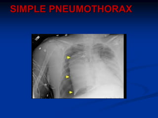
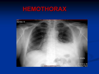
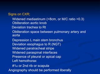
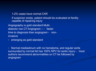
1) Chest trauma is predominantly caused by motor vehicle accidents and falls. The most common injuries are chest wall trauma and hemothorax.
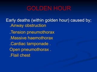
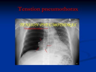
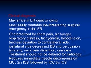
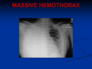
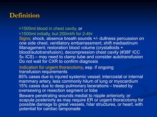
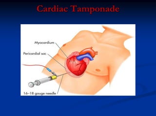
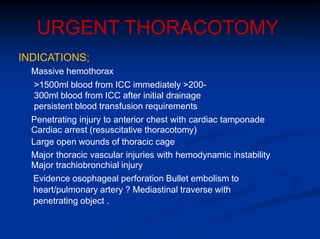
2) Early deaths from chest trauma are often caused by airway obstruction, tension pneumothorax, massive hemothorax, or cardiac tamponade.
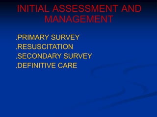
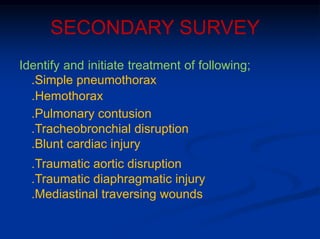
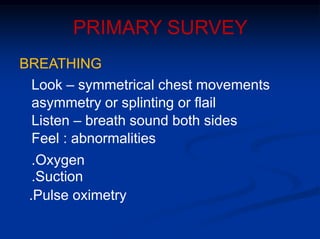
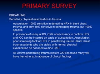
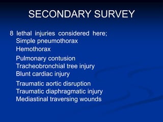
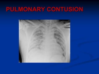
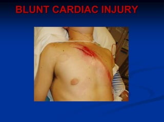
3) Initial assessment focuses on the ABCs with stabilization of life-threatening injuries like tension pneumothorax the top priority. Secondary surveys then identify and treat other injuries like simple pneumothorax, pulmonary contusion, and blunt cardiac injury.
Trauma is a leading cause of death and disability. Chest injuries from blunt or penetrating trauma can cause life-threatening conditions like tension pneumothorax, massive hemothorax, and pericardial tamponade if not recognized and treated promptly. The primary survey focuses on identifying and correcting issues like airway obstruction, hypoxemia, and hypovolemia. Chest x-rays and further tests in secondary survey can identify additional injuries like simple pneumothorax, pulmonary contusion, and cardiac injuries that require specific management. Proper evaluation and treatment of chest trauma aims to prevent complications and reduce mortality from uncontrolled hemorrhage, hypoxia, or delayed interventions.
The document provides an overview of immediately life-threatening chest injuries. It classifies chest injuries as either immediately life-threatening or potentially life-threatening. The six immediately life-threatening conditions discussed in detail are airway obstruction, tension pneumothorax, open pneumothorax, massive hemothorax, pericardial tamponade, and tracheobronchial injuries. For each condition, the document outlines clinical presentation and emergency management strategies. It also discusses emergency department thoracotomy and provides indications and contraindications for its use.
Emergency anaesthetic management of extensive thoracic traumaHossam atef
A document on thoracic trauma outlines several types of injuries that can occur from blunt or penetrating chest trauma, including injuries to the chest wall, pleural space, lungs, heart, great vessels, tracheobronchial tree, esophagus, and diaphragm. It emphasizes the importance of establishing airway and ventilation, maintaining circulation and treating life-threatening injuries like tension pneumothorax through needle decompression or immediate surgery. Resuscitation of patients with severe thoracic trauma requires addressing airway, breathing, circulation and neurological status before diagnostic evaluation and priority surgical interventions.
A 28-year-old male was brought to the emergency room after a motor vehicle accident. He complained of chest pain, a forehead wound, and right forearm pain. Examination found he was conscious with stable vital signs. Chest x-ray revealed an abnormal aortic knob and widened mediastinum. CT angiogram confirmed a traumatic aortic tear. Despite treatment, his condition deteriorated with low blood pressure and increased pain. Aortography then definitively diagnosed aortic rupture, which requires urgent surgical repair for survival.
Thoracic trauma can result in serious injuries and accounts for over 25% of trauma deaths, with blunt trauma making up 70% of chest injuries mostly from motor vehicle accidents. Common thoracic injuries include rib fractures, pneumothorax, hemothorax, and injuries to the heart and great vessels which require stabilization of the airway, breathing, and circulation followed by diagnostic imaging and treatment of life-threatening injuries. Management involves establishing ABCs, treating tension pneumothorax and sucking chest wounds, and having a high index of suspicion to identify specific injuries like aortic injuries that require further evaluation.
A motorcyclist was in a head-on collision and presented with difficulty breathing, severe chest pain, restlessness and smell of alcohol. Examination found dyspnea, tachycardia, distended neck veins, left tracheal deviation, hypotension and cyanosis, and a hyper-resonant right chest with absent breath sounds. Chest X-ray showed a right tension pneumothorax. Needle decompression was performed followed by tube thoracostomy, resulting in improved breathing, comfort, and oxygen saturation.
A stab wound victim presented with subcutaneous emphysema, tachycardia and distended neck veins. Chest X-ray revealed a left hemopneumothorax
This document provides information on key topics in the management of chest trauma:
- Tension pneumothorax is a medical emergency diagnosed clinically and treated with needle or tube thoracostomy without delay.
- Hemothorax over 1.5L or drainage over 200mL/hr requires tube thoracostomy or thoracotomy for repair of bleeding.
- Flail chest is treated with oxygenation, fluid restriction, and analgesia to improve ventilation.
- Blunt cardiac injury can cause hypotension and dysrhythmias diagnosed by echocardiogram or troponin levels.
This document provides information on key topics in the management of chest trauma:
- Tension pneumothorax is a medical emergency diagnosed clinically and treated with needle or tube thoracostomy without delay.
- Hemothorax over 1.5L or drainage over 200mL/hr requires tube thoracostomy or thoracotomy. Cardiac tamponade is diagnosed clinically and with ultrasound.
- Pulmonary contusions are managed with oxygenation, fluids restriction, and analgesia. Blunt cardiac injuries may cause hypotension or arrhythmias.
chest trauma is one of the leading cause of death in poly trauma patients. ER doctor should be aware of how to suspect and how to deal with life threatening conditions resulting from chest trauma
1. The document provides guidelines for evaluating and treating chest trauma, with a focus on the ABCs (airway, breathing, circulation). It discusses assessing and securing the airway, treating tension pneumothorax, hemothorax, and cardiac tamponade.
2. The secondary survey involves using imaging like CXR, CT, and ultrasound to identify injuries like aortic tears, tracheal lacerations, and pulmonary contusions. Procedures like bronchoscopy and thoracotomy may be needed. Outcomes of various injuries are discussed.
3. Complications of chest trauma like empyema are outlined. Treatment depends on imaging and test findings. Pneumatoceles may become infected and
Penetrating thoracic trauma can range from stable patients with few complaints to hemodynamically unstable patients requiring immediate intervention. Potential injuries include injuries to the chest wall, lungs, heart, great vessels, esophagus, and diaphragm. Life-threatening injuries that require rapid assessment and treatment include tension pneumothorax, open pneumothorax, massive hemothorax, cardiac tamponade, and massive hemothorax. Initial management involves the ABCs with consideration of adjuncts like chest tubes and consideration of urgent thoracotomy for control of bleeding from major vessels or repair of cardiac injuries.
Chest trauma can cause significant morbidity and mortality if not managed promptly and effectively. The document discusses the pathophysiology of chest trauma from both blunt and penetrating mechanisms. It emphasizes the importance of the primary survey approach, including assessing the airway, breathing, and circulation (ABCs). For patients who are unstable, interventions like intubation, chest tube insertion, and fluid resuscitation may be required during the initial assessment to stabilize their condition.
This document provides an overview of chest trauma and its management. It discusses that chest trauma accounts for 25% of all trauma deaths and is the second leading cause of trauma deaths after head injuries. It then covers the epidemiology, types, causes, pathophysiology and initial assessment of chest injuries. It describes important life-threatening injuries to assess for such as tension pneumothorax, massive hemothorax, flail chest and cardiac tamponade. It outlines the questions to ask and signs to look for when initially evaluating a patient with chest trauma. It also discusses monitoring the patient, management approaches, and complications to watch out for with chest trauma patients.
This presentation discusses chest trauma. It begins with an introduction noting that the chest contains vital organs and thoracic trauma is a common cause of death. [It then provides details on the anatomy of the chest and classifications of blunt vs penetrating trauma.] It identifies the "Deadly Dozen" as the most life-threatening chest injuries requiring immediate treatment, including tension pneumothorax, massive hemothorax, and flail chest. [It also discusses several potentially life-threatening injuries requiring secondary survey, such as tracheobronchial disruptions.] Throughout, it emphasizes the importance of strict adherence to ATLS protocols to identify and treat deadly chest injuries.
The document discusses various types of chest trauma, including:
1. Thoracic wall injuries such as rib fractures, sternal fractures, and flail chest which involve damage to the chest wall bones and muscles.
2. Lung injuries such as pneumothorax, hemothorax, pulmonary contusion, and pulmonary laceration which involve damage to the lungs themselves.
3. The etiology, pathophysiology, diagnosis and management of different chest injuries are explained in detail with examples provided. Chest trauma can range from minor to life-threatening depending on the extent of injuries to thoracic structures.
This document discusses the management of chest injuries. It begins with an introduction stating that chest trauma is a significant cause of morbidity and mortality worldwide. It then covers the epidemiology, relevant anatomy, causes, pathophysiology, investigations, management, and complications of various chest injuries. The pathophysiology section describes the mechanisms and types of injuries that can occur to the chest wall, pleura, lungs, heart, and major vessels. Management involves following ATLS protocols, administering analgesics and antibiotics, and performing procedures such as tube thoracostomy or thoracotomy when needed to treat injuries such as hemothorax, flail chest, or cardiac tamponade. Complications include wound infections, dehiscence,
This document discusses various types of thoracic trauma, including injuries to the chest wall, lungs, heart and great vessels. It describes evaluation and management of specific injuries such as rib fractures, flail chest, pneumothorax and hemothorax. Treatment options include chest tube insertion, ventilation support, epidural analgesia, thoracotomy and thoracoscopy. Complications like pulmonary contusion, hematoma and acute respiratory distress syndrome are also covered. The document provides detailed clinical guidance for treating thoracic trauma injuries.
Chest trauma can involve injuries to the chest wall or internal organs. The primary survey focuses on life-threatening injuries like tension pneumothorax, massive hemothorax, or cardiac tamponade. Tension pneumothorax requires immediate needle decompression while massive hemothorax may require a chest tube or thoracotomy. Cardiac tamponade is treated with pericardiocentesis if due to blunt trauma or urgent surgery if from penetrating trauma. The secondary survey entails a full examination to identify injuries like lung contusions or aortic disruption that require further treatment or monitoring.
Chest trauma can cause injuries to the chest wall, heart, lungs and blood vessels in the chest. Common injuries include rib fractures, flail chest, pneumothorax, hemothorax and cardiac tamponade. Initial management involves stabilizing the patient, assessing for life-threatening injuries like tension pneumothorax, and treating hypoxia and hypotension. Further management may include chest tube insertion, thoracotomy or other surgeries depending on the specific injuries present. Complications can include respiratory failure, pneumonia and acute respiratory distress syndrome. Proper evaluation, timely intervention and supportive care are needed to optimize outcomes from chest trauma.
Chest injuries account for 25% of severe injuries and are usually caused by road traffic accidents. The primary survey focuses on the airway, breathing, circulation, disability and exposure to identify and treat immediate life threats like tension pneumothorax. Secondary survey involves a full history, examination and investigations like CXR and CT scan to evaluate injuries. Most chest injuries are managed conservatively but conditions like massive hemothorax or cardiac tamponade may require chest tube insertion or thoracotomy.
Rehabilitation of patient with pleural effusionAdemola Adeyemo
1) Pulmonary embolism occurs when a blood clot blocks an artery in the lungs, and can cause pleural effusions in about 30% of cases. Physiotherapy is an important part of managing patients with pleural effusions secondary to pulmonary embolism.
2) Physiotherapy includes techniques like incentive spirometry, chest physiotherapy, and exercises to improve cardiopulmonary function and endurance. Drainage of fluid from chest tubes is also facilitated.
3) As the patient's condition improves with physiotherapy, their ability to exercise intensifies and shortness of breath decreases, with the goal of restoring independence and fitness.
This document discusses principles of managing chest trauma, including sucking chest wounds, chest tube insertion and management, chest bleeding, pneumothorax, hemothorax, flail chest, aortic disruption, cardiac injury, diaphragmatic rupture, esophageal injury, pulmonary contusion, and indications for emergency thoracotomy. Key points include how to manage a sucking chest wound with an occlusive dressing and chest tube, signs of tension pneumothorax, indications for chest tube placement, and situations where emergency thoracotomy may be necessary to control hemorrhage.
Thoracic trauma accounts for 25% of all injury-related deaths and is a contributory factor in 50% more deaths. While most thoracic injuries can be treated without surgery, it is important to recognize life-threatening injuries like tension pneumothorax, massive hemothorax, and cardiac tamponade during the initial assessment. Adjuncts like chest x-rays, ultrasound, and tube thoracostomy can help identify injuries, while interventions like needle decompression and tube insertion treat immediate life threats. A full secondary survey evaluates injuries like rib fractures, lung contusions, and injuries to the heart and great vessels that require monitoring or further investigation.
Chest injuries ranks 3rd after head injuries and extremity injuries in a case of multisystem trauma.It is of two types blunt chest trauma and peneterating chest trauma.The main cause of blunt chest trauma is road side accidents due to vehicles. Peneterating chest trauma is more dangerous and is common in war injuries and civilian terroism.In this ppp I have discussed some useful uncommon and important aspects of chest injuries
Chest injuries are a leading cause of trauma deaths. Immediate life-threatening chest injuries include airway obstruction, tension pneumothorax, open pneumothorax, massive hemothorax, flail chest, and cardiac tamponade. These injuries can cause death within seconds if not recognized and treated promptly. Potentially life-threatening injuries include cardiac contusion, aortic disruption, diaphragmatic rupture, esophageal injury, pulmonary contusion, and tracheobronchial injuries. Prompt assessment and management following ATLS guidelines is essential to stabilize vital functions and prevent complications such as respiratory failure, shock, and death from chest injuries.
Chest trauma is one of the leading causes of death worldwide. It can be classified as either blunt trauma or penetrating trauma. Blunt chest trauma is more common and results from forces like compression or impacts. Common injuries include rib fractures, sternal fractures, pulmonary contusions, pneumothorax, hemothorax, and flail chest. Diagnostic tests include chest x-rays and CT scans. Treatment depends on the severity and type of injury but generally involves pain management, breathing treatments, and surgery to repair damage and drain fluids if needed.
This document discusses shock in trauma and provides an overview of key topics including:
- The pathophysiology of shock involves inadequate oxygen delivery leading to cellular dysfunction.
- Hypovolemic shock from blood or fluid loss is a leading cause and requires rapid fluid resuscitation.
- A thorough physical exam and diagnostic tests are needed to identify the source of bleeding.
- Early intervention and resuscitation are important to reverse the effects of shock and reduce mortality.
This document provides an overview of evaluating orthopedic x-rays using the ABCs approach - Assessing Adequacy, Alignment, Bones, Cartilage, and Soft Tissues. It describes examining x-rays for the appropriate number of views and penetration, alignment of bones, any fractures or distortions of bones, widening of joint spaces, and soft tissue swelling. Examples are given of using this approach to identify fractures. Key terms for describing fractures such as open vs closed, location, fracture line, and relationship of fragments are also defined.
1) Trauma is a leading cause of non-obstetric mortality in pregnant patients. Common causes include motor vehicle accidents, domestic violence, falls, and penetrating injuries.
2) Pregnant patients experience many physiological changes including increased blood volume, cardiac output, and respiration that must be considered in their trauma management. Anatomical changes like uterine size and position can also impact injuries.
3) Diagnostic studies like ultrasound and CT scans are important for evaluation while minimizing radiation exposure to the fetus. Continuous fetal heart monitoring can identify non-reassuring signs. Surgical interventions may be needed for unstable mothers or fetuses.
1- Initial Assessment and Management of the Trauma Patient.pptxAsgraf
The document provides an overview of the initial assessment and management of trauma patients, covering the primary and secondary surveys with a focus on evaluating airway, breathing, circulation, disability, and exposure/environment, as well as discussing specific injuries and treatments for conditions like tension pneumothorax, hemorrhagic shock, and cardiac tamponade.
The Glasgow Coma Scale (GCS) was developed to assess neurological function and brain injury severity. It evaluates eye opening, verbal response, and motor response on a scale of 3 to 15, with lower scores indicating more severe brain injury. The GCS is useful for prognosis, monitoring changes, and assessing the need for airway support. A modified Children's Coma Scale was also developed for those too young to communicate verbally.
This document provides guidance on trauma procedures for a patient presenting with a stab wound and signs of alcohol intoxication. It outlines steps for securing the airway with endotracheal intubation or cricothyroidotomy if needed. It also describes performing a tube thoracostomy for a hemopneumothorax, obtaining central venous access, arterial blood gas sampling, and placing a nasogastric tube. Key steps and considerations are outlined for each procedure along with potential complications. The document emphasizes the importance of practice to gain skill in lifesaving surgical procedures.
The document discusses the initial assessment and management of pediatric trauma. It notes that while ABCDE priorities are the same as adults, children have unique anatomic and physiologic characteristics that require consideration, such as a larger head, softer bones, and narrower airway. It emphasizes the importance of recognizing potential abuse injuries and preventing injuries through strategies like bicycle helmet promotion.
Chest trauma can be life-threatening and accounts for 25% of all trauma deaths. It includes injuries from blunt forces like rib fractures or penetrating injuries from stab wounds. Without treatment, injuries can lead to hypoxia and hypotension from issues like pneumothorax, hemothorax, or flail chest. Proper management requires following ABC (airway, breathing, circulation) to ensure oxygen delivery and addressing specific injuries like tension pneumothorax through needle decompression or chest tube insertion.
FAST (Focused Assessment with Sonography in Trauma) is an ultrasound technique used to quickly detect the presence of free fluid in the abdomen or chest resulting from trauma. It can visualize fluid in seven dependent areas of the abdomen and the pericardial sac. FAST has sensitivity of 88-91.7% and specificity of 94.7-99% for detecting fluid. A positive FAST means fluid is detected in one of the areas scanned, while a negative FAST means no fluid is seen. FAST provides a rapid initial assessment and can help guide trauma management, but CT is more sensitive for characterizing injuries. Limitations include difficulty with obese patients, bowel gas, or retroperitoneal injuries.
The document provides an overview of the approach to musculoskeletal injuries in the emergency department. It discusses that musculoskeletal problems comprise approximately 11% of emergency department visits. The physician needs a systematic approach to orthopedic complaints that can range from minor sprains to life-threatening trauma. Commonly seen injuries include soft tissue injuries, fractures, dislocations, and infections. Less common but important injuries include spinal injuries, crush injuries, and compartment syndrome. The document then discusses fractures, pelvic fractures, long bone fractures, open fractures, deep venous thrombosis, crush injuries, compartment syndrome, and spinal injuries. It emphasizes the importance of thorough evaluation, splinting, consultation, and managing complications for these types of musculoskeletal emergencies.
Spinal cord injuries are commonly caused by motor vehicle accidents, falls, and sports. The cervical spine is most commonly injured, especially between C5-C6. Plain films and advanced imaging can help evaluate injuries. Flexion injuries risk paralysis and require stabilization to prevent further injury. Compression fractures are generally stable with rest, while burst fractures may require surgery if unstable.
This document discusses the medical management of acute coronary syndrome (ACS). It notes that over 4 million patients are admitted each year with unstable angina or acute myocardial infarction, and over 900,000 undergo percutaneous coronary intervention. ACS refers to a spectrum of conditions ranging from unstable angina to Q-wave and non-Q-wave myocardial infarction that are characterized by a disrupted atherosclerotic plaque. For treatment of unstable angina, the document recommends anticoagulant therapy with heparin or low-molecular weight heparin to reduce risks of myocardial infarction and death.
This document discusses acute coronary syndrome (ACS), including unstable angina (UA), non-ST elevation myocardial infarction (NSTEMI), and ST elevation myocardial infarction (STEMI). It defines ACS and describes how the conditions are classified based on ECG and cardiac enzyme levels. It also discusses risk stratification in NSTEMI and typical management approaches for NSTEMI, UA, and STEMI. The document concludes with a case study describing a patient presenting with chest pain.
This document provides an overview of coronary artery disease, myocardial infarction, and related cardiac pharmacology. It discusses the etiology and risk factors of coronary artery disease, describes different types of angina pectoris, and outlines goals and medications for treating angina. It also covers atherosclerosis, statins, calcium channel blockers, beta blockers, ACE inhibitors, angiotensin receptor blockers, digoxin, and other cardiac medications. The document defines myocardial infarction and discusses gender differences, EKG changes, types, risks, effects, complications, diagnosis, and goals of treatment for MI.
The document provides an overview of burns, including epidemiology, classification, severity, treatment and complications. It notes that around 500,000 people in the US are treated annually for burns, which can be caused by heat, fires, scalding or chemicals. Burn extent is determined using the Rule of 9s or Lund & Browder chart and severity is classified from first to fourth degree burns based on depth of tissue damage. Treatment depends on severity but may include fluid resuscitation, wound care, surgery and infection prevention. Complications can include infection, respiratory distress and contractures.
The document discusses burn classifications and management. It describes the three main types of burns as thermal, chemical, and electrical. Thermal burns are the most common and caused by heat sources. Burn depth is classified as superficial, partial thickness, or full thickness. Initial burn management focuses on the ABCs - securing the airway, assessing breathing and circulation. Fluid resuscitation is crucial to prevent shock in large burns. Ongoing nursing care involves wound care, infection prevention and managing complications.
Care of the Burn Patient provides guidelines for treating burn injuries. It outlines conducting a primary and secondary survey to assess airway, breathing, circulation, disability, and exposure. This includes estimating burn size using the Rule of Nines or Lund & Browder chart for children. It also describes fluid resuscitation using the Parkland formula to replace fluid losses from capillary leakage in burned tissue. Proper burn management focuses on stopping the burning process, providing oxygen, intravenous fluids, pain control, and preventing infection.
Histololgy of Female Reproductive System.pptxAyeshaZaid1
Dive into an in-depth exploration of the histological structure of female reproductive system with this comprehensive lecture. Presented by Dr. Ayesha Irfan, Assistant Professor of Anatomy, this presentation covers the Gross anatomy and functional histology of the female reproductive organs. Ideal for students, educators, and anyone interested in medical science, this lecture provides clear explanations, detailed diagrams, and valuable insights into female reproductive system. Enhance your knowledge and understanding of this essential aspect of human biology.
Integrating Ayurveda into Parkinson’s Management: A Holistic ApproachAyurveda ForAll
Explore the benefits of combining Ayurveda with conventional Parkinson's treatments. Learn how a holistic approach can manage symptoms, enhance well-being, and balance body energies. Discover the steps to safely integrate Ayurvedic practices into your Parkinson’s care plan, including expert guidance on diet, herbal remedies, and lifestyle modifications.
TEST BANK For An Introduction to Brain and Behavior, 7th Edition by Bryan Kol...rightmanforbloodline
TEST BANK For An Introduction to Brain and Behavior, 7th Edition by Bryan Kolb, Ian Q. Whishaw, Verified Chapters 1 - 16, Complete Newest Versio
TEST BANK For An Introduction to Brain and Behavior, 7th Edition by Bryan Kolb, Ian Q. Whishaw, Verified Chapters 1 - 16, Complete Newest Version
Rasamanikya is a excellent preparation in the field of Rasashastra, it is used in various Kushtha Roga, Shwasa, Vicharchika, Bhagandara, Vatarakta, and Phiranga Roga. In this article Preparation& Comparative analytical profile for both Formulationon i.e Rasamanikya prepared by Kushmanda swarasa & Churnodhaka Shodita Haratala. The study aims to provide insights into the comparative efficacy and analytical aspects of these formulations for enhanced therapeutic outcomes.
Here is the updated list of Top Best Ayurvedic medicine for Gas and Indigestion and those are Gas-O-Go Syp for Dyspepsia | Lavizyme Syrup for Acidity | Yumzyme Hepatoprotective Capsules etc
TEST BANK For Community Health Nursing A Canadian Perspective, 5th Edition by...Donc Test
TEST BANK For Community Health Nursing A Canadian Perspective, 5th Edition by Stamler, Verified Chapters 1 - 33, Complete Newest Version Community Health Nursing A Canadian Perspective, 5th Edition by Stamler, Verified Chapters 1 - 33, Complete Newest Version Community Health Nursing A Canadian Perspective, 5th Edition by Stamler Community Health Nursing A Canadian Perspective, 5th Edition TEST BANK by Stamler Test Bank For Community Health Nursing A Canadian Perspective, 5th Edition Pdf Chapters Download Test Bank For Community Health Nursing A Canadian Perspective, 5th Edition Pdf Download Stuvia Test Bank For Community Health Nursing A Canadian Perspective, 5th Edition Study Guide Test Bank For Community Health Nursing A Canadian Perspective, 5th Edition Ebook Download Stuvia Test Bank For Community Health Nursing A Canadian Perspective, 5th Edition Questions and Answers Quizlet Test Bank For Community Health Nursing A Canadian Perspective, 5th Edition Studocu Test Bank For Community Health Nursing A Canadian Perspective, 5th Edition Quizlet Test Bank For Community Health Nursing A Canadian Perspective, 5th Edition Stuvia Community Health Nursing A Canadian Perspective, 5th Edition Pdf Chapters Download Community Health Nursing A Canadian Perspective, 5th Edition Pdf Download Course Hero Community Health Nursing A Canadian Perspective, 5th Edition Answers Quizlet Community Health Nursing A Canadian Perspective, 5th Edition Ebook Download Course hero Community Health Nursing A Canadian Perspective, 5th Edition Questions and Answers Community Health Nursing A Canadian Perspective, 5th Edition Studocu Community Health Nursing A Canadian Perspective, 5th Edition Quizlet Community Health Nursing A Canadian Perspective, 5th Edition Stuvia Community Health Nursing A Canadian Perspective, 5th Edition Test Bank Pdf Chapters Download Community Health Nursing A Canadian Perspective, 5th Edition Test Bank Pdf Download Stuvia Community Health Nursing A Canadian Perspective, 5th Edition Test Bank Study Guide Questions and Answers Community Health Nursing A Canadian Perspective, 5th Edition Test Bank Ebook Download Stuvia Community Health Nursing A Canadian Perspective, 5th Edition Test Bank Questions Quizlet Community Health Nursing A Canadian Perspective, 5th Edition Test Bank Studocu Community Health Nursing A Canadian Perspective, 5th Edition Test Bank Quizlet Community Health Nursing A Canadian Perspective, 5th Edition Test Bank Stuvia
TEST BANK For Basic and Clinical Pharmacology, 14th Edition by Bertram G. Kat...rightmanforbloodline
TEST BANK For Basic and Clinical Pharmacology, 14th Edition by Bertram G. Katzung, Verified Chapters 1 - 66, Complete Newest Version.
Osteoporosis - Definition , Evaluation and Management .pdfJim Jacob Roy
Osteoporosis is an increasing cause of morbidity among the elderly.
In this document , a brief outline of osteoporosis is given , including the risk factors of osteoporosis fractures , the indications for testing bone mineral density and the management of osteoporosis
2. Trauma is predominantly blunt, and primarily result
of MVA and falls 8-10% chest trauma is penetrating
Chest wall trauma is most frequent injury after blunt
thoracic trauma
Chest trauma is predictor of more serious injury
elsewhere
Most chest injuries managed with observation,
analgesia, thoracentesis, and respiratory support
15-20% require thoracotomy
Operation rate <5% in blunt trauma and has poorer
outcome
Overall thoracic trauma mortality 10%
Accounts for ¼ trauma deaths
3. GOLDEN HOUR
Early deaths (within golden hour) caused by;
.Airway obstruction
.Tension pneumothorax
.Massive haemothorax
.Cardiac tamponade .
Open pneumothorax .
.Flail chest
8. PRIMARY SURVEY
AIRWAY MAINTENANCE
.Chin lift
.Jaw thrust
.Oropharyngeal airway
.Nasopharyngeal airway .Definitive
airway - ETT - Surgical airway
.Prehospital ETT confers no survival
advantage over BVM and slightly increases
prehospital time .
9. PRIMARY SURVEY
C-SPINE
CT significantly out performs, plain X-ray as
screening tool for very high risk C-spine injury
CT should be initial screening tool in those with
significantly depressed mental status Plain x-
ray still recommended as initial screening test
for low risk patients
10. PRIMARY SURVEY
BREATHING
Look – symmetrical chest movements
asymmetry or splinting or flail
Listen – breath sound both sides
Feel : abnormalities
.Oxygen
.Suction
.Pulse oximetry
11. PRIMARY SURVEY
BREATHING
Sensitivity physical examination in trauma
Auscultation 100% sensitive in detecting HPX in blunt chest
trauma, and only 50% sensitive in penetrating trauma, but 100%
specific
In presence of unequal BS, CXR unnecessary to confirm HPX,
and ICC can be inserted on basis of auscultation. Auscultation
poor screening tool for HPX in penetrating trauma .Blunt chest
trauma patients who are stable with normal physical
examination do not need routine CXR
All victims penetrating trauma require CXR because many will
have hemothorax in absence of clinical findings.
13. Clinical diagnosis
May arrive in ER dead or dying
Most easily treatable life-threatening surgical
emergency in the ER
Characterized by chest pain, air hunger,
respiratory distress, tachycardia, hypotension,
tracheal deviation to contralateral side,
ipsilateral side decreased BS and percussion
tympany, neck vein distention, cyanosis
Treatment should not be delayed for radiology
Requires immediate needle decompression
MCL 2nd ICS followed by ICC 5th ICS
14. OPEN PNEUMOTHORAX
If opening in chest wall 2/3 diameter
trachea, air passes preferentially through
defect with each respiratory effort
16. 2 or more # ribs in 2 or more places
Severe disruption normal chest wall movement with paradoxical
movements
If significant underlying lung injury exists, serious hypoxia may result
Diagnosis made by clinical examination, CXR, CT
ABG required to assess need for mechanical ventilation
TREATMENT
humidified O2, analgesia, judicious fluids, mechanical ventilation
(PEEP, SIMV, PSV), bronchoscopic aspiration, chest physiotherapy,
+/- IV Atb
Indication for mechanical ventilation; Pa02<60mmHg,
PaC02>50mmHg, assoc severe trauma with LOC +/- shock, airway
obstruction or repeated atelectasis
Surgical stabilization – indicated if prolonged ventilatory assistance
expected; benefits include - less ventilatory support and pneumonia,
shorter ICU stay, cost effective, improved FVC, faster return to work
18. >1500ml blood in chest cavity, or
<1500ml initially, but 200ml/h for 2-4hr
Signs; shock, absence breath sounds +/- dullness percussion on
one side chest, ventilatory embarrassment, shift mediastinum
Management; restoration blood volume (crystalloids +
blood/autotransfusion), decompression chest cavity (#38F ICC
5th ICS) – may need to clamp tube and consider autotransfusion
Do not wait for CXR to confirm diagnosis
Indication for urgent thoracotomy, esp. if ongoing
transfusion requirements
85% cases due to injured systemic vessel; intercostal or internal
mammary artery, less commonly hilum of lung or myocardium
15% cases due to deep pulmonary lacerations – treated by
oversewing or resection segment or lobe
Beware penetrating wounds medial to nipple anteriorly, or
scapula posteriorly as may require ER or urgent thoracotomy for
possible damage to great vessels, hilar structures, or heart, with
potential for cardiac tamponade
Definition
20. Pericardium fills with blood from heart,
pericardial vessels, or great vessels
15-20ml may be enough to cause
haemodynamic compromise
Most commonly from penetrating injuries
Any patient with penetrating thoracic injury
has cardiac injury until proven otherwise
Wounded heart will follow on of two courses;
exsanguination or tamponade (80%)
Exsanguination group - most die in field or in
ER – if survive show signs hemorrhagic
shock
Definition
21. PRESENTATION
Beck’s triad (over 90% cases)
.venous pressure elevation •
.decline arterial pressure •
.muffled heart sounds
(hypotension may be due to hypovolaemia, neck veins
may not be distended due to hypovolaemia) May be
restless and anxious and refuse to lie supine Pulsus
paradoxis >10mm Hg (10% cases) Kusssmaul’s sign
– rise in venous pressure with inspiration
PEA in absence of hypovolaemia or tension
pneumothorax suggest cardiac tamponade
22. Pericardiocentesis indicated if high index suspicion and
failure to respond to resuscitation for hemorrhagic shock
All patients with positive pericardiocentesis require open
thoracotomy.
Management
24. ER THORACOTOMY
Dismal survival
Best outcomes with ;
• Isolated chest injury
• Single vs multiple chest injuries
• Penetrating injury
• Stab rather than gunshot
• Right sided, single chamber heart injuries
• Major factor in outcome is presenting physiology - Better
outcome if vital signs present on admission
• If BP present on arrival ER – survival advantage if
thoracotomy performed in theatre
• Prehospital intubation
• Cardiac tamponade
• Presence mentation in field or on arrival in ER
25. ER THORACOTOMY
Six potential therapeutic goals;
Control bleeding (cardiac or thoracic)
Effective cardiac compressions
Relief pericardial tamponade
Cross clamp lung hilum after suspected air
embolism
Cross clamp descending aorta for lower torso
hemorrhage control
Confirm ETT placement
26. URGENT THORACOTOMY
INDICATIONS;
Massive hemothorax
>1500ml blood from ICC immediately >200-
300ml blood from ICC after initial drainage
persistent blood transfusion requirements
Penetrating injury to anterior chest with cardiac tamponade
Cardiac arrest (resuscitative thoracotomy)
Large open wounds of thoracic cage
Major thoracic vascular injuries with hemodynamic instability
Major trachiobronchial injury
Evidence osophageal perforation Bullet embolism to
heart/pulmonary artery ? Mediastinal traverse with
penetrating object .
30. Second most common injury from chest trauma after #ribs
Upto 50% occult and not seen on initial CXR 20% have
accompanying hemothorax From both penetrating and
blunt trauma Signs; decreased BS on affected side
hyperesonance on affected side Erect CXR required for
diagnosis Managed by ICC 5th ICS (28Fr)
ICC is safest option, irrespective of size, because unpredictable
progression to tension pneumothorax
Observation +/- aspiration risky! and 10% fail conservative Rx
Always needs ICC if undergoing transport, GA, or PPV( 40%
increase size)
Small bore pleural catheters assoc with increased incidence
recurrence and complications
32. Blunt or penetrating trauma
Bleeding from; lung laceration, intercostal
vessel, internal mammary artery, T-spine #
Usually self-limiting
if large enough to appear on CXR needs ICC
ICC reduces late incidence of empyema or
fibrothorax.
34. Most common potentially lethal chest injury
Damage to parenchyma from passage shock wave
through tissue
CXR – may be normal, but usually shows opacity in lung
field, lags 12-24hr behind clinical picture
Respiratory failure subtle and develops over time. ABG
worsens at day 2-3 as oedema increases, careful
monitoring and reevaluation. Intubation and ventilation
if Sao2<90% especially if co- morbidities, or being
transported
In multi-injured trauma patient pulmonary contusion may
be superimposed by sepsis, SIRS, MODS, ARDS
Definition
36. TRACHEOBRONCHIAL TREE INJURY
Uncommon injury (<2% major thoracic injuries)
Often overlooked on initial assessment In blunt
trauma the majority injuries occur within 2.5cm
carina
Most die at scene
If survive - high mortality from associated injuries
SIGNS;
hemoptysis, subcutaneous emphysema, tension
pneumothorax with mediastinal shift, progressive
mediastinal air
Suspect if persistent large air leak after tube
thoracostomy or if surgical emphysema
37. TRACHEOBRONCHIAL TREE INJURY
Mor than one ICC may be needed to overcome leak
and expand the lung.
Confirmed on bronchoscopy
Opposite main stem bronchial intubation may be
temporarily required
TREATMENT;
Distal bronchus – usually close with chest tube drainage
alone within days
Proximal bronchus – posterolateral thoracotomy – repair
bronchus with monofilament if possible, otherwise
segmentectomy or lobectomy required
39. BLUNT CARDIAC INJURY
Suspect in any patient who sustains high energy impact to
thorax
May cause; myocardial contusion, cardiac chamber rupture,
valvular disruption
Assoc injuries common – sternal #, #ribs, flail chest, seatbelt
sign over precordium
May be asymptomatic
Chamber rupture – most die in field, typically presents with
cardiac tamponade (mostly R side) – presentation and treatment
similar to penetrating injuries
Myocardial contusion may present with chest discomfort,
hypotension, conduction abnormalities, valvular dysfunction,
papillary muscle rupture, raised CVP (due to RV dysfunction)
40. BLUNT CARDIAC INJURY
DIAGNOSIS – goal is to identify patients who need treatment (few)
ECG
variable, multiple PVC’s, sinus tacchycardia, AF, BBB usually R, ST
segment ∆ (ST or T-wave ∆ more specific than arrhythmia) negative
ECG predicts absence of contusion, and even more strongly suggests
patient not likely to suffer cardiac complication Troponin
no correlation with level and severity injury, reports vary as to
whether it adds any more info to ECG
Transthoracic Echo
as part of FAST in all patients with truncal trauma in some centres
indicated if clinical, biochemical, ECG abnormality, wall motion
abnormality, thrombi, pericardial effusion
Less commonly – radionuclide angiography, TOE
41. BLUNT CARDIAC TRAUMA
If ECG, Troponin, and FAST normal, and no clinical
signs or cardiac dysfunction – do not pursue diagnosis
further
Second troponin and ECG 8hr after injury required to
reliably exclude significant myocardial injury
If clinical, ECG, or chemical evidence of cardiac
contusion, then monitor in HDU/CCU/ICU until resolution
clinical signs and return ECG/Troponin to normal
Unstable patients admitted to ICU
Overall prognosis after cardiac contusion is good
43. Common cause of sudden death after MVA or fall from height –
80-85% mortality before reaching hospital
If survive tend to have incomplete laceration near ligamentum
arteriosum at aortic isthmus (95%), just distal to LSCA – fixed
nature at this point
All survivors have contained hematoma -intact adventitial layer
or contained mediastinal hematoma prevent immediate death If
survive to hospital – 30% mortality in 6hr, 40-50% mortality in
24hr
Persistent or recurrent hypotension is usually due to other cause
If ruptures into L chest is fatal within minutes
Specific signs and symptoms frequently absent Diagnosis
made by high index suspicion, mechanism injury
(deceleration), characteristic radiological findings, TOE, CT
angiogram, arteriography
44. Signs on CXR;
Widened mediastinum (>8cm, or M/C ratio >0.3)
Obliteration aortic knob
Deviation trachea to Rt
Obliteration space between pulmonary artery and
aorta
Depression L main stem bronchus
Deviation esophagus to R (NGT)
Widened paratracheal stripe
Widened paraspinal interfaces
Presence of pleural or apical cap
Left hemothorax
#1st or 2nd rib or scapula
Angiography should be performed liberally
45. 1-2% cases have normal CXR
If suspicion exists, patient should be evaluated at facility
capable of repairing injury
Angiography is gold standard Multi-
detector row CT Angiogram – • faster
time to diagnosis than angiogram • non-
invasive
• emerging as gold standard
• Normal mediastinum with no hematoma, and regular aorta
surrounded by normal fat has 100% NPV for aortic injury • most
authors recommend abnormalities on CT be followed by
angiogram
47. TRAUMATIC DIAPHRAGMATIC
INJURY
Diaphragmatic injury most common injury requiring
surgery after blunt trauma
More commonly diagnosed on L, but near equal
incidence
Diagnosed by physical examination, CXR – elevated
or indistinct hemidiaphragm, bowel, stomach, NGT in
chest (20% CXR normal)
CT scan, US
Liver obliterates defect or protects it on R side – more
difficult to detect initially, 75% delayed diagnosis, MRI
helpful in R sided injuries
Elevated R hemi diaphragm may be only finding on
CXR
48. TRAUMATIC
DIAPHRAGMATIC INJURY
Blunt trauma produces radial tears that lead to herniation
Penetrating trauma produces small perforations that
often take time, even years to develop
If injury suspected on left side – NGT should be inserted
Upper GIT contrast study necessary if diagnosis unclear
Laparoscopy or Thoracoscopy may be useful if diagnosis
unclear
Laparotomy for other causes may reveal diaphragmatic
injury
49. TRAUMATIC
DIAPHRAGMATIC INJURY
TREATMENT
All diaphragmatic injuries should be repaired even if
small and of no apparent importance, due to risk
herniation and incarceration
Preferred closure is direct repair with interrupted non-
absorbable sutures
Synthetic materials may be required to close large
defects
50. MEDIASTINAL TRAVERSING
WOUNDS
Penetrating objects that traverse mediastinum may
injure heart, great vessels, tracheobronchial tree,
esophagus
Diagnosis made when examination or CXR reveals
entrance wound in one hemithorax and exit wound or
missile lodged in opposite side or mediastinum If
hemodynamically abnormal consider exsanguinating
thoracic hemorrhage, tension pneumothorax,
pericardial tamponade Bilateral ICC’s necessary
Indications for urgent thoracotomy similar for massive
hemothorax
Both sides may need thoracotomy but start with side
with most blood
52. OSOPHAGEAL RUPTURE
Most commonly penetrating
Blunt trauma causes forceful expulsion stomach contents into
esophagus – linear tear in lower esophagus which may leak into
mediastinum
Mediastinitis with immediate or delayed rupture into pleural
space causes empyema
Mortality >20%
Prompt recognition and initiation treatment mandatory
Delays in treatment assoc with higher morbidity and mortality
Primary closure within 24hr best outcome
Needs to be excluded in all patients with traversing mediastinal
injuries
May present with painful swallowing, hematemesis,
subcutaneous emphysema, SOB, fever
53. OESOPHAGEAL RUPTURE
Consider in all patients with;
• L pneumothorax or hemothorax without rib #
• Blow to sternum or epigastrium with pain +/- shock inconsistent with
apparent injury
• Food or particulate matter in ICC
• Mediastinal air on CXR
• Persistent pneumothorax
Confirm with esophagogram or esophagoscopy
Esophagogram difficult in intoxicated, combative, unprotected
airway, poor view cervical esophagus if intubated Endoscopy
– sens and spec 90-100%, performed in ER or at bedside
Endoscopy all patients with signs and symptoms or suspicious
nature of injury
May be role for VAT in diagnosis and treatment
55. Most common injured part of chest
Pain impairs ventilation and effective coughing which
may lead to atelectasis and pneumonia especially
with pre-existing lung disease
# scapula, 1st or 2nd rib may have severe associated
injuries with mortality up to 35%
Ribs 4-9 sustain majority of blunt trauma
More force required in younger patients
# ribs 10-12 should raise suspicion for hepatosplenic
injury
56. Likelihood of hepatosplenic injury;
10-16% rib #’ have concomitant intra-abdominal
injury to liver or spleen
Liver injury increased with low rib # (8-12), R
sided rib #
Splenic injury increased with low rib #.lt sided.
57. IMAGING
CXR has poor sensitivity, and rarely influences management #
anterior cartilages or separation costochondral junctions may
not be seen on CXR
Special rib view Xrays unnecessary as may not ∆ management
Ultrasound reveals rib # more accurately than X-ray and clinical
acumen, specificity 85%
USS superior to CT scan without radiation dose Follow-up
USS at 3/52 post injury is gold standard May impact on
patients with higher levels of activity such as manual
workers or athletes wrt return to work and physical activity
58. SIGNS
localized pain, tenderness, and crepitus
TREATMENT
Goal of treatment is to exclude underlying more serious injury, and
ensure adequate analgesia
Analgesia with; PO and IV analgesics, PCA, intercostal block,
epidural
Oxygen
Physiotherapy
SURGERY
Surgical stabilization may reduce pain and ventilator requirements
Methods ; fixator plates, intramedullary wires or nails, struts
Indications ; select group with flail chest not weaning from
mechanical ventilation after 5-7/7 with only moderate extrathoracic
injuries and no HI, pulmonary contusion, or ARDS.
60. Usually result of direct blow May be
accompanied by pulmonary
contusion
Consider blunt cardiac injury with all
sternal #
Does not necessarily require monitoring
Operative repair needed . posterior
sternoclavicular dislocation may cause
SVC obstruction needs reduction.