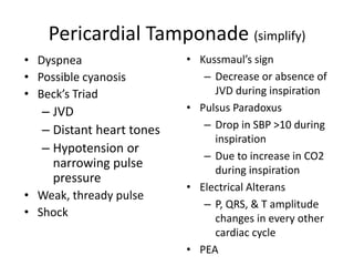
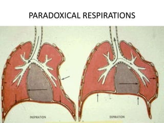
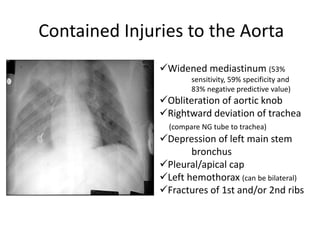
This document discusses various types of chest trauma, including penetrating and blunt injuries. It covers mechanisms of injury, specific injuries like rib fractures, pneumothorax, hemothorax, flail chest, and cardiac injuries. Management of life-threatening injuries like tension pneumothorax, pericardial tamponade, and tracheal disruptions are outlined. Imaging findings and treatments for many chest trauma injuries are also presented.
This document discusses the management of thoracic trauma. It begins with an introduction covering causes of thoracic trauma and objectives. It then covers anatomy and physiology of the thorax in detail. Next, it discusses pathophysiology of various types of thoracic injuries from blunt and penetrating trauma such as pneumothorax, hemothorax, pulmonary contusion, myocardial contusion, pericardial tamponade, aortic rupture and others. It concludes with sections on initial assessment and management of thoracic trauma patients.
Thoracic injuries can be immediately life-threatening, potentially life-threatening, or non-life-threatening. Immediately life-threatening injuries include tension pneumothorax, massive hemothorax, and cardiac tamponade which require urgent treatment to prevent death. Potentially life-threatening injuries like aortic injury or cardiac injury may not initially present severe symptoms but can worsen without treatment. The majority of thoracic injuries are non-life-threatening such as rib fractures and pulmonary contusions which are managed with pain control, pulmonary toilet, and observation.
Chest injuries are common and can be life-threatening. The document discusses various types of chest injuries including blunt trauma, penetrating trauma, and compression injuries. It covers anatomy of the chest, definitions, causes, symptoms, management, and potential complications of specific injuries like pneumothorax, hemothorax, flail chest, and cardiac tamponade. Immediate life-threatening injuries require stabilization of the airway, breathing, and circulation before further treatment.
This document discusses thoracic injuries, including the structures at risk, common injury types, signs and symptoms, and assessment. Key points include:
- The heart, blood vessels, lungs, esophagus and trachea are vital structures at risk from thoracic trauma. Injuries can include pneumothorax, hemothorax, pulmonary contusion, and cardiac trauma.
- Signs of life-threatening injuries include decreased breath sounds, chest pain on breathing, subcutaneous emphysema, tracheal shifting, and hypotension. Tension pneumothorax can rapidly lead to respiratory failure.
- Assessment of thoracic trauma patients involves rapid trauma survey checking breath sounds, jugular vein
Chest injuries account for 25% of severe injuries and are usually caused by road traffic accidents. The primary survey focuses on the airway, breathing, circulation, disability and exposure to identify and treat immediate life threats like tension pneumothorax. Secondary survey involves a full history, examination and investigations like CXR and CT scan to evaluate injuries. Most chest injuries are managed conservatively but conditions like massive hemothorax or cardiac tamponade may require chest tube insertion or thoracotomy.
This document provides an overview of chest trauma and its management. It discusses that chest trauma accounts for 25% of all trauma deaths and is the second leading cause of trauma deaths after head injuries. It then covers the epidemiology, types, causes, pathophysiology and initial assessment of chest injuries. It describes important life-threatening injuries to assess for such as tension pneumothorax, massive hemothorax, flail chest and cardiac tamponade. It outlines the questions to ask and signs to look for when initially evaluating a patient with chest trauma. It also discusses monitoring the patient, management approaches, and complications to watch out for with chest trauma patients.
Thoracic trauma accounts for 25% of all injury-related deaths and is a contributory factor in 50% more deaths. While most thoracic injuries can be treated without surgery, it is important to recognize life-threatening injuries like tension pneumothorax, massive hemothorax, and cardiac tamponade during the initial assessment. Adjuncts like chest x-rays, ultrasound, and tube thoracostomy can help identify injuries, while interventions like needle decompression and tube insertion treat immediate life threats. A full secondary survey evaluates injuries like rib fractures, lung contusions, and injuries to the heart and great vessels that require monitoring or further investigation.
The document discusses trauma to the thorax and abdomen. It begins with an introduction to trauma epidemiology and classifications. It then covers early management principles including the primary and secondary surveys. Specific thoracic injuries discussed include pneumothorax, hemothorax, flail chest, and cardiac tamponade. Abdominal injuries addressed include injuries to solid organs like the spleen and liver as well as hollow organ injuries. Diagnostic tools and management strategies are provided for each type of injury.
Thoracic trauma can result from blunt or penetrating injuries and often involves rib fractures, pneumothorax, hemothorax, or injuries to the heart or lungs. Assessment focuses on life-threatening injuries like tension pneumothorax, massive hemothorax, or pericardial tamponade. Management priorities are airway control, oxygen therapy, needle decompression for tension pneumothorax, chest tube insertion for pneumothorax or hemothorax, and rapid transport to a trauma center.
Chest injuries are a leading cause of trauma deaths. Immediate life-threatening chest injuries include airway obstruction, tension pneumothorax, open pneumothorax, massive hemothorax, flail chest, and cardiac tamponade. These injuries can cause death within seconds if not recognized and treated promptly. Potentially life-threatening injuries include cardiac contusion, aortic disruption, diaphragmatic rupture, esophageal injury, pulmonary contusion, and tracheobronchial injuries. Prompt assessment and management following ATLS guidelines is essential to stabilize vital functions and prevent complications such as respiratory failure, shock, and death from chest injuries.
Chest trauma can cause injuries to the chest wall, heart, lungs and blood vessels in the chest. Common injuries include rib fractures, flail chest, pneumothorax, hemothorax and cardiac tamponade. Initial management involves stabilizing the patient, assessing for life-threatening injuries like tension pneumothorax, and treating hypoxia and hypotension. Further management may include chest tube insertion, thoracotomy or other surgeries depending on the specific injuries present. Complications can include respiratory failure, pneumonia and acute respiratory distress syndrome. Proper evaluation, timely intervention and supportive care are needed to optimize outcomes from chest trauma.
This document provides an overview of the approach to evaluating and managing chest pain. It discusses the anatomy and pathophysiology of chest pain, including the differences between somatic and visceral pain. Common causes of chest pain are reviewed, including acute coronary syndrome (ACS), pulmonary embolism, thoracic aortic dissection, and others. Risk stratification tools for ACS are described. The document then outlines the stepwise approach to a patient with chest pain, including history, physical exam, ECG, imaging, labs, and potential treatments.
Thoracic injuries can cause serious complications by compromising the airway, ventilation, and circulation. Key threats include tension pneumothorax, massive hemothorax, and cardiac tamponade. Proper assessment is important to identify these life-threatening injuries through primary and secondary surveys using inspection, auscultation, palpation, and adjuncts like ultrasound. Early interventions such as needle decompression, chest tube insertion, and surgery can help manage injuries and prevent further deterioration.
This document provides information on the anatomy of the chest and causes, symptoms, diagnosis, and treatment of various types of chest injuries. It discusses conditions like airway obstruction, flail chest, sucking chest wounds, hemothorax, tension pneumothorax, and cardiac tamponade. For each condition, it outlines the pathophysiology, diagnostic indicators, and emergency treatment procedures like needle decompression and pericardiocentesis. The document emphasizes the importance of stabilizing life-threatening conditions in the primary survey and provides details on emergency department management of chest trauma.
Chest trauma can cause serious injury and is a common cause of disability and mortality after head and spinal cord injuries. Mechanisms of injury include blunt trauma, crush injuries, penetrating wounds, burns, and inhalation of foreign objects. Specific injuries include rib fractures, lung injuries, pneumothoraces, hemothoraces, aortic ruptures, and diaphragm injuries. Diagnosis involves history, physical exam noting diminished breath sounds and tracheal deviation, chest X-ray to detect fractures and lung abnormalities, and CT scan which aids in precise diagnosis of injuries.
- Blunt and penetrating chest trauma are leading causes of trauma death, accounting for approximately 25% of trauma fatalities in the US each year.
- Common injuries from chest trauma include rib fractures, flail chest, pulmonary contusion, pneumothorax, hemothorax, and cardiac/vascular injuries. Aggressive management is needed to support ventilation and treat underlying injuries.
- Tension pneumothorax is a life-threatening emergency requiring immediate needle decompression to relieve pressure on the heart and great vessels. Other critical treatments include securing the airway, administering oxygen, performing needle decompression if indicated, giving IV fluids, and rapid transport to a trauma center.
This document provides an overview of chest trauma, including the anatomy of the chest, causes of chest injuries, types of chest injuries, and treatments. It begins with the objectives and anatomy of the thorax. The main causes of chest trauma are then discussed as blunt trauma, penetrating trauma, and compression injuries. Various chest injuries are described in detail such as rib fractures, flail chest, pneumothorax, hemothorax, pulmonary contusion, and pericardial tamponade. The treatments for each type of injury are also outlined. The document provides a comprehensive review of chest trauma for medical professionals.
Chest trauma can represent a major burden and lead to significant morbidity and mortality. Chest injuries require prompt evaluation and management to address life-threatening injuries like tension pneumothorax, hemothorax, and cardiac injuries. The chest is divided into the thoracic cavity containing the lungs, heart, and great vessels which are vulnerable to injury from blunt or penetrating trauma. Common injuries include rib fractures, flail chest, pulmonary contusions, and pneumothorax. Immediate airway control and treatment of life-threatening injuries is essential, followed by management of potential injuries and complications to optimize outcomes.
This document provides guidance on evaluating and managing patients presenting with chest pain. It outlines the general approach which includes ensuring patient stability, providing oxygen and analgesia. It describes performing a focused history and physical exam, as well as relevant investigations. It discusses life-threatening causes of chest pain including acute coronary syndrome, cardiac tamponade, pulmonary embolism, tension pneumothorax, and aortic dissection. For each condition, it details symptoms, signs, diagnostic tests, and general treatment approaches.
This document discusses the management of various chest injuries including hemothorax, pneumothorax, injuries to blood vessels, heart, lungs, esophagus, chest wall and diaphragm. Key points include: indications for thoracotomy include significant hemorrhage or certain imaging findings; endovascular stenting is the mainstay treatment for aortic injuries; penetrating cardiac injuries are usually repaired operatively while blunt injuries are monitored; tracheobronchial injuries are repaired by debridement and anastomosis when possible; chest wall injuries are usually treated non-operatively. Life-threatening injuries like tension pneumothorax, massive hemothorax and cardiac tamponade require immediate needle decompression,
Chest injuries ranks 3rd after head injuries and extremity injuries in a case of multisystem trauma.It is of two types blunt chest trauma and peneterating chest trauma.The main cause of blunt chest trauma is road side accidents due to vehicles. Peneterating chest trauma is more dangerous and is common in war injuries and civilian terroism.In this ppp I have discussed some useful uncommon and important aspects of chest injuries
Chest trauma accounts for 25% of all trauma deaths and can have serious pathological consequences if not treated promptly. The most common types of chest trauma are blunt injuries such as rib fractures and penetrating injuries such as stab wounds. Proper diagnosis and treatment of injuries is important to stabilize the patient and prevent complications such as tension pneumothorax, cardiac tamponade, and aortic rupture which can quickly lead to death if not addressed.
- Ischemic heart disease is caused by a reduced blood supply to the heart muscle due to coronary artery atherosclerosis. The heart requires constant blood flow to function properly but atherosclerotic plaques can block this blood flow.
- Symptoms range from stable angina (predictable chest pain) to unstable angina (increasing chest pain) to myocardial infarction (heart attack caused by severe blockage or clot).
- Risk factors include high blood pressure, high cholesterol, smoking, obesity, and lack of exercise. Proper management can reduce the risk of further heart problems.
This document discusses various types of chest trauma, including pneumothorax, hemothorax, flail chest, diaphragmatic injury, aortic tear, esophageal rupture, pulmonary contusion, and cardiac tamponade. It provides details on the procedures used to treat chest trauma, such as intercostal drains and tracheostomy. Chest trauma can be caused by blunt or penetrating mechanisms and accounts for 20-25% of trauma deaths. Clinical features, investigations, and management are described for each type of chest injury.
1. The document discusses the approach to evaluating and categorizing different types of chest pain, including acute coronary syndrome.
2. Key factors in the initial evaluation of chest pain include obtaining a detailed history, performing a physical exam, 12-lead EKG, and cardiac biomarker testing to help determine if the chest pain is caused by life-threatening conditions like myocardial infarction.
3. Patients are categorized as having possible acute ischemia, probable acute ischemia, or myocardial infarction based on their history, symptoms, physical exam findings, EKG results, and cardiac enzyme levels in order to determine the urgency of further testing and treatment.
This document provides an overview of thoracic trauma anatomy, physiology, and management. It describes the structures of the mediastinum and vasculature. Common thoracic injuries are classified and their pathophysiology, signs, and treatment are outlined. These include pneumothorax, hemothorax, flail chest, and cardiac tamponade. The primary and secondary surveys for evaluating thoracic trauma are reviewed, including inspection, palpation, percussion, and auscultation of the chest.
Chest Trauma - Medical and Surgical Treatment.pptxAitzazIjaz1
Chest trauma can occur from blunt or penetrating mechanisms and is classified as such. Signs and symptoms depend on the specific injury but may include pain, difficulty breathing, and decreased breath sounds. Common injuries include rib fractures, flail chest, and pneumothorax. A tension pneumothorax is a medical emergency requiring immediate needle decompression to relieve pressure on the heart and blood vessels. Chest tube placement is the definitive treatment for many chest injuries.
baru donny Sensitivitas dan Spesifisitas Ankle Brachial Index (ABI)-1.pptxDonnyWahyuPratomo
ABI dan PWHD memiliki sensitivitas dan spesifisitas yang tinggi dalam mendiagnosis PAD dibandingkan dengan angiografi sebagai tes emas. Kombinasi kedua tes ini memberikan hasil yang lebih baik daripada tes tunggal dengan sensitivitas 100% dan spesifisitas 80%. Penelitian ini menunjukkan bahwa ABI dan PWHD dapat digunakan secara luas untuk skrining awal PAD karena mudah dan non-invasif.
ATLS membahas tiga puncak utama dalam penanganan trauma, yaitu detik-menit untuk penanganan darurat seperti laserasi otak, menit-jam untuk cedera seperti EDH dan SDH, dan lebih dari seminggu untuk komplikasi seperti sepsis dan gagal organ. Dokumen ini juga menjelaskan tahapan penilaian awal (primary dan secondary survey) serta penanganan berbagai cedera seperti kepala, dada, tulang belakang, sesuai ped
Malformasi anorektal merupakan kelainan bawaan dimana terjadi kegagalan perkembangan anus atau rektum secara normal pada janin. Terjadi akibat kegagalan pertumbuhan septum urorektal sehingga kloaka tidak terbagi dengan sempurna menjadi sinus urogenital dan rektum. Penatalaksanaannya meliputi kolostomi sementara, dilatasi anus, dan operasi posterosagital anorectoplasty. Prognosis pasien cenderung baik dengan pendekatan
1. The document provides a table summarizing research activities of permanent lecturers in the Study Program of Surgery including the name of lecturer, title of research, integrated subject, form of integration, year of research, level of research, and alignment with roadmap.
2. It lists 12 research activities conducted by lecturers from the Division of Orthopedic Surgery, 3 from the Division of Digestive Surgery, 3 from the Division of Oncology Surgery, 2 from the Division of Vascular Surgery, and 2 from the Division of Urology Surgery.
3. The research topics covered include hip geometry, lumbar herniated nucleus pulposus, spine injury stabilization, surgical site
Nano-gold for Cancer Therapy chemistry investigatory projectSIVAVINAYAKPK
chemistry investigatory project
The development of nanogold-based cancer therapy could revolutionize oncology by providing a more targeted, less invasive treatment option. This project contributes to the growing body of research aimed at harnessing nanotechnology for medical applications, paving the way for future clinical trials and potential commercial applications.
Cancer remains one of the leading causes of death worldwide, prompting the need for innovative treatment methods. Nanotechnology offers promising new approaches, including the use of gold nanoparticles (nanogold) for targeted cancer therapy. Nanogold particles possess unique physical and chemical properties that make them suitable for drug delivery, imaging, and photothermal therapy.
Pictorial and detailed description of patellar instability with sign and symptoms and how to diagnose , what investigations you should go with and how to approach with treatment options . I have presented this slide in my 2nd year junior residency in orthopedics at LLRM medical college Meerut and got good reviews for it
After getting it read you will definitely understand the topic.
NAVIGATING THE HORIZONS OF TIME LAPSE EMBRYO MONITORING.pdfRahul Sen
Time-lapse embryo monitoring is an advanced imaging technique used in IVF to continuously observe embryo development. It captures high-resolution images at regular intervals, allowing embryologists to select the most viable embryos for transfer based on detailed growth patterns. This technology enhances embryo selection, potentially increasing pregnancy success rates.
5-hydroxytryptamine or 5-HT or Serotonin is a neurotransmitter that serves a range of roles in the human body. It is sometimes referred to as the happy chemical since it promotes overall well-being and happiness.
It is mostly found in the brain, intestines, and blood platelets.
5-HT is utilised to transport messages between nerve cells, is known to be involved in smooth muscle contraction, and adds to overall well-being and pleasure, among other benefits. 5-HT regulates the body's sleep-wake cycles and internal clock by acting as a precursor to melatonin.
It is hypothesised to regulate hunger, emotions, motor, cognitive, and autonomic processes.
Travel Clinic Cardiff: Health Advice for International TravelersNX Healthcare
Travel Clinic Cardiff offers comprehensive travel health services, including vaccinations, travel advice, and preventive care for international travelers. Our expert team ensures you are well-prepared and protected for your journey, providing personalized consultations tailored to your destination. Conveniently located in Cardiff, we help you travel with confidence and peace of mind. Visit us: www.nxhealthcare.co.uk
Breast cancer: Post menopausal endocrine therapyDr. Sumit KUMAR
Breast cancer in postmenopausal women with hormone receptor-positive (HR+) status is a common and complex condition that necessitates a multifaceted approach to management. HR+ breast cancer means that the cancer cells grow in response to hormones such as estrogen and progesterone. This subtype is prevalent among postmenopausal women and typically exhibits a more indolent course compared to other forms of breast cancer, which allows for a variety of treatment options.
Diagnosis and Staging
The diagnosis of HR+ breast cancer begins with clinical evaluation, imaging, and biopsy. Imaging modalities such as mammography, ultrasound, and MRI help in assessing the extent of the disease. Histopathological examination and immunohistochemical staining of the biopsy sample confirm the diagnosis and hormone receptor status by identifying the presence of estrogen receptors (ER) and progesterone receptors (PR) on the tumor cells.
Staging involves determining the size of the tumor (T), the involvement of regional lymph nodes (N), and the presence of distant metastasis (M). The American Joint Committee on Cancer (AJCC) staging system is commonly used. Accurate staging is critical as it guides treatment decisions.
Treatment Options
Endocrine Therapy
Endocrine therapy is the cornerstone of treatment for HR+ breast cancer in postmenopausal women. The primary goal is to reduce the levels of estrogen or block its effects on cancer cells. Commonly used agents include:
Selective Estrogen Receptor Modulators (SERMs): Tamoxifen is a SERM that binds to estrogen receptors, blocking estrogen from stimulating breast cancer cells. It is effective but may have side effects such as increased risk of endometrial cancer and thromboembolic events.
Aromatase Inhibitors (AIs): These drugs, including anastrozole, letrozole, and exemestane, lower estrogen levels by inhibiting the aromatase enzyme, which converts androgens to estrogen in peripheral tissues. AIs are generally preferred in postmenopausal women due to their efficacy and safety profile compared to tamoxifen.
Selective Estrogen Receptor Downregulators (SERDs): Fulvestrant is a SERD that degrades estrogen receptors and is used in cases where resistance to other endocrine therapies develops.
Combination Therapies
Combining endocrine therapy with other treatments enhances efficacy. Examples include:
Endocrine Therapy with CDK4/6 Inhibitors: Palbociclib, ribociclib, and abemaciclib are CDK4/6 inhibitors that, when combined with endocrine therapy, significantly improve progression-free survival in advanced HR+ breast cancer.
Endocrine Therapy with mTOR Inhibitors: Everolimus, an mTOR inhibitor, can be added to endocrine therapy for patients who have developed resistance to aromatase inhibitors.
Chemotherapy
Chemotherapy is generally reserved for patients with high-risk features, such as large tumor size, high-grade histology, or extensive lymph node involvement. Regimens often include anthracyclines and taxanes.
Know the difference between Endodontics and Orthodontics.Gokuldas Hospital
Your smile is beautiful.
Let’s be honest. Maintaining that beautiful smile is not an easy task. It is more than brushing and flossing. Sometimes, you might encounter dental issues that need special dental care. These issues can range anywhere from misalignment of the jaw to pain in the root of teeth.
Are you looking for a long-lasting solution to your missing tooth?
Dental implants are the most common type of method for replacing the missing tooth. Unlike dentures or bridges, implants are surgically placed in the jawbone. In layman’s terms, a dental implant is similar to the natural root of the tooth. It offers a stable foundation for the artificial tooth giving it the look, feel, and function similar to the natural tooth.
How to Control Your Asthma Tips by gokuldas hospital.Gokuldas Hospital
Respiratory issues like asthma are the most sensitive issue that is affecting millions worldwide. It hampers the daily activities leaving the body tired and breathless.
The key to a good grip on asthma is proper knowledge and management strategies. Understanding the patient-specific symptoms and carving out an effective treatment likewise is the best way to keep asthma under control.
STUDIES IN SUPPORT OF SPECIAL POPULATIONS: GERIATRICS E7shruti jagirdar
Unit 4: MRA 103T Regulatory affairs
This guideline is directed principally toward new Molecular Entities that are
likely to have significant use in the elderly, either because the disease intended
to be treated is characteristically a disease of aging ( e.g., Alzheimer's disease) or
because the population to be treated is known to include substantial numbers of
geriatric patients (e.g., hypertension).
Histololgy of Female Reproductive System.pptxAyeshaZaid1
Dive into an in-depth exploration of the histological structure of female reproductive system with this comprehensive lecture. Presented by Dr. Ayesha Irfan, Assistant Professor of Anatomy, this presentation covers the Gross anatomy and functional histology of the female reproductive organs. Ideal for students, educators, and anyone interested in medical science, this lecture provides clear explanations, detailed diagrams, and valuable insights into female reproductive system. Enhance your knowledge and understanding of this essential aspect of human biology.
PGx Analysis in VarSeq: A User’s PerspectiveGolden Helix
Since our release of the PGx capabilities in VarSeq, we’ve had a few months to gather some insights from various use cases. Some users approach PGx workflows by means of array genotyping or what seems to be a growing trend of adding the star allele calling to the existing NGS pipeline for whole genome data. Luckily, both approaches are supported with the VarSeq software platform. The genotyping method being used will also dictate what the scope of the tertiary analysis will be. For example, are your PGx reports a standalone pipeline or would your lab’s goal be to handle a dual-purpose workflow and report on PGx + Diagnostic findings.
The purpose of this webcast is to:
Discuss and demonstrate the approaches with array and NGS genotyping methods for star allele calling to prep for downstream analysis.
Following genotyping, explore alternative tertiary workflow concepts in VarSeq to handle PGx reporting.
Moreover, we will include insights users will need to consider when validating their PGx workflow for all possible star alleles and options you have for automating your PGx analysis for large number of samples. Please join us for a session dedicated to the application of star allele genotyping and subsequent PGx workflows in our VarSeq software.
2. Mechanism of Injury
Penetrating Trauma
– Low Energy
• Arrows, knives, handguns
• Injury caused by direct contact
– High Energy
• Military, hunting rifles & high powered hand guns
• Extensive injury due to high pressure
3. • Either:
– direct blow (e.g. rib fracture)
– deceleration injury
– compression injury
• Rib fracture is the most common sign of blunt thoracic
trauma
• Fracture of scapula, sternum, or first rib suggests
massive force of injury
• Age Factors
• Pediatric Thorax: More cartilage = Absorbs forces
• Geriatric Thorax: Calcification & osteoporosis = More fracture
Blunt injuries
8. Tension Pneumothorax
– Ventile phenomenon
– Build up of air under
pressure in the thorax.
– Excessive pressure
reduces effectiveness
of respiration
– Air is unable to escape
from inside the pleural
space
– Progression of Simple
(closed) or Open
Pneumothorax
14. SUCKING CHEST WOUND
• Upon exhaling, air in
the chest escapes
through the flutter-type
valve created by taping
3 sides only
• With inhaling, the patch
should suck against the
skin, preventing air
entry
15. – Restriction to cardiac filling caused by blood or
other fluid within the pericardium
– Occurs in <2% of all serious chest trauma
• However, very high mortality
– Results from tear in the coronary artery or
penetration of myocardium
• Blood seeps into pericardium and is unable to escape
• 200-300 ml of blood can restrict effectiveness of
cardiac contractions
– Removing as little as 20 ml can provide relief
Pericardial Tamponade
16. • Dyspnea
• Possible cyanosis
• Beck’s Triad
– JVD
– Distant heart tones
– Hypotension or
narrowing pulse
pressure
• Weak, thready pulse
• Shock
Pericardial Tamponade (simplify)
• Kussmaul’s sign
– Decrease or absence of
JVD during inspiration
• Pulsus Paradoxus
– Drop in SBP >10 during
inspiration
– Due to increase in CO2
during inspiration
• Electrical Alterans
– P, QRS, & T amplitude
changes in every other
cardiac cycle
• PEA
21. Crucial 1° Survey Differential Dx:
Cardiac Tamponade vs. Tension
Pneumothorax
Clinical Sign Cardiac
Tamponade
Tension
Pneumothorax
Blood Pressure
Cardiac Tones
Breath Sounds
Neck Veins
Respirations
Treatment
Low (PEA) Low
Muffled Normal
Normal Absent - collapsed side
Distended (flat
in hypovolemia)
Flat
± Normal Tachypnea
Needle/drain
pericardium
Needle/tube chest
22. Hemothorax
• Hemothorax
– Accumulation of blood in the pleural space
– Serious hemorrhage may accumulate 1,500 mL of blood
• Mortality rate of 75%
• Each side of thorax may hold up to 3,000 mL
• MASSIVE (criteria)
– Blood loss in thorax causes a decrease in tidal volume
• Ventilation/Perfusion Mismatch & Shock
– Typically accompanies pneumothorax
• Hemopneumothorax
23. • Blunt or penetrating chest
trauma
• Shock
– Dyspnea
– Tachycardia
– Tachypnea
– Diaphoresis
– Hypotension massive
• Dull to percussion over injured
side
• Treatment
Chest tube insertion & consult
Hemothorax (simplify)
26. Flail chest
• Multiple rib fractures produce a mobile
fragment which moves paradoxically with
respiration
• Significant force required
• Usually diagnosed clinically
• Treatment
– ABC
– Analgesia
– Fixation : internal &/ external
29. Tracheobronchial Injury
– MOI
• Blunt trauma
• Penetrating trauma
– 50% of patients with injury die within 1 hr of injury
– Disruption can occur anywhere in tracheobronchial tree
– Signs & Symptoms
• Dyspnea
• Cyanosis
• Hemoptysis
• Massive subcutaneous emphysema
• Suspect/Evaluate for other closed chest trauma
35. • Blunt or penetrating trauma (extrinsic compression from
hematoma)
– Intra/extra thoracic location (supraglotic, glotic, subglotic
• Presentation
• Massive, sometimes uncontrollable air leak
– Stridor, acute respiratory distress, Δ voice
– Neck, upper chest subcutaneous emphysema – often
– massive and disfiguring
• Acutely manage with deep intubation (beyond injury), scope,
sometimes tracheostomy
Tracheal Disruption
36. – Restriction to cardiac filling caused by blood or
other fluid within the pericardium
– Occurs in <2% of all serious chest trauma
• However, very high mortality
– Results from tear in the coronary artery or
penetration of myocardium
• Blood seeps into pericardium and is unable to escape
• 200-300 ml of blood can restrict effectiveness of
cardiac contractions
– Removing as little as 20 ml can provide relief
Pericardial Tamponade
37. • Dyspnea
• Possible cyanosis
• Beck’s Triad
– JVD
– Distant heart tones
– Hypotension or
narrowing pulse
pressure
• Weak, thready pulse
• Shock
Pericardial Tamponade (simplify)
• Kussmaul’s sign
– Decrease or absence of
JVD during inspiration
• Pulsus Paradoxus
– Drop in SBP >10 during
inspiration
– Due to increase in CO2
during inspiration
• Electrical Alterans
– P, QRS, & T amplitude
changes in every other
cardiac cycle
• PEA
50. Immediate Life Threatening Thoracic
Injuries: Aortic Disruption
• Most common at ligamentum
arteriosum but can be
multiple (pendulum effect)
• ~⅓ fatal on site due to free
rupture (uncontained)
• Hypotension, exsanguination
• MVA, falls from height
51. Contained Injuries to the Aorta
Widened mediastinum (53%
sensitivity, 59% specificity and
83% negative predictive value)
Obliteration of aortic knob
Rightward deviation of trachea
(compare NG tube to trachea)
Depression of left main stem
bronchus
Pleural/apical cap
Left hemothorax (can be bilateral)
Fractures of 1st and/or 2nd ribs
53. Contained Injuries to the Aorta
• Not a source of multiple hypotensive episodes in
survivors - look for other injuries
• Salvageable tear when hematoma contained
• ~⅓ die per 24 hours without treatment
• Widened mediastinum very unreliable sign on
portable x-ray
• TEE, helical contrast CT scan, MRI, aortogram
• Consider percutaneous stent placement
• Address after life threatening injuries stabilized
54. Summary
• Life ending thoracic injuries are common
• Survival depends on proper and immediate
diagnosis and appropriate management
• ED thoracotomy can save lives but expected
survivorship is <10%
• Don’t forget ABC’s of trauma and damage
control principles