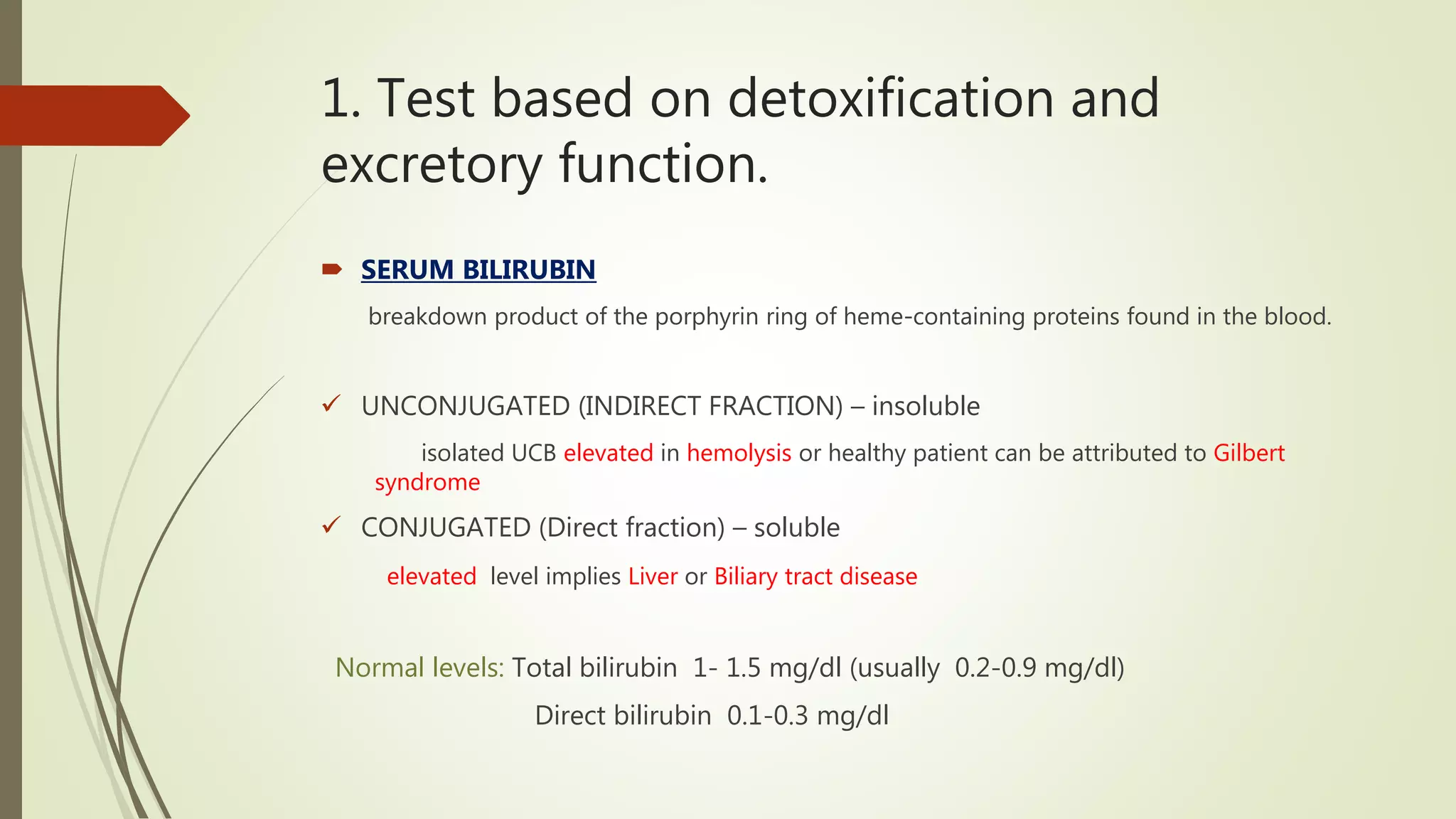
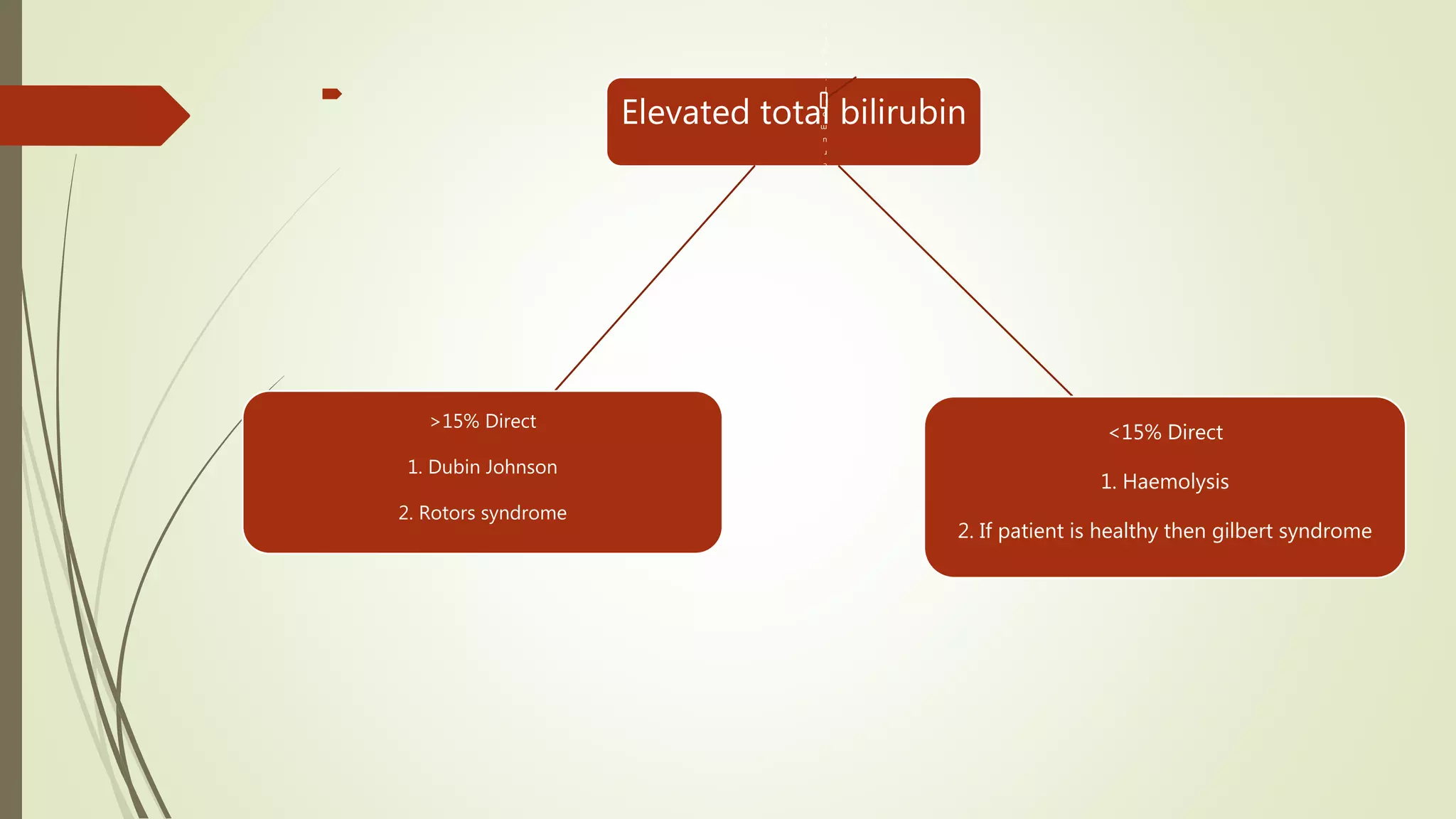
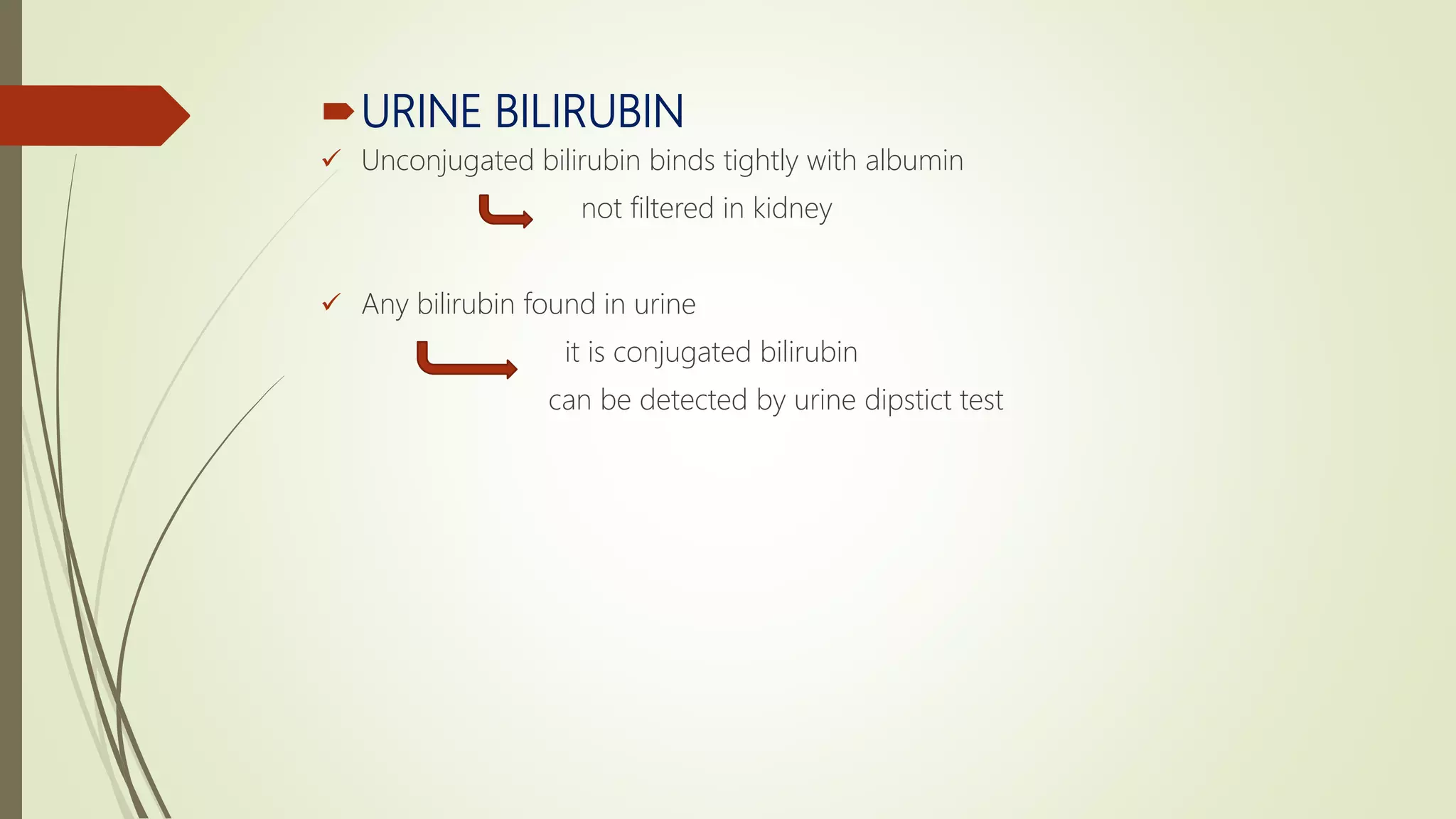
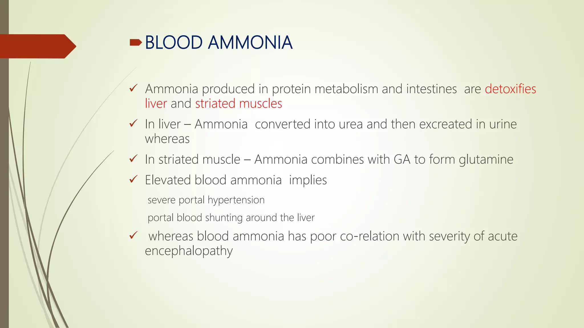
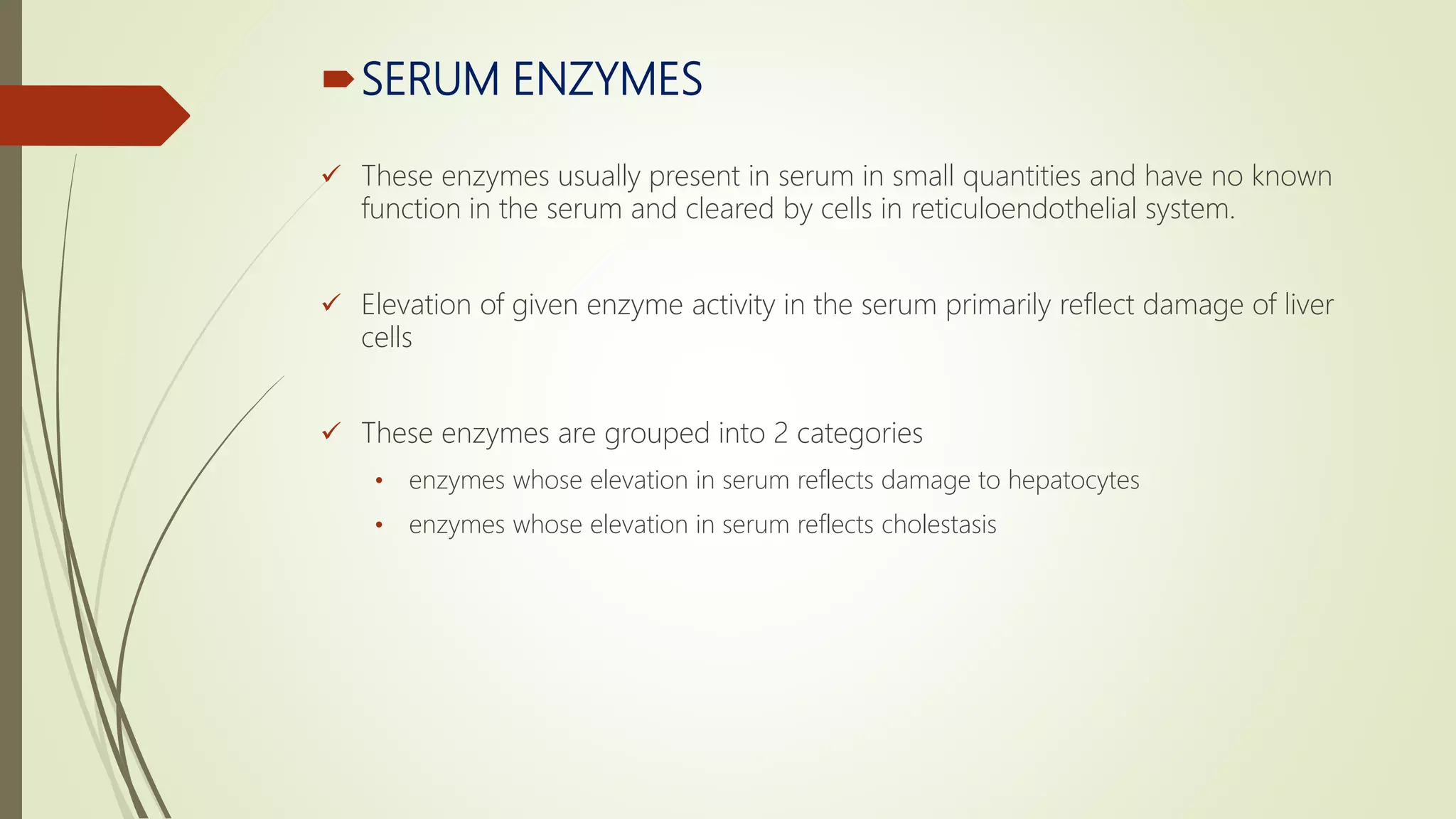
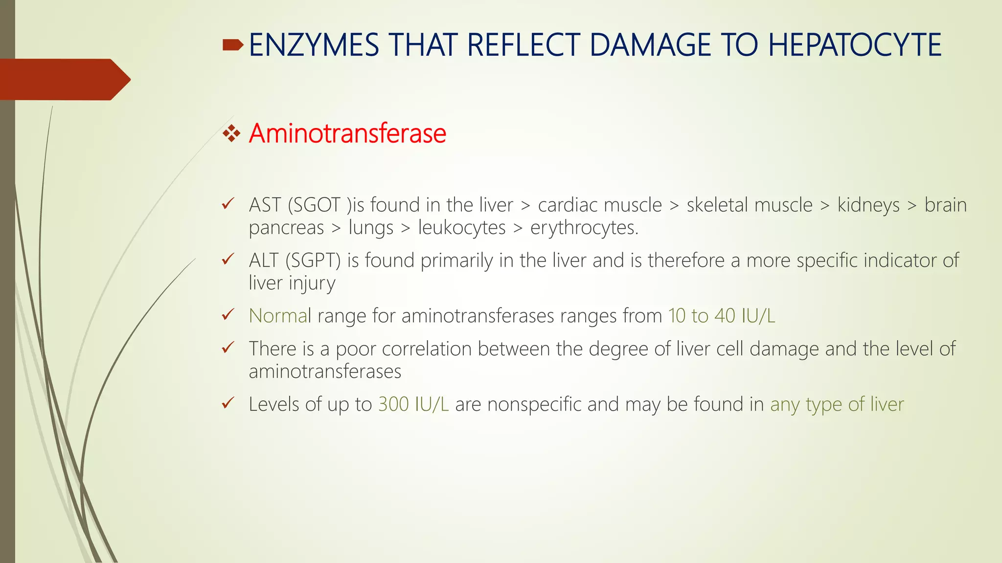
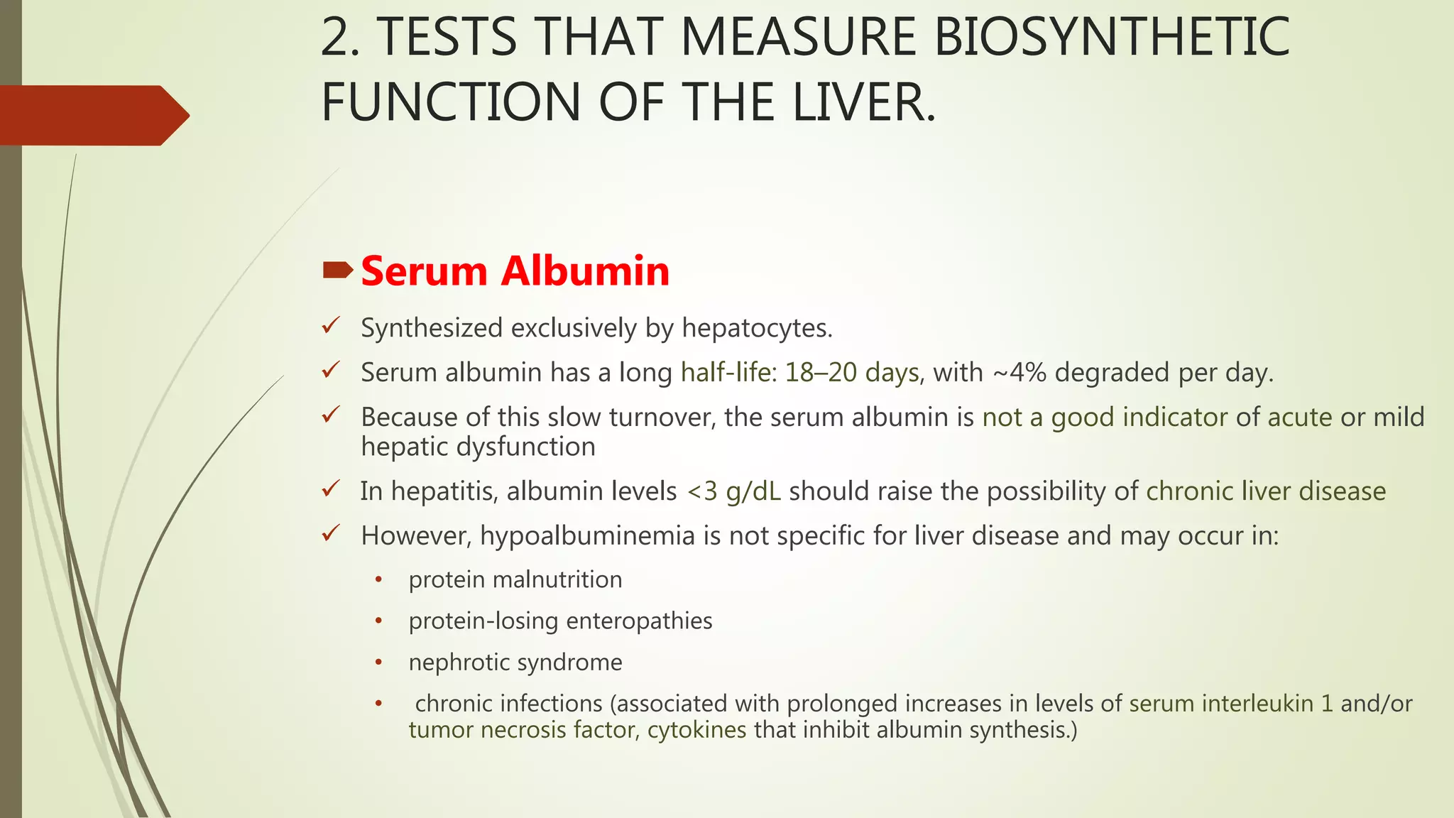
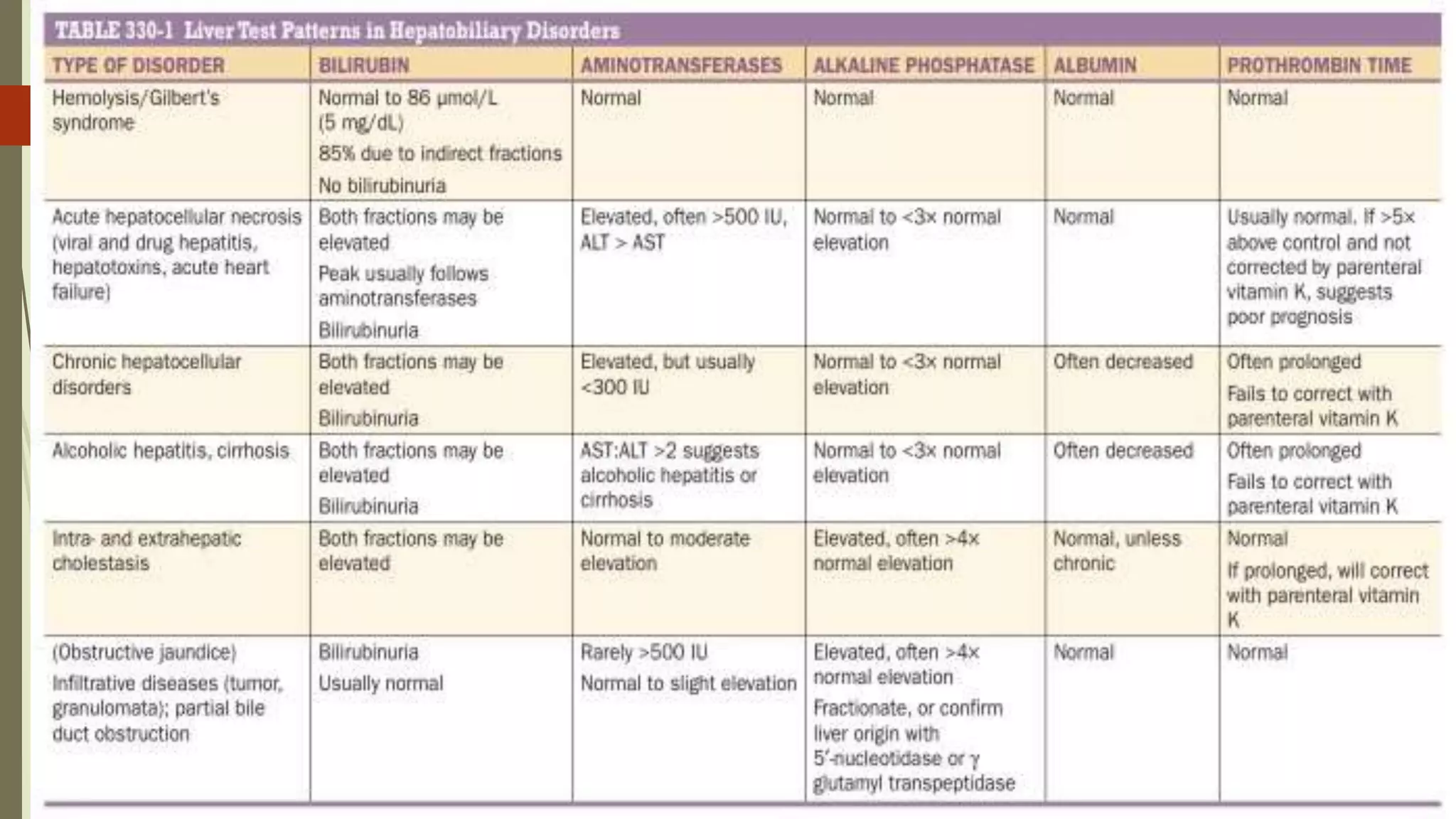
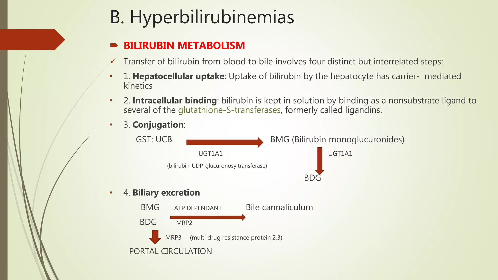
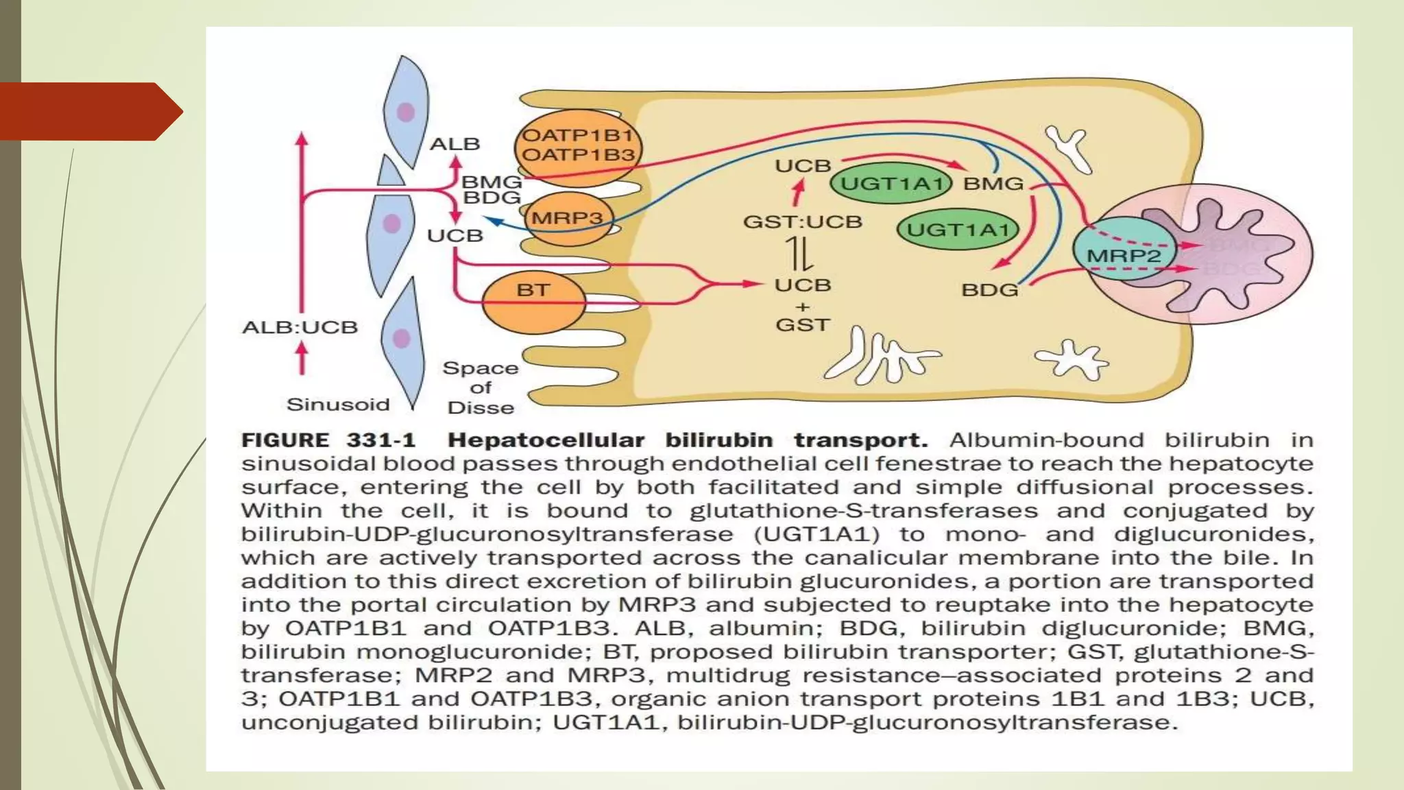
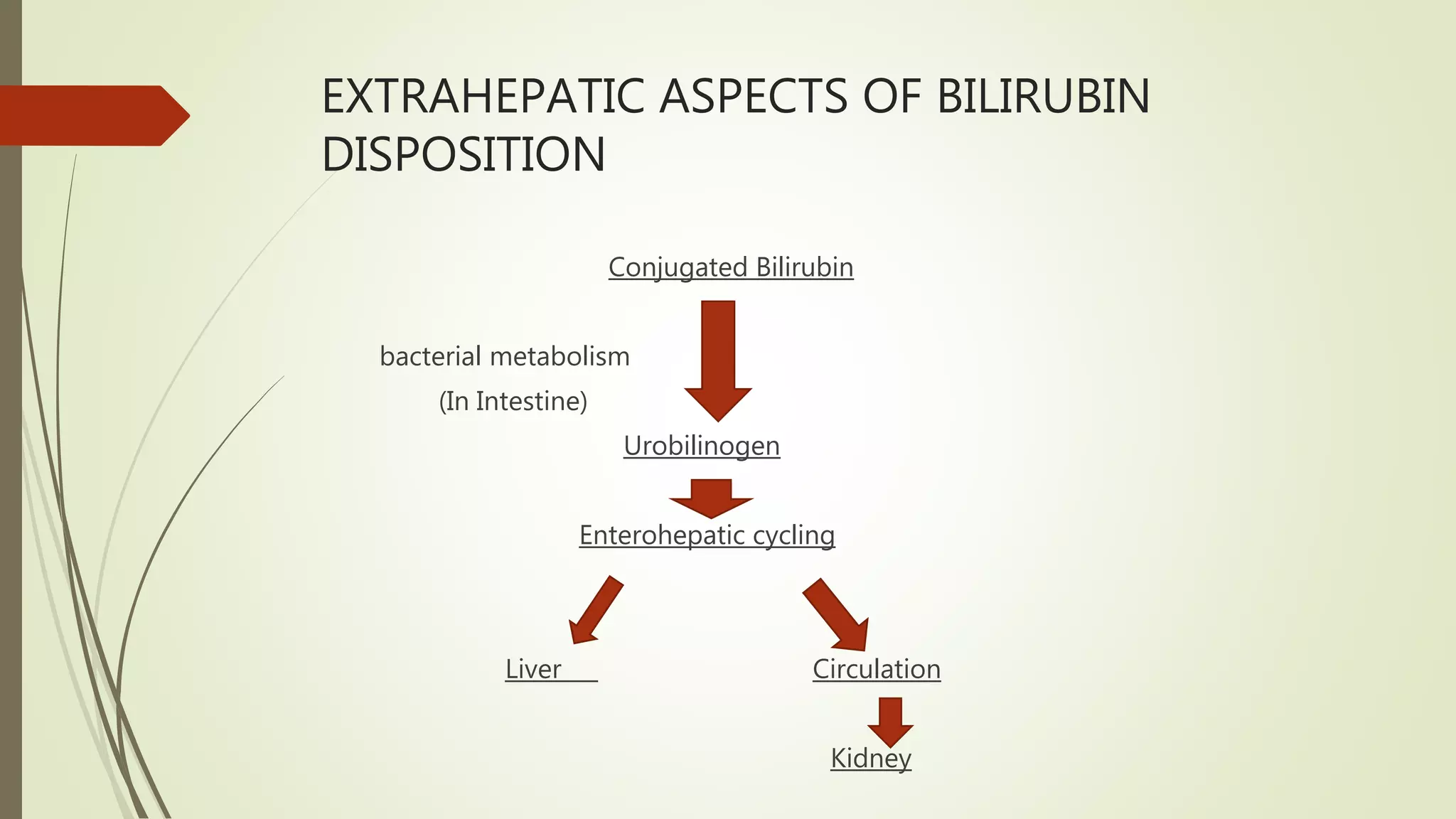
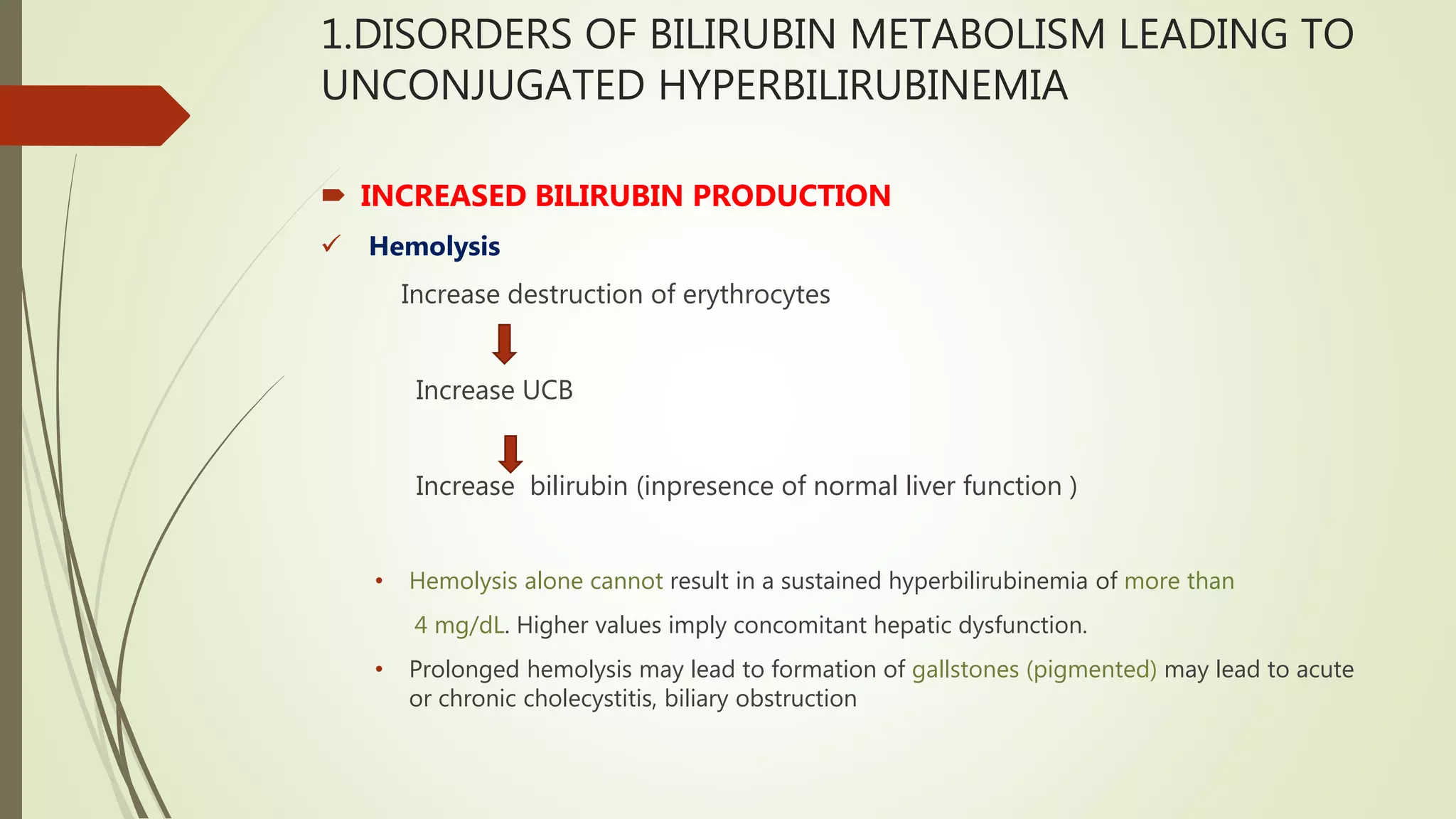
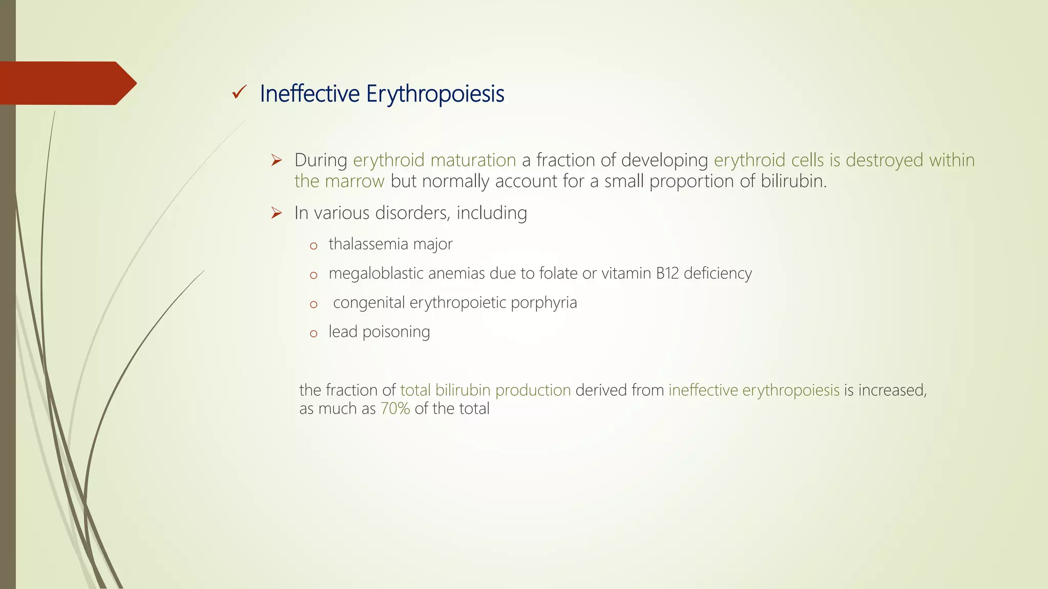
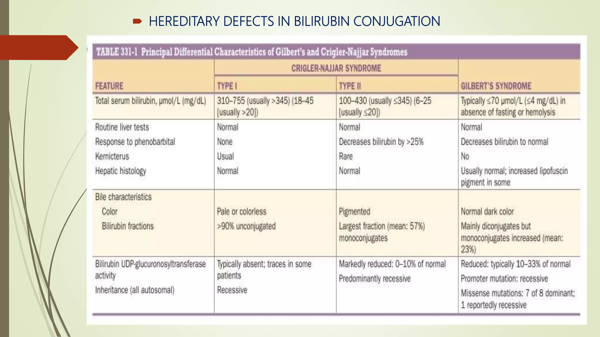
EVALUATION OF LIVER
FUNCTION AND
THE HYPERBILIRUBINEMIAS
Dr. UJJWAL DEEP
JR1 (DNB INTERNAL MEDICINE)
A. EVALUATION OF LIVER FUNCTION
 Liver function tests can be used to
 detect the presence of liver disease
 distinguish among different types of liver disorders
 gauge the extent of known liver damage
 follow the response to treatment.
1. Test based on detoxification and
excretory function.
 SERUM BILIRUBIN
breakdown product of the porphyrin ring of heme-containing proteins found in the blood.
 UNCONJUGATED (INDIRECT FRACTION) – insoluble
isolated UCB elevated in hemolysis or healthy patient can be attributed to Gilbert
syndrome
 CONJUGATED (Direct fraction) – soluble
elevated level implies Liver or Biliary tract disease
Normal levels: Total bilirubin 1- 1.5 mg/dl (usually 0.2-0.9 mg/dl)
Direct bilirubin 0.1-0.3 mg/dl
 leva
Elevated total bilirubin
S
e
r
u
m
b
i
l
i
r
u
i
n
<15% Direct
1. Haemolysis
2. If patient is healthy then gilbert syndrome
>15% Direct
1. Dubin Johnson
2. Rotors syndrome
URINE BILIRUBIN
 Unconjugated bilirubin binds tightly with albumin
not filtered in kidney
 Any bilirubin found in urine
it is conjugated bilirubin
can be detected by urine dipstict test
BLOOD AMMONIA
 Ammonia produced in protein metabolism and intestines are detoxifies
liver and striated muscles
 In liver – Ammonia converted into urea and then excreated in urine
whereas
 In striated muscle – Ammonia combines with GA to form glutamine
 Elevated blood ammonia implies
severe portal hypertension
portal blood shunting around the liver
 whereas blood ammonia has poor co-relation with severity of acute
encephalopathy
SERUM ENZYMES
 These enzymes usually present in serum in small quantities and have no known
function in the serum and cleared by cells in reticuloendothelial system.
 Elevation of given enzyme activity in the serum primarily reflect damage of liver
cells
 These enzymes are grouped into 2 categories
• enzymes whose elevation in serum reflects damage to hepatocytes
• enzymes whose elevation in serum reflects cholestasis
ENZYMES THAT REFLECT DAMAGE TO HEPATOCYTE
 Aminotransferase
 AST (SGOT )is found in the liver > cardiac muscle > skeletal muscle > kidneys > brain
pancreas > lungs > leukocytes > erythrocytes.
 ALT (SGPT) is found primarily in the liver and is therefore a more specific indicator of
liver injury
 Normal range for aminotransferases ranges from 10 to 40 IU/L
 There is a poor correlation between the degree of liver cell damage and the level of
aminotransferases
 Levels of up to 300 IU/L are nonspecific and may be found in any type of liver
 Aminotransferases >1000 IU/L—occur almost exclusively in disorders associated
with extensive hepatocellular injury such as
(1) viral hepatitis,
(2) ischemic liver injury (prolonged hypotension or acute heart failure)
(3) toxin- or drug-induced liver injury
 If ALT is higher or equal to AST – it reflects Acute hepatocellular injury
 AST:ALT <1 – reflects chronic viral hepatitis, Non alcoholic fatty liver
 AST :ALT >1 - reflects cirrhosis
 AST :ALT >2:1 Alcoholic liver disease
>3:1
 ENZYMES THAT REFLECT CHOLESTASIS
 Alkaline phosphatase more specific for cholestasis
 5′-nucleotidase
 γ-glutamyl transpeptidase (GGT) – identify occult alcohol use
 Alkaline phosphatase
 consists of many distinct isoenzymes found in the liver, bone, placenta, and, less
commonly, in the small intestine
 Mildly elevated alkaline phosphatase (1–1.5 times normal):
• Patients over age 60 yrs
• In individuals with blood types O and B can have an elevation of the serum alkaline
phosphatase after eating a fatty meal due to the influx of intestinal alkaline phosphatase
into the blood
• children and adolescents undergoing rapid bone growth
• In normal pregnancies due to the influx of placental alkaline phosphatase
 Elevations greater than four times normal :
• cholestatic liver disorders
• infiltrative liver diseases such as cancer and amyloidosis,
• bone conditions characterized by rapid bone turnover (e.g., Paget’s disease)
 If an elevated serum alkaline phosphatase is the only abnormal finding
identification of the source of elevated isoenzymes can be approached in two
ways :
• First, and most precise, is the fractionation of the alkaline phosphatase by electrophoresis.
• The second, best substantiated, and most available approach involves the measurement of
serum 5′-nucleotidase or GGT. These enzymes are rarely elevated in conditions other than liver
disease.
 Cause isolated elevations of the alkaline phosphatase include
• Hodgkin’s disease
• diabetes
• hyperthyroidism
• congestive heart failure
• amyloidosis
• inflammatory bowel disease.
2. TESTS THAT MEASURE BIOSYNTHETIC
FUNCTION OF THE LIVER.
Serum Albumin
 Synthesized exclusively by hepatocytes.
 Serum albumin has a long half-life: 18–20 days, with ~4% degraded per day.
 Because of this slow turnover, the serum albumin is not a good indicator of acute or mild
hepatic dysfunction
 In hepatitis, albumin levels <3 g/dL should raise the possibility of chronic liver disease
 However, hypoalbuminemia is not specific for liver disease and may occur in:
• protein malnutrition
• protein-losing enteropathies
• nephrotic syndrome
• chronic infections (associated with prolonged increases in levels of serum interleukin 1 and/or
tumor necrosis factor, cytokines that inhibit albumin synthesis.)
Serum Globulins
 Group of proteins made up of:
• γ globulins (immunoglobulins) produced by B lymphocytes
• α and β globulins produced primarily in hepatocytes.
 γ globulins are increased in
• chronic liver disease, such as chronic hepatitis and cirrhosis
due to the increased synthesis of antibodies, some of which are directed against
intestinal bacteria. This occurs because the cirrhotic liver fails to clear bacterial antigens
that normally reach the liver through the hepatic circulation
 Diffuse polyclonal increases in
• IgG levels are common in autoimmune hepatitis
• IgM levels are common in primary biliary cirrhosis
• IgA levels occur in alcoholic liver disease
COAGULATION FACTORS
 With the exception of factor VIII, which is produced by vascular
cells, the blood clotting factors are made exclusively in hepatocytes
 Their serum half-lives are much shorter than albumin, ranging from 6 h
factor VII to 5 days for fibrinogen
 Hence, it is the single best acute measure of hepatic synthetic function
helpful in both diagnosis and assessing the prognosis of acute
parenchymal liver disease
 Serum prothrombin time which collectively measures factors II, V, VII, and
X
B. Hyperbilirubinemias
 BILIRUBIN METABOLISM
 Transfer of bilirubin from blood to bile involves four distinct but interrelated steps:
• 1. Hepatocellular uptake: Uptake of bilirubin by the hepatocyte has carrier- mediated
kinetics
• 2. Intracellular binding: bilirubin is kept in solution by binding as a nonsubstrate ligand to
several of the glutathione-S-transferases, formerly called ligandins.
• 3. Conjugation:
GST: UCB BMG (Bilirubin monoglucuronides)
UGT1A1 UGT1A1
(bilirubin-UDP-glucuronosyltransferase)
BDG
• 4. Biliary excretion
BMG ATP DEPENDANT Bile cannaliculum
BDG MRP2
MRP3 (multi drug resistance protein 2,3)
PORTAL CIRCULATION
EXTRAHEPATIC ASPECTS OF BILIRUBIN
DISPOSITION
Conjugated Bilirubin
bacterial metabolism
(In Intestine)
Urobilinogen
Enterohepatic cycling
Liver Circulation
Kidney
1.DISORDERS OF BILIRUBIN METABOLISM LEADING TO
UNCONJUGATED HYPERBILIRUBINEMIA
 INCREASED BILIRUBIN PRODUCTION
 Hemolysis
Increase destruction of erythrocytes
Increase UCB
Increase bilirubin (inpresence of normal liver function )
• Hemolysis alone cannot result in a sustained hyperbilirubinemia of more than
4 mg/dL. Higher values imply concomitant hepatic dysfunction.
• Prolonged hemolysis may lead to formation of gallstones (pigmented) may lead to acute
or chronic cholecystitis, biliary obstruction
 Ineffective Erythropoiesis
 During erythroid maturation a fraction of developing erythroid cells is destroyed within
the marrow but normally account for a small proportion of bilirubin.
 In various disorders, including
o thalassemia major
o megaloblastic anemias due to folate or vitamin B12 deficiency
o congenital erythropoietic porphyria
o lead poisoning
the fraction of total bilirubin production derived from ineffective erythropoiesis is increased,
as much as 70% of the total
 DECREASED HEPATIC BILIRUBIN CLEARANCE
 Decreased Hepatic Uptake
 Gilbert’s syndrome (GS)
 drugs
• flavaspidic acid
• Novobiocin
• rifampin
• cholecystographic contrast agent
 Impaired Conjugation
A. PHYSIOLOGIC NEONATAL JAUNDICE
• Immediately after birth many hepatic physiologic processes are incompletely developed
• Levels of UGT1A1 are low, and alternative excretory pathways allow passage of
bilirubin into the gut
• most neonates develop mild unconjugated hyperbilirubinemia between days 2 and 5 after
birth with Peak levels (5–10 mg/dL) and decline to normal adult concentrations within 2
• Prematurity, often associated with more profound immaturity of hepatic function and
hemolysis, can result in higher levels of unconjugated hyperbilirubinemia
• levels upto 20 mg/dL puts the infant at risk for bilirubin encephalopathy, or kernicterus
• Under these circumstances, bilirubin crosses an immature blood-brain barrier and
precipitates in the basal ganglia and other areas of the brain
B.ACQUIRED CONJUGATION DEFECTS
• Observed in
o advanced hepatitis or cirrhosis
o drugs, including pregnanediol, novobiocin, chloramphenicol, gentamicin, and atazanavir
• Breast milk jaundice
o Bilirubin conjugation may be inhibited by certain fatty acids that are present in breast milk
 HEREDITARY DEFECTS IN BILIRUBIN CONJUGATION
 CN IIdiffers from CN-I in several specific ways :
 Average bilirubin concentrations are lower in CN-II
 CN-II is only infrequently associated with kernicterus
 Bile is deeply colored, and bilirubin glucuronides are present, with a striking,
characteristic increase in the proportion of monoglucuronides
 UGT1A1 in liver is usually present at reduced levels (typically ≤10% of normal)
 while typically detected in infancy, hyperbilirubinemia was not recognized in
cases until later in life
2.DISORDERS OF BILIRUBIN METABOLISM
LEADING TO MIXED OR PREDOMINANTLY
CONJUGATED HYPERBILIRUBINEMIA
 Acquired liver disease
• acute hepatitis
• common bile duct stone
there are usually elevations in the serum concentrations of both conjugated and
unconjugated bilirubin.
 Although biliary tract obstruction or hepatocellular cholestatic injury may present
on occasion with a predominantly conjugated hyperbilirubinemia
 FAMILIAL DEFECTS IN HEPATIC EXCRETORY FUNCTION
THANK YOU

Evaluation of liver function and hyperbilirubinemias

  • 1.
    EVALUATION OF LIVER FUNCTIONAND THE HYPERBILIRUBINEMIAS Dr. UJJWAL DEEP JR1 (DNB INTERNAL MEDICINE)
  • 2.
    A. EVALUATION OFLIVER FUNCTION  Liver function tests can be used to  detect the presence of liver disease  distinguish among different types of liver disorders  gauge the extent of known liver damage  follow the response to treatment.
  • 3.
    1. Test basedon detoxification and excretory function.  SERUM BILIRUBIN breakdown product of the porphyrin ring of heme-containing proteins found in the blood.  UNCONJUGATED (INDIRECT FRACTION) – insoluble isolated UCB elevated in hemolysis or healthy patient can be attributed to Gilbert syndrome  CONJUGATED (Direct fraction) – soluble elevated level implies Liver or Biliary tract disease Normal levels: Total bilirubin 1- 1.5 mg/dl (usually 0.2-0.9 mg/dl) Direct bilirubin 0.1-0.3 mg/dl
  • 4.
     leva Elevated totalbilirubin S e r u m b i l i r u i n <15% Direct 1. Haemolysis 2. If patient is healthy then gilbert syndrome >15% Direct 1. Dubin Johnson 2. Rotors syndrome
  • 5.
    URINE BILIRUBIN  Unconjugatedbilirubin binds tightly with albumin not filtered in kidney  Any bilirubin found in urine it is conjugated bilirubin can be detected by urine dipstict test
  • 6.
    BLOOD AMMONIA  Ammoniaproduced in protein metabolism and intestines are detoxifies liver and striated muscles  In liver – Ammonia converted into urea and then excreated in urine whereas  In striated muscle – Ammonia combines with GA to form glutamine  Elevated blood ammonia implies severe portal hypertension portal blood shunting around the liver  whereas blood ammonia has poor co-relation with severity of acute encephalopathy
  • 7.
    SERUM ENZYMES  Theseenzymes usually present in serum in small quantities and have no known function in the serum and cleared by cells in reticuloendothelial system.  Elevation of given enzyme activity in the serum primarily reflect damage of liver cells  These enzymes are grouped into 2 categories • enzymes whose elevation in serum reflects damage to hepatocytes • enzymes whose elevation in serum reflects cholestasis
  • 8.
    ENZYMES THAT REFLECTDAMAGE TO HEPATOCYTE  Aminotransferase  AST (SGOT )is found in the liver > cardiac muscle > skeletal muscle > kidneys > brain pancreas > lungs > leukocytes > erythrocytes.  ALT (SGPT) is found primarily in the liver and is therefore a more specific indicator of liver injury  Normal range for aminotransferases ranges from 10 to 40 IU/L  There is a poor correlation between the degree of liver cell damage and the level of aminotransferases  Levels of up to 300 IU/L are nonspecific and may be found in any type of liver
  • 9.
     Aminotransferases >1000IU/L—occur almost exclusively in disorders associated with extensive hepatocellular injury such as (1) viral hepatitis, (2) ischemic liver injury (prolonged hypotension or acute heart failure) (3) toxin- or drug-induced liver injury  If ALT is higher or equal to AST – it reflects Acute hepatocellular injury  AST:ALT <1 – reflects chronic viral hepatitis, Non alcoholic fatty liver  AST :ALT >1 - reflects cirrhosis  AST :ALT >2:1 Alcoholic liver disease >3:1
  • 10.
     ENZYMES THATREFLECT CHOLESTASIS  Alkaline phosphatase more specific for cholestasis  5′-nucleotidase  γ-glutamyl transpeptidase (GGT) – identify occult alcohol use  Alkaline phosphatase  consists of many distinct isoenzymes found in the liver, bone, placenta, and, less commonly, in the small intestine  Mildly elevated alkaline phosphatase (1–1.5 times normal): • Patients over age 60 yrs • In individuals with blood types O and B can have an elevation of the serum alkaline phosphatase after eating a fatty meal due to the influx of intestinal alkaline phosphatase into the blood • children and adolescents undergoing rapid bone growth • In normal pregnancies due to the influx of placental alkaline phosphatase
  • 11.
     Elevations greaterthan four times normal : • cholestatic liver disorders • infiltrative liver diseases such as cancer and amyloidosis, • bone conditions characterized by rapid bone turnover (e.g., Paget’s disease)  If an elevated serum alkaline phosphatase is the only abnormal finding identification of the source of elevated isoenzymes can be approached in two ways : • First, and most precise, is the fractionation of the alkaline phosphatase by electrophoresis. • The second, best substantiated, and most available approach involves the measurement of serum 5′-nucleotidase or GGT. These enzymes are rarely elevated in conditions other than liver disease.  Cause isolated elevations of the alkaline phosphatase include • Hodgkin’s disease • diabetes • hyperthyroidism • congestive heart failure • amyloidosis • inflammatory bowel disease.
  • 12.
    2. TESTS THATMEASURE BIOSYNTHETIC FUNCTION OF THE LIVER. Serum Albumin  Synthesized exclusively by hepatocytes.  Serum albumin has a long half-life: 18–20 days, with ~4% degraded per day.  Because of this slow turnover, the serum albumin is not a good indicator of acute or mild hepatic dysfunction  In hepatitis, albumin levels <3 g/dL should raise the possibility of chronic liver disease  However, hypoalbuminemia is not specific for liver disease and may occur in: • protein malnutrition • protein-losing enteropathies • nephrotic syndrome • chronic infections (associated with prolonged increases in levels of serum interleukin 1 and/or tumor necrosis factor, cytokines that inhibit albumin synthesis.)
  • 13.
    Serum Globulins  Groupof proteins made up of: • γ globulins (immunoglobulins) produced by B lymphocytes • α and β globulins produced primarily in hepatocytes.  γ globulins are increased in • chronic liver disease, such as chronic hepatitis and cirrhosis due to the increased synthesis of antibodies, some of which are directed against intestinal bacteria. This occurs because the cirrhotic liver fails to clear bacterial antigens that normally reach the liver through the hepatic circulation  Diffuse polyclonal increases in • IgG levels are common in autoimmune hepatitis • IgM levels are common in primary biliary cirrhosis • IgA levels occur in alcoholic liver disease
  • 14.
    COAGULATION FACTORS  Withthe exception of factor VIII, which is produced by vascular cells, the blood clotting factors are made exclusively in hepatocytes  Their serum half-lives are much shorter than albumin, ranging from 6 h factor VII to 5 days for fibrinogen  Hence, it is the single best acute measure of hepatic synthetic function helpful in both diagnosis and assessing the prognosis of acute parenchymal liver disease  Serum prothrombin time which collectively measures factors II, V, VII, and X
  • 16.
    B. Hyperbilirubinemias  BILIRUBINMETABOLISM  Transfer of bilirubin from blood to bile involves four distinct but interrelated steps: • 1. Hepatocellular uptake: Uptake of bilirubin by the hepatocyte has carrier- mediated kinetics • 2. Intracellular binding: bilirubin is kept in solution by binding as a nonsubstrate ligand to several of the glutathione-S-transferases, formerly called ligandins. • 3. Conjugation: GST: UCB BMG (Bilirubin monoglucuronides) UGT1A1 UGT1A1 (bilirubin-UDP-glucuronosyltransferase) BDG • 4. Biliary excretion BMG ATP DEPENDANT Bile cannaliculum BDG MRP2 MRP3 (multi drug resistance protein 2,3) PORTAL CIRCULATION
  • 18.
    EXTRAHEPATIC ASPECTS OFBILIRUBIN DISPOSITION Conjugated Bilirubin bacterial metabolism (In Intestine) Urobilinogen Enterohepatic cycling Liver Circulation Kidney
  • 19.
    1.DISORDERS OF BILIRUBINMETABOLISM LEADING TO UNCONJUGATED HYPERBILIRUBINEMIA  INCREASED BILIRUBIN PRODUCTION  Hemolysis Increase destruction of erythrocytes Increase UCB Increase bilirubin (inpresence of normal liver function ) • Hemolysis alone cannot result in a sustained hyperbilirubinemia of more than 4 mg/dL. Higher values imply concomitant hepatic dysfunction. • Prolonged hemolysis may lead to formation of gallstones (pigmented) may lead to acute or chronic cholecystitis, biliary obstruction
  • 20.
     Ineffective Erythropoiesis During erythroid maturation a fraction of developing erythroid cells is destroyed within the marrow but normally account for a small proportion of bilirubin.  In various disorders, including o thalassemia major o megaloblastic anemias due to folate or vitamin B12 deficiency o congenital erythropoietic porphyria o lead poisoning the fraction of total bilirubin production derived from ineffective erythropoiesis is increased, as much as 70% of the total
  • 21.
     DECREASED HEPATICBILIRUBIN CLEARANCE  Decreased Hepatic Uptake  Gilbert’s syndrome (GS)  drugs • flavaspidic acid • Novobiocin • rifampin • cholecystographic contrast agent  Impaired Conjugation A. PHYSIOLOGIC NEONATAL JAUNDICE • Immediately after birth many hepatic physiologic processes are incompletely developed • Levels of UGT1A1 are low, and alternative excretory pathways allow passage of bilirubin into the gut • most neonates develop mild unconjugated hyperbilirubinemia between days 2 and 5 after birth with Peak levels (5–10 mg/dL) and decline to normal adult concentrations within 2
  • 22.
    • Prematurity, oftenassociated with more profound immaturity of hepatic function and hemolysis, can result in higher levels of unconjugated hyperbilirubinemia • levels upto 20 mg/dL puts the infant at risk for bilirubin encephalopathy, or kernicterus • Under these circumstances, bilirubin crosses an immature blood-brain barrier and precipitates in the basal ganglia and other areas of the brain B.ACQUIRED CONJUGATION DEFECTS • Observed in o advanced hepatitis or cirrhosis o drugs, including pregnanediol, novobiocin, chloramphenicol, gentamicin, and atazanavir • Breast milk jaundice o Bilirubin conjugation may be inhibited by certain fatty acids that are present in breast milk
  • 23.
     HEREDITARY DEFECTSIN BILIRUBIN CONJUGATION
  • 24.
     CN IIdiffersfrom CN-I in several specific ways :  Average bilirubin concentrations are lower in CN-II  CN-II is only infrequently associated with kernicterus  Bile is deeply colored, and bilirubin glucuronides are present, with a striking, characteristic increase in the proportion of monoglucuronides  UGT1A1 in liver is usually present at reduced levels (typically ≤10% of normal)  while typically detected in infancy, hyperbilirubinemia was not recognized in cases until later in life
  • 25.
    2.DISORDERS OF BILIRUBINMETABOLISM LEADING TO MIXED OR PREDOMINANTLY CONJUGATED HYPERBILIRUBINEMIA  Acquired liver disease • acute hepatitis • common bile duct stone there are usually elevations in the serum concentrations of both conjugated and unconjugated bilirubin.  Although biliary tract obstruction or hepatocellular cholestatic injury may present on occasion with a predominantly conjugated hyperbilirubinemia
  • 26.
     FAMILIAL DEFECTSIN HEPATIC EXCRETORY FUNCTION
  • 27.