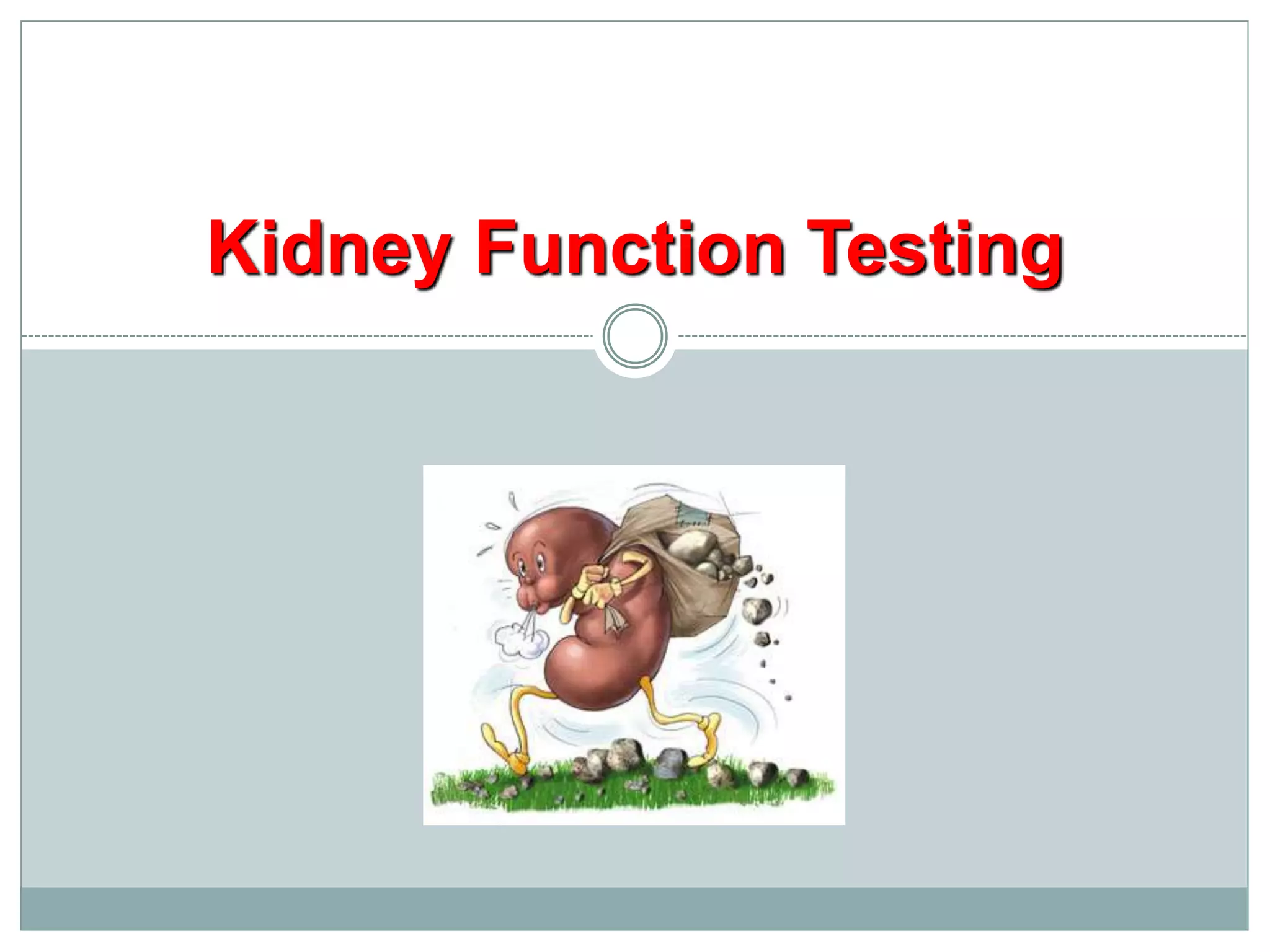
Kidney Function Testing
An Introduction to the Urinary System
Produces urine
Transports urine
towards bladder
Temporarily store
urine
Conducts urine
to exterior
The Function of Urinary System
 Excretion & Elimination:
removal of organic wastes products
from body fluids (urea, creatinine,
uric acid)
 Homeostatic regulation:
Water -Salt Balance
Acid - base Balance
 Enocrine function:
Hormones
A)
B)
C)
Kidney – basic data
 Urine excreted daily in adults: cca 1.5L
 Kidney only ca 1% of total body weight, despite it
 The renal blood flow= 20% of cardiac output
 Plasma renal flow= PRF ca 600 mL/Min./1.73 M2
 Reflects two processes
 Ultrafiltration (GFR): 180 L/day
 Reabsorption: >99% of the amount filtered
Renal threshold
 Renal threshold of a substance is the concentration
in blood beyond which it is excreted in urine
 Renal threshold for glucose is 180mg/dL
 Tubular maximum (Tm): maximum capacity of the
kidneys to absorb a particular substance
 Tm for glucose is 350 mg/min
Kidney Function
A plumbers view
Filter
Processor
Input
Arterial
Output
Venous
Output
Urine
How do you know it’s broken?
 Decreased urine
production
 Clinical symptoms
 Tests
Filter
Processor
Input
Arterial
Output
Venous
Output
Urine
Where can it break?
 Pre-renal
 Renal
(intrarenal)
 Post-renal
(obstruction)
Filter
Processor
Input
Arterial
Output
Venous
Output
Urine
Causes of kidney functional disorders
 Pre-renal e.g.
decreased
intravascular volum
 Renal e.g. acute
tubular necrosis
 Postrenal e.g. ureteral
obstruction
Signs and Symptoms of Renal Failure
 Symptoms of Uraemia (nausea, vomiting,
lethargy)
 Disorders of Micturation (frequency, nocturia,
dysuria)
 Disorders of Urine volume (polyuria, oliguria,
anuria)
 Alterations in urine composition (haematuria,
proteinuria, bacteriuria, leukocytouria, calculi)
 Pain
 Oedema (hypoalbuminaemia, salt and water
retention)
Why Test Renal Function?
 To identify renal dysfunction.
 To diagnose renal disease.
 To monitor disease progress.
 To monitor response to treatment.
 To assess changes in function that may impact on
therapy (e.g. Digoxin, chemotherapy).
When should you assess renal
function?
 Older age
 Family history of Chronic Kidney disease (CKD)
 Decreased renal mass
 Low birth weight
 Diabetes Mellitus (DM)
 Hypertension (HTN)
 Autoimmune disease
 Systemic infections
 Urinary tract infections (UTI)
 Nephrolithiasis
 Obstruction to the lower urinary tract
 Drug toxicity
Biochemical Tests of Renal Function
 Measurement of GFR
 Clearance tests
 Plasma creatinine
 Urea, uric acid and β2-microglobulin
 Renal tubular function tests
 Osmolality measurements
 Specific proteinurea
 Glycouria
 Aminoaciduria
 Urinalysis
 Appearance
 Specific gravity and osmolality
 pH
 osmolality
 Glucose
 Protein
 Urinary sediments
Biochemical Tests of Renal Function
Measurement of GFR
Clearance tests
Plasma creatinine
Urea, uric acid and β2-microglobulin
In acute and chronic renal failure, there is effectively a loss of
function of whole nephrons
 Filtration is essential to the formation of urine  tests of
glomerular function are almost always required in the
investigation and management of any patient with renal disease.
The most frequently used tests are those that assess either the
GFR or the integrity of the glomerular filtration barrier.
Biochemical Tests of renal function
GFR can be estimated by measuring the urinary excretion of a substance that is completely filtered
from the blood by the glomeruli and it is not secreted, reabsorbed or metabolized by the renal
tubules.
 Clearance is defined as the (hypothetical) quantity of blood or plasma completely cleared of a
substance per unit of time.
 Clearance of substances that are filtered exclusively or predominantly by the glomeruli but
neither reabsorbed nor secreted by other regions of the nephron can be used to measure GFR.
 Inulin
The Volume of blood from which inulin is cleared or completely removed in one minute is
known as the inulin clearance and is equal to the GFR.
Measurement of inulin clearance requires the infusion of inulin into the blood and is not
suitable for routine clinical use
GFR =
(U V)
P
inulin
inulin

V is not urine volume, it is urine flow rate
Measurement of glomerular filtration rate
Biochemical Tests of Renal Function
Measurement of GFR
Clearance tests
Plasma creatinine
Urea, uric acid and β2-microglobulin
1 to 2% of muscle creatine spontaneously converts to creatinine
daily and released into body fluids at a constant rate.
Endogenous creatinine produced is proportional to muscle
mass, it is a function of total muscle mass the production
varies with age and sex
 Dietary fluctuations of creatinine intake cause only minor
variation in daily creatinine excretion of the same person.
 Creatinine released into body fluids at a constant rate and its
plasma levels maintained within narrow limits  Creatinine
clearance may be measured as an indicator of GFR.
Creatinine
The most frequently used clearance test is based on the
measurement of creatinine.
 Small quantity of creatinine is reabsorbed by the tubules and
other quantities are actively secreted by the renal tubules  So
creatinine clearance is approximately 7% greater than inulin
clearance.
The difference is not significant when GFR is normal but when
the GFR is low (less 10 ml/min), tubular secretion makes the
major contribution to creatinine excretion and the creatinine
clearance significantly overestimates the GFR.
Creatinine clearance and clinical utility
An estimate of the GFR can be calculated from the creatinine content of a 24-hour
urine collection, and the plasma concentration within this period.
The volume of urine is measured, urine flow rate is calculated (ml/min) and the
assay for creatinine is performed on plasma and urine to obtain the concentration in
mg per dl or per ml.
Creatinine clearance in adults is normally about of 120 ml/min,
The accurate measurement of creatinine clearance is difficult, especially in outpatients,
since it is necessary to obtain a complete and accurately timed sample of urine
Creatinine clearance clinical utility
The 'clearance' of creatinine from plasma is directly related to the
GFR if:
The urine volume is collected accurately
There are no ketones or heavy proteinuria present to interfere
with the creatinine determination.
It should be noted that the GFR decline with age (to a greater extent
in males than in females) and this must be taken into account when
interpreting results.
Creatinine clearance and clinical utility
Use of Formulae to Predict
Clearance
• Formulae have been derived to predict Creatinine
Clearance (CC) from Plasma creatinine.
• Plasma creatinine derived from muscle mass which is
related to body mass, age, sex.
• Cockcroft & Gault Formula
CC = k[(140-Age) x weight(Kg))] / serum Creatinine (µmol/L)
k = 1.224 for males & 1.04 for females
• Modifications required for children & obese subjects
• Can be modified to use Surface area
Biochemical Tests of Renal Function
Measurement of GFR
Clearance tests
Plasma creatinine
Urea, uric acid and β2-microglobulin
Catabolism of proteins and nucleic acids results in formation of
so called nonprotein nitrogenous compounds.
Protein
 Proteolysis, principally enzymatic
Amino acids
 Transamination and oxidative deamination
Ammonia
 Enzymatic synthesis in the “urea cycle”
Urea
Measurement of nonprotein nitrogen-
containing compounds
Urea is the major nitrogen-containing metabolic product of protein
catabolism in humans,
 Its elimination in the urine represents the major route for nitrogen
excretion.
 More than 90% of urea is excreted through the kidneys, with losses
through the GIT and skin
 Urea is filtered freely by the glomeruli
 Plasma urea concentration is often used as an index of renal glomerular
function
 Urea production is increased by a high protein intake and it is decreased
in patients with a low protein intake or in patients with liver disease.
Plasma Urea
Many renal diseases with various glomerular, tubular, interstitial or vascular damage can
cause an increase in plasma urea concentration.
The reference interval for serum urea of healthy adults is 5-39 mg/dl. Plasma
concentrations also tend to be slightly higher in males than females. High protein diet causes
significant increases in plasma urea concentrations and urinary excretion.
Measurement of plasma creatinine provides a more accurate assessment than urea
because there are many factors that affect urea level.
Nonrenal factors can affect the urea level (normal adults is level 5-39 mg/dl) like:
Mild dehydration,
high protein diet,
increased protein catabolism, muscle wasting as in starvation,
reabsorption of blood proteins after a GIT haemorrhage,
treatment with cortisol or its synthetic analogous
Plasma Urea
Clinical Significance
 States associated with elevated levels of urea in blood
are referred to as uremia or azotemia.
 Causes of urea plasma elevations:
 Prerenal: renal hypoperfusion
 Renal: acute tubular necrosis
 Postrenal: obstruction of urinary flow
 In human, uric acid is the major product of the catabolism of the purine
nucleosides, adenosine and guanosine.
 Purines are derived from catabolism of dietary nucleic acid (nucleated cells,
like meat) and from degradation of endogenous nucleic acids.
 Overproduction of uric acid may result from increased synthesis of purine
precursors.
 In humans, approximately 75% of uric acid excreted is lost in the urine;
most of the reminder is secreted into the GIT
Uric acid
Renal handling of uric acid is complex and involves four sequential steps:
Glomerular filtration of virtually all the uric acid in capillary plasma
entering the glomerulus.
Reabsorption in the proximal convoluted tubule of about 98 to 100%
of filtered uric acid.
Subsequent secretion of uric acid into the lumen of the distal portion
of the proximal tubule.
Further reabsorption in the distal tubule.
 Hyperuricemia is defined by serum or plasma uric acid concentrations higher
than 7.0 mg/dl (0.42mmol/L) in men or greater than 6.0 mg/dl (0.36mmol/L)
in women
Uric acid
β2-microglobulin is a small peptide (molecular weight 11.8 kDa),
It is present on the surface of most cells and in low concentrations in the
plasma.
It is completely filtered by the glomeruli and is reabsorbed and catabolized
by proximal tubular cells.
The plasma concentration of β2-microglobulin is a good index of GFR in
normal people, being unaffected by diet or muscle mass.
It is increased in certain malignancies and inflammatory diseases.
Since it is normally reabsorbed and catabolized in the tubules, measurement
of β2-microglobulin excretion provides a sensitive method of assessing
tubular integrity.
Plasma β2-microglobulin
Role of Biochemical Testing
 Presentation of patients: -
 Routine urinalysis
 Symptom or physical sign
 Systemic disease with known renal component.
 Effective management of renal disease depends upon
establishing a definitive diagnosis: -
 Detailed clinical history
 Diagnostic imaging and biopsy
 Role of biochemistry: -
 Rarely establishes the cause
 Screening for damage
 Monitoring progression.
Creatinine Clearance
 Timed urine collection for creatinine
measurement (usually 24h)
 Blood sample taken within the period of
collection.
 Normal range = 120-145ml/min
Problems: -
 Practical problems of accurate urine collection
and volume measurement.
 Within subject variability = 11%
Plasma Creatinine Concentration
Difficulties: -
 Concentration depends on balance between
input and output.
 Production determined by muscle mass which
is related to age, sex and weight.
 High between subject variability but low
within subject.
 Concentration inversely related to GFR.
 Small changes in creatinine within and around the
reference limits = large changes in GFR.
Kidney
Output
Plasma
Pool
Content
Creatinine
Input
Normal
Muscle
Mass
Normal
Kidneys
Diseased
Kidneys
Normal
Muscle
Mass
Diseased
Kidneys
Normal
Kidneys
Increased
Muscle
Mass
Reduced
Muscle
Mass
Effect of Muscle Mass on Serum Creatinine
Acute Renal Failure
Metabolic features:
 Retention of:
 Urea & creatinine
 Na & water
 potassium with hyper-
kalaemia
 Acid with metabolic
acidosis
Classification of Causes:
 Pre-renal
 reduced perfusion
 Renal
 inflammation
 infiltration
 toxicity
 Post-renal
 obstruction
Pre-renal versus intrinsic ARF
Test Result
Pre-renal Renal
Urea & Creatinine Disproportionate
rise in Urea
Tend to rise
together
Protein in urine Uncommon Present on
dipstick
testing
T H A N K Y O U
V . V E N A K T A K A R T H I K

Kidney function test

  • 1.
  • 2.
    An Introduction tothe Urinary System Produces urine Transports urine towards bladder Temporarily store urine Conducts urine to exterior
  • 3.
    The Function ofUrinary System  Excretion & Elimination: removal of organic wastes products from body fluids (urea, creatinine, uric acid)  Homeostatic regulation: Water -Salt Balance Acid - base Balance  Enocrine function: Hormones A) B) C)
  • 4.
    Kidney – basicdata  Urine excreted daily in adults: cca 1.5L  Kidney only ca 1% of total body weight, despite it  The renal blood flow= 20% of cardiac output  Plasma renal flow= PRF ca 600 mL/Min./1.73 M2  Reflects two processes  Ultrafiltration (GFR): 180 L/day  Reabsorption: >99% of the amount filtered
  • 5.
    Renal threshold  Renalthreshold of a substance is the concentration in blood beyond which it is excreted in urine  Renal threshold for glucose is 180mg/dL  Tubular maximum (Tm): maximum capacity of the kidneys to absorb a particular substance  Tm for glucose is 350 mg/min
  • 6.
    Kidney Function A plumbersview Filter Processor Input Arterial Output Venous Output Urine
  • 7.
    How do youknow it’s broken?  Decreased urine production  Clinical symptoms  Tests Filter Processor Input Arterial Output Venous Output Urine
  • 8.
    Where can itbreak?  Pre-renal  Renal (intrarenal)  Post-renal (obstruction) Filter Processor Input Arterial Output Venous Output Urine
  • 9.
    Causes of kidneyfunctional disorders  Pre-renal e.g. decreased intravascular volum  Renal e.g. acute tubular necrosis  Postrenal e.g. ureteral obstruction
  • 10.
    Signs and Symptomsof Renal Failure  Symptoms of Uraemia (nausea, vomiting, lethargy)  Disorders of Micturation (frequency, nocturia, dysuria)  Disorders of Urine volume (polyuria, oliguria, anuria)  Alterations in urine composition (haematuria, proteinuria, bacteriuria, leukocytouria, calculi)  Pain  Oedema (hypoalbuminaemia, salt and water retention)
  • 11.
    Why Test RenalFunction?  To identify renal dysfunction.  To diagnose renal disease.  To monitor disease progress.  To monitor response to treatment.  To assess changes in function that may impact on therapy (e.g. Digoxin, chemotherapy).
  • 12.
    When should youassess renal function?  Older age  Family history of Chronic Kidney disease (CKD)  Decreased renal mass  Low birth weight  Diabetes Mellitus (DM)  Hypertension (HTN)  Autoimmune disease  Systemic infections  Urinary tract infections (UTI)  Nephrolithiasis  Obstruction to the lower urinary tract  Drug toxicity
  • 13.
    Biochemical Tests ofRenal Function  Measurement of GFR  Clearance tests  Plasma creatinine  Urea, uric acid and β2-microglobulin  Renal tubular function tests  Osmolality measurements  Specific proteinurea  Glycouria  Aminoaciduria  Urinalysis  Appearance  Specific gravity and osmolality  pH  osmolality  Glucose  Protein  Urinary sediments
  • 14.
    Biochemical Tests ofRenal Function Measurement of GFR Clearance tests Plasma creatinine Urea, uric acid and β2-microglobulin
  • 15.
    In acute andchronic renal failure, there is effectively a loss of function of whole nephrons  Filtration is essential to the formation of urine  tests of glomerular function are almost always required in the investigation and management of any patient with renal disease. The most frequently used tests are those that assess either the GFR or the integrity of the glomerular filtration barrier. Biochemical Tests of renal function
  • 16.
    GFR can beestimated by measuring the urinary excretion of a substance that is completely filtered from the blood by the glomeruli and it is not secreted, reabsorbed or metabolized by the renal tubules.  Clearance is defined as the (hypothetical) quantity of blood or plasma completely cleared of a substance per unit of time.  Clearance of substances that are filtered exclusively or predominantly by the glomeruli but neither reabsorbed nor secreted by other regions of the nephron can be used to measure GFR.  Inulin The Volume of blood from which inulin is cleared or completely removed in one minute is known as the inulin clearance and is equal to the GFR. Measurement of inulin clearance requires the infusion of inulin into the blood and is not suitable for routine clinical use GFR = (U V) P inulin inulin  V is not urine volume, it is urine flow rate Measurement of glomerular filtration rate
  • 17.
    Biochemical Tests ofRenal Function Measurement of GFR Clearance tests Plasma creatinine Urea, uric acid and β2-microglobulin
  • 18.
    1 to 2%of muscle creatine spontaneously converts to creatinine daily and released into body fluids at a constant rate. Endogenous creatinine produced is proportional to muscle mass, it is a function of total muscle mass the production varies with age and sex  Dietary fluctuations of creatinine intake cause only minor variation in daily creatinine excretion of the same person.  Creatinine released into body fluids at a constant rate and its plasma levels maintained within narrow limits  Creatinine clearance may be measured as an indicator of GFR. Creatinine
  • 19.
    The most frequentlyused clearance test is based on the measurement of creatinine.  Small quantity of creatinine is reabsorbed by the tubules and other quantities are actively secreted by the renal tubules  So creatinine clearance is approximately 7% greater than inulin clearance. The difference is not significant when GFR is normal but when the GFR is low (less 10 ml/min), tubular secretion makes the major contribution to creatinine excretion and the creatinine clearance significantly overestimates the GFR. Creatinine clearance and clinical utility
  • 20.
    An estimate ofthe GFR can be calculated from the creatinine content of a 24-hour urine collection, and the plasma concentration within this period. The volume of urine is measured, urine flow rate is calculated (ml/min) and the assay for creatinine is performed on plasma and urine to obtain the concentration in mg per dl or per ml. Creatinine clearance in adults is normally about of 120 ml/min, The accurate measurement of creatinine clearance is difficult, especially in outpatients, since it is necessary to obtain a complete and accurately timed sample of urine Creatinine clearance clinical utility
  • 21.
    The 'clearance' ofcreatinine from plasma is directly related to the GFR if: The urine volume is collected accurately There are no ketones or heavy proteinuria present to interfere with the creatinine determination. It should be noted that the GFR decline with age (to a greater extent in males than in females) and this must be taken into account when interpreting results. Creatinine clearance and clinical utility
  • 22.
    Use of Formulaeto Predict Clearance • Formulae have been derived to predict Creatinine Clearance (CC) from Plasma creatinine. • Plasma creatinine derived from muscle mass which is related to body mass, age, sex. • Cockcroft & Gault Formula CC = k[(140-Age) x weight(Kg))] / serum Creatinine (µmol/L) k = 1.224 for males & 1.04 for females • Modifications required for children & obese subjects • Can be modified to use Surface area
  • 23.
    Biochemical Tests ofRenal Function Measurement of GFR Clearance tests Plasma creatinine Urea, uric acid and β2-microglobulin
  • 24.
    Catabolism of proteinsand nucleic acids results in formation of so called nonprotein nitrogenous compounds. Protein  Proteolysis, principally enzymatic Amino acids  Transamination and oxidative deamination Ammonia  Enzymatic synthesis in the “urea cycle” Urea Measurement of nonprotein nitrogen- containing compounds
  • 25.
    Urea is themajor nitrogen-containing metabolic product of protein catabolism in humans,  Its elimination in the urine represents the major route for nitrogen excretion.  More than 90% of urea is excreted through the kidneys, with losses through the GIT and skin  Urea is filtered freely by the glomeruli  Plasma urea concentration is often used as an index of renal glomerular function  Urea production is increased by a high protein intake and it is decreased in patients with a low protein intake or in patients with liver disease. Plasma Urea
  • 26.
    Many renal diseaseswith various glomerular, tubular, interstitial or vascular damage can cause an increase in plasma urea concentration. The reference interval for serum urea of healthy adults is 5-39 mg/dl. Plasma concentrations also tend to be slightly higher in males than females. High protein diet causes significant increases in plasma urea concentrations and urinary excretion. Measurement of plasma creatinine provides a more accurate assessment than urea because there are many factors that affect urea level. Nonrenal factors can affect the urea level (normal adults is level 5-39 mg/dl) like: Mild dehydration, high protein diet, increased protein catabolism, muscle wasting as in starvation, reabsorption of blood proteins after a GIT haemorrhage, treatment with cortisol or its synthetic analogous Plasma Urea
  • 27.
    Clinical Significance  Statesassociated with elevated levels of urea in blood are referred to as uremia or azotemia.  Causes of urea plasma elevations:  Prerenal: renal hypoperfusion  Renal: acute tubular necrosis  Postrenal: obstruction of urinary flow
  • 28.
     In human,uric acid is the major product of the catabolism of the purine nucleosides, adenosine and guanosine.  Purines are derived from catabolism of dietary nucleic acid (nucleated cells, like meat) and from degradation of endogenous nucleic acids.  Overproduction of uric acid may result from increased synthesis of purine precursors.  In humans, approximately 75% of uric acid excreted is lost in the urine; most of the reminder is secreted into the GIT Uric acid
  • 29.
    Renal handling ofuric acid is complex and involves four sequential steps: Glomerular filtration of virtually all the uric acid in capillary plasma entering the glomerulus. Reabsorption in the proximal convoluted tubule of about 98 to 100% of filtered uric acid. Subsequent secretion of uric acid into the lumen of the distal portion of the proximal tubule. Further reabsorption in the distal tubule.  Hyperuricemia is defined by serum or plasma uric acid concentrations higher than 7.0 mg/dl (0.42mmol/L) in men or greater than 6.0 mg/dl (0.36mmol/L) in women Uric acid
  • 30.
    β2-microglobulin is asmall peptide (molecular weight 11.8 kDa), It is present on the surface of most cells and in low concentrations in the plasma. It is completely filtered by the glomeruli and is reabsorbed and catabolized by proximal tubular cells. The plasma concentration of β2-microglobulin is a good index of GFR in normal people, being unaffected by diet or muscle mass. It is increased in certain malignancies and inflammatory diseases. Since it is normally reabsorbed and catabolized in the tubules, measurement of β2-microglobulin excretion provides a sensitive method of assessing tubular integrity. Plasma β2-microglobulin
  • 31.
    Role of BiochemicalTesting  Presentation of patients: -  Routine urinalysis  Symptom or physical sign  Systemic disease with known renal component.  Effective management of renal disease depends upon establishing a definitive diagnosis: -  Detailed clinical history  Diagnostic imaging and biopsy  Role of biochemistry: -  Rarely establishes the cause  Screening for damage  Monitoring progression.
  • 32.
    Creatinine Clearance  Timedurine collection for creatinine measurement (usually 24h)  Blood sample taken within the period of collection.  Normal range = 120-145ml/min Problems: -  Practical problems of accurate urine collection and volume measurement.  Within subject variability = 11%
  • 33.
    Plasma Creatinine Concentration Difficulties:-  Concentration depends on balance between input and output.  Production determined by muscle mass which is related to age, sex and weight.  High between subject variability but low within subject.  Concentration inversely related to GFR.  Small changes in creatinine within and around the reference limits = large changes in GFR.
  • 34.
  • 35.
    Acute Renal Failure Metabolicfeatures:  Retention of:  Urea & creatinine  Na & water  potassium with hyper- kalaemia  Acid with metabolic acidosis Classification of Causes:  Pre-renal  reduced perfusion  Renal  inflammation  infiltration  toxicity  Post-renal  obstruction
  • 36.
    Pre-renal versus intrinsicARF Test Result Pre-renal Renal Urea & Creatinine Disproportionate rise in Urea Tend to rise together Protein in urine Uncommon Present on dipstick testing
  • 37.
    T H AN K Y O U V . V E N A K T A K A R T H I K