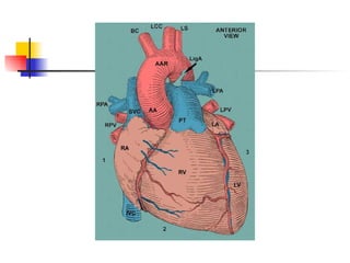
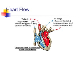
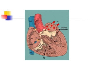
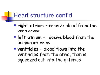
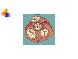
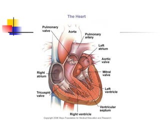
Chapter 9 The Mammalian Heart
mammalian heart The mammalian heart has a mass of about 300 g (size of fist) composed of cardiac muscle What’s unique about cardiac muscle?
cardiac muscle cardiac muscle made of interconnecting cells plasma membranes very tight to facilitate passing of waves of electrical excitation nucleated cells with striated fibers
Heart Flow
Heart structure largest arching blood vessel –  aorta aorta branches, upwards towards the head and the mainflow doubling downwards to the rest of the body (descending aorta) pulmonary artery  – blood vessel leaving the heart  with two branches leading to each lung
Heart structure cont’d pulmonary veins  – bring blood from the left and right lungs vena cava  – two large veins merge, bringing blood downwards from the head (superior vc) and upwards (inferior vc) from the rest of the body coronary arteries  - branch from the aorta delivering oxygenated blood
 
 
Heart structure cont’d septum  – wall of muscle that divides left side and right sides of the heart, blood cannot pass though the septum four chambers  – two on the left, two on the right atrium (auricle) –  upper chamber each side, both receive blood from the veins
 
Heart structure cont’d right atrium –  receive blood from the vena cavae left atrium –  receive blood from the pulmonary veins ventricles –  blood flows into the ventricles from the atria, then is squeezed out into the arteries
 
atrio-ventricular valves mitral or bicuspid  – between the left atrium and ventricle tricuspid  – between right atrium and ventricle
 
The Cardiac Cycle cardiac cycle –  sequence of events that makes up one heart beat normal heart pulse rate –  70 beats per minute
atrial systole atrial spaces fill with blood and the muscles of the atrial walls contract low pressure on this contraction forces blood through the atrio-ventricular valves semilunar valves prevent backflow atrial muscle walls are thin
 
ventricular systole   0.1 sec after the atria contract the ventricle contract lasts about 0.3 sec ventricles thick muscle ventricles squeeze inward on the blood increasing the pressure, pushing it out of the heart
ventricular systole   blood leaves the ventricles through the aorta and pulmonary artery pressure in the ventricle becomes greater than the atria and pushes the atrio-ventricular valves shut,  papillary muscle  – attached to the valves by tendons ( Chordae tendineae) , prevents the valves from being forced inside out
 
ventricular diastole ventricle muscle relax pressure in the ventricles drops blood in the arteries puts pressure on the cusps of the semilunar valves forcing them shut preventing backflow
diastole   whole of the heart muscle relaxes even though the pressure of the blood in the veins is low, the blood fills the atria as their thin walls distend
diastole some blood can trickle through the atrio-ventricular valves into the ventricles atrial muscle contract and the cycle begins again
Why is the left ventricular wall thicker than the right? The left ventricle must develop sufficient force to push blood around the rest of the body The right ventricle pushes blood to the lungs, this requires much less pressure, therefore the right ventricle wall is thinner
Control of the Heart Beat myogenic  – naturally contracts and relaxes without receiving nerve impulses cardiac cells grown in oxygenated nutrient solution will rhythmically contract and relax all by themselves What if all the cardiac cells of the heart contracted at their own rhythms?
Control of the Heart Beat Heart has its own built-in controlling and coordinating system Sinoatrial node   – SAN – pacemaker  – specialized patch of muscle in the wall of the right atrium Muscle cells of the SAN  – set the rhythm for all the other cardiac cells
Control of the Heart Beat SAN muscles  – contract slightly faster than the rest of the heart muscle Set up a wave of electrical activity  – wave spreads out rapidly over the whole atrial walls Atrial wall cardiac muscle  – responds to this excitation wave by contracting at the same rhythm as the SAN
Control of the Heart Beat Both atria  – muscle cells almost contract simultaneously As the wave spreads  Atrio-ventricular node  – AVN – patch of conducting fibers in the septum - the AVN picks up the excitation wave as it spreads across the atria Atrioventricular fibrous tissue -  Band of fibers between the atria and ventricles that do not conduct the excitation wave
Purkyne tissue (Purkinje fibers) after a delay of 0.1 s, the excitation wave is passed on to a bunch of conducting fibers that run down the septum, the  Purkyne tissue Purkyne tissue –  transmits the excitation wave rapidly to the base of the septum where it spreads out through the ventricle walls The excitation –  causes the ventricle walls to contract from the bottom up, squeezing blood upwards and into the arteries
 
Healthy Heart atria contract then the ventricles, from the bottom up Lub-dub What if the coordination of contraction goes wrong?
 
Fibrillation Fibrillation –  heart flutters rather than contracting as a whole and relaxing as a whole Must be treated instantly or  could be fatal Electric shock  often used
Electrocardiograms (ECG) Electrocardiograms (ECG) –  graph of voltage against time P –  represents the wave of excitation sweeping over the atrial walls Q, R, & S  – represent the wave of excitation in the ventricle walls T  – indicates the recovery of the ventricle walls
Electrocardiograms (ECG)
How to Read an EKG Strip EKG paper is a grid where time is measured along the horizontal axis. * Each small square is 1 mm in length and represents 0.04 seconds. * Each larger square is 5 mm in length and represents 0.2 seconds.
Voltage is measured along the vertical axis. * 10 mm is equal to 1mV in voltage. * The diagram below illustrates the configuration of EKG graph paper and where to measure the components of the EKG wave form
 
Heart rate Heart rate can be easily calculated from the EKG strip: * When the rhythm is regular, the heart rate is 300 divided by the number of large squares between the QRS complexes. For example, if there are 4 large squares between regular QRS complexes, the heart rate is 75 (300/4=75).
Heart rate * The second method can be used with an irregular rhythm to estimate the rate. Count the number of R waves in a 6 second strip and multiply by 10. For example, if there are 7 R waves in a 6 second strip, the heart rate is 70 (7x10=70).
This dysrhythmia results in the absence of cardiac output. Almost always occurs with serious heart disease, especially acute MI. The course of treatment for ventricular fibrillation includes: * immediate defibrillation and ACLS protocols.
Atrial fibrillation may occur paroxysmally, but it often becomes chronic. It is usually associated with COPD, CHF or other heart disease. Treatment includes: * Digoxin, diltiazem, or other anti-dysrhythmic medications to control the AV conduction rate and assist with conversion back to normal sinus rhythm. * Cardioversion (shocking simultaneously with the QRS) may also be necessary to terminate this rhythm.

Chapter 9

  • 1.
    Chapter 9 TheMammalian Heart
  • 2.
    mammalian heart Themammalian heart has a mass of about 300 g (size of fist) composed of cardiac muscle What’s unique about cardiac muscle?
  • 3.
    cardiac muscle cardiacmuscle made of interconnecting cells plasma membranes very tight to facilitate passing of waves of electrical excitation nucleated cells with striated fibers
  • 4.
  • 5.
    Heart structure largestarching blood vessel – aorta aorta branches, upwards towards the head and the mainflow doubling downwards to the rest of the body (descending aorta) pulmonary artery – blood vessel leaving the heart with two branches leading to each lung
  • 6.
    Heart structure cont’dpulmonary veins – bring blood from the left and right lungs vena cava – two large veins merge, bringing blood downwards from the head (superior vc) and upwards (inferior vc) from the rest of the body coronary arteries - branch from the aorta delivering oxygenated blood
  • 7.
  • 8.
  • 9.
    Heart structure cont’dseptum – wall of muscle that divides left side and right sides of the heart, blood cannot pass though the septum four chambers – two on the left, two on the right atrium (auricle) – upper chamber each side, both receive blood from the veins
  • 10.
  • 11.
    Heart structure cont’dright atrium – receive blood from the vena cavae left atrium – receive blood from the pulmonary veins ventricles – blood flows into the ventricles from the atria, then is squeezed out into the arteries
  • 12.
  • 13.
    atrio-ventricular valves mitralor bicuspid – between the left atrium and ventricle tricuspid – between right atrium and ventricle
  • 14.
  • 15.
    The Cardiac Cyclecardiac cycle – sequence of events that makes up one heart beat normal heart pulse rate – 70 beats per minute
  • 16.
    atrial systole atrialspaces fill with blood and the muscles of the atrial walls contract low pressure on this contraction forces blood through the atrio-ventricular valves semilunar valves prevent backflow atrial muscle walls are thin
  • 17.
  • 18.
    ventricular systole 0.1 sec after the atria contract the ventricle contract lasts about 0.3 sec ventricles thick muscle ventricles squeeze inward on the blood increasing the pressure, pushing it out of the heart
  • 19.
    ventricular systole blood leaves the ventricles through the aorta and pulmonary artery pressure in the ventricle becomes greater than the atria and pushes the atrio-ventricular valves shut, papillary muscle – attached to the valves by tendons ( Chordae tendineae) , prevents the valves from being forced inside out
  • 20.
  • 21.
    ventricular diastole ventriclemuscle relax pressure in the ventricles drops blood in the arteries puts pressure on the cusps of the semilunar valves forcing them shut preventing backflow
  • 22.
    diastole whole of the heart muscle relaxes even though the pressure of the blood in the veins is low, the blood fills the atria as their thin walls distend
  • 23.
    diastole some bloodcan trickle through the atrio-ventricular valves into the ventricles atrial muscle contract and the cycle begins again
  • 24.
    Why is theleft ventricular wall thicker than the right? The left ventricle must develop sufficient force to push blood around the rest of the body The right ventricle pushes blood to the lungs, this requires much less pressure, therefore the right ventricle wall is thinner
  • 25.
    Control of theHeart Beat myogenic – naturally contracts and relaxes without receiving nerve impulses cardiac cells grown in oxygenated nutrient solution will rhythmically contract and relax all by themselves What if all the cardiac cells of the heart contracted at their own rhythms?
  • 26.
    Control of theHeart Beat Heart has its own built-in controlling and coordinating system Sinoatrial node – SAN – pacemaker – specialized patch of muscle in the wall of the right atrium Muscle cells of the SAN – set the rhythm for all the other cardiac cells
  • 27.
    Control of theHeart Beat SAN muscles – contract slightly faster than the rest of the heart muscle Set up a wave of electrical activity – wave spreads out rapidly over the whole atrial walls Atrial wall cardiac muscle – responds to this excitation wave by contracting at the same rhythm as the SAN
  • 28.
    Control of theHeart Beat Both atria – muscle cells almost contract simultaneously As the wave spreads Atrio-ventricular node – AVN – patch of conducting fibers in the septum - the AVN picks up the excitation wave as it spreads across the atria Atrioventricular fibrous tissue - Band of fibers between the atria and ventricles that do not conduct the excitation wave
  • 29.
    Purkyne tissue (Purkinjefibers) after a delay of 0.1 s, the excitation wave is passed on to a bunch of conducting fibers that run down the septum, the Purkyne tissue Purkyne tissue – transmits the excitation wave rapidly to the base of the septum where it spreads out through the ventricle walls The excitation – causes the ventricle walls to contract from the bottom up, squeezing blood upwards and into the arteries
  • 30.
  • 31.
    Healthy Heart atriacontract then the ventricles, from the bottom up Lub-dub What if the coordination of contraction goes wrong?
  • 32.
  • 33.
    Fibrillation Fibrillation – heart flutters rather than contracting as a whole and relaxing as a whole Must be treated instantly or could be fatal Electric shock often used
  • 34.
    Electrocardiograms (ECG) Electrocardiograms(ECG) – graph of voltage against time P – represents the wave of excitation sweeping over the atrial walls Q, R, & S – represent the wave of excitation in the ventricle walls T – indicates the recovery of the ventricle walls
  • 35.
  • 36.
    How to Readan EKG Strip EKG paper is a grid where time is measured along the horizontal axis. * Each small square is 1 mm in length and represents 0.04 seconds. * Each larger square is 5 mm in length and represents 0.2 seconds.
  • 37.
    Voltage is measuredalong the vertical axis. * 10 mm is equal to 1mV in voltage. * The diagram below illustrates the configuration of EKG graph paper and where to measure the components of the EKG wave form
  • 38.
  • 39.
    Heart rate Heartrate can be easily calculated from the EKG strip: * When the rhythm is regular, the heart rate is 300 divided by the number of large squares between the QRS complexes. For example, if there are 4 large squares between regular QRS complexes, the heart rate is 75 (300/4=75).
  • 40.
    Heart rate *The second method can be used with an irregular rhythm to estimate the rate. Count the number of R waves in a 6 second strip and multiply by 10. For example, if there are 7 R waves in a 6 second strip, the heart rate is 70 (7x10=70).
  • 41.
    This dysrhythmia resultsin the absence of cardiac output. Almost always occurs with serious heart disease, especially acute MI. The course of treatment for ventricular fibrillation includes: * immediate defibrillation and ACLS protocols.
  • 42.
    Atrial fibrillation mayoccur paroxysmally, but it often becomes chronic. It is usually associated with COPD, CHF or other heart disease. Treatment includes: * Digoxin, diltiazem, or other anti-dysrhythmic medications to control the AV conduction rate and assist with conversion back to normal sinus rhythm. * Cardioversion (shocking simultaneously with the QRS) may also be necessary to terminate this rhythm.