The cardiac cycle refers to the alternating contraction and relaxation of the heart's chambers during one heartbeat. It involves two main phases: systole, when the ventricles contract, and diastole, when the ventricles relax and refill with blood. The cycle is precisely coordinated by electrical signals from the heart's conduction system and involves the opening and closing of valves between the chambers.


![AV valves* Semi lunar valves† Status of ventricles
and atria
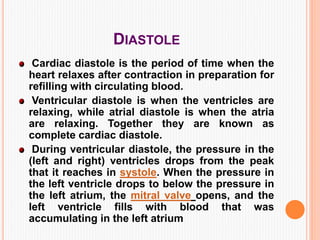
1. earlydiastole open closed
• whole heart is relaxed
• ventricles are expanding
and filling
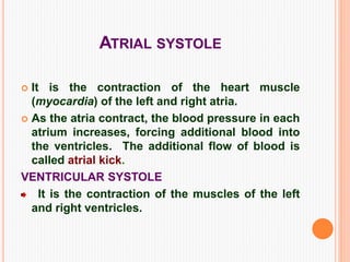
2. atria systole open closed
• atria contract and pump
blood
• additional 10–40% filling
of ventricles[2]
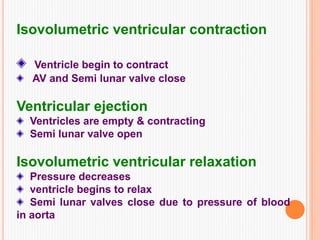
3. isovolumic ventricular
contraction
closed closed
•
ventricular myocytes begi
n to contract
• ventricle volume
unchanged
4. ventricular ejection closed open
• ventricles fully contract
• pump blood to rest of
body
5. Isovolumic ventricular
relaxation
closed closed
• ventricles relax
• ventricle volume
unchanged
• atria expand and are
filling](https://image.slidesharecdn.com/cardiaccycle-151031080543-lva1-app6892/85/Cardiac-cycle-10-320.jpg)