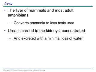
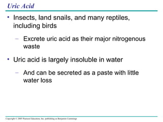
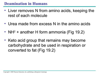
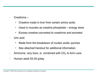
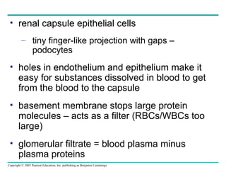
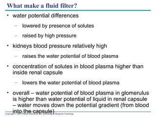
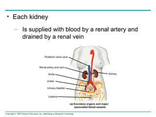
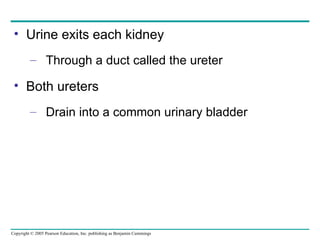
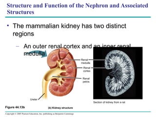
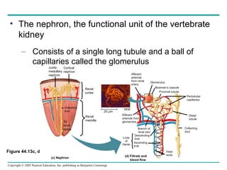
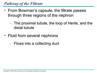
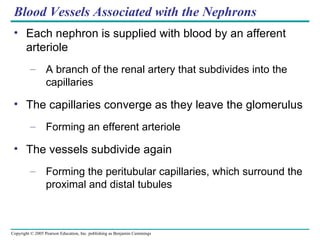
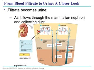
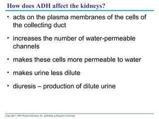
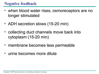
Control,  Coordination and Homeostasis Overview: A balancing act The physiological systems of animals Operate in a fluid environment The relative concentrations of water and solutes in this environment Must be maintained within fairly narrow limits
Freshwater animals Show adaptations that reduce water uptake and conserve solutes Desert and marine animals face desiccating environments With the potential to quickly deplete the body water Figure 44.1
Osmoregulation Regulates solute concentrations and balances the gain and loss of water Excretion Gets rid of metabolic wastes
Osmoregulation balances the uptake and loss of water and solutes Osmoregulation is based largely on controlled movement of solutes Between internal fluids and the external environment
Osmosis Cells require a balance Between osmotic gain and loss of water Water uptake and loss Are balanced by various mechanisms of osmoregulation in different environments
Homeostasis Maintaining a stable internal environment Temperature Amount of water Amount of glucose How is this accomplished?
Feedback Mechanisms Negative  (most) Receptors  (sensors) and effectors (produce a response) Input –  reduces output Positive Receptors  Input – increases output
Accomplished by Transport Epithelia Transport epithelia Are specialized cells that regulate solute movement Are essential components of osmotic regulation and metabolic waste disposal Are arranged into complex tubular networks
An animal’s nitrogenous wastes reflect its phylogeny and habitat The type and quantity of an animal’s waste products May have a large impact on its water balance
Among the most important wastes Are the nitrogenous breakdown products of proteins and nucleic acids Figure 44.8 Proteins Nucleic acids Amino acids Nitrogenous bases – N H 2 Amino groups Most aquatic animals, including most bony fishes Mammals, most amphibians, sharks, some bony fishes Many reptiles (including birds), insects, land snails Ammonia Urea Uric acid N H 3 N H 2 N H 2 O C C C N C O N H H C O N C H N O H
Forms of Nitrogenous Wastes Different animals Excrete nitrogenous wastes in different forms
Ammonia Animals that excrete nitrogenous wastes as ammonia Need access to lots of water Release it across the whole body surface or through the gills
Urea The liver of mammals and most adult amphibians Converts ammonia to less toxic urea Urea is carried to the kidneys, concentrated And excreted with a minimal loss of water
Uric Acid Insects, land snails, and many reptiles, including birds Excrete uric acid as their major nitrogenous waste Uric acid is largely insoluble in water And can be secreted as a paste with little water loss
Deamination in Humans Liver removes N from amino acids, keeping the rest of each molecule Urea made from excess N in the amino acids NH 2  + another H form ammonia (Fig 19.2) Keto acid group that remains may become  carbohydrate and be used in respiration or converted to fat (Fig 19.2)
Creatinine –  Creatine made in liver from certain amino acids Used in muscles as creatine phosphate – energy store Excess creatine converted to creatinine and excreted Uric acid Made from the breakdown of nucleic acids- purines See attached handout for additional information Ammonia, very toxic, is  combined with CO 2  to form urea Human adult 25-30 g/day
Uric Acid is a Byproduct of Normal Body Functions Uric acid, C 5 H 4 N 4 O 3 , is the byproduct of the purines; guanine and adenine. Purine is a muscle protein that enters the body either through dietary intake (about 30 percent) or from the breakdown of the body's own cells during cell turnover (about 70 percent).
Humans Cannot Metabolize Uric Acid, So It Is Excreted Humans lack the enzyme (uricase) that breaks down uric acid. So uric acid is excreted by the kidneys and the intestines.  Under normal circumstances about 70 percent of uric acid is excreted in the urine, with the intestines passing the remaining 30 percent. However, when renal function is insufficient, a greater percentage is excreted via the intestines.  Uric acid is relatively insoluble. So, when concentrations exceed normal levels, it may crystallize in the joints, the urinary tract or under the skin  - gout
Excretory Processes Most excretory systems Produce urine by refining a filtrate derived from body fluids Filtration.  The excretory tubule collects a filtrate from the blood. Water and solutes are forced by blood pressure across the  selectively permeable membranes of a cluster of capillaries and  into the excretory tubule. Reabsorption.  The transport epithelium reclaims valuable substances  from the filtrate and returns them to the body fluids. Secretion.  Other substances, such as toxins and excess ions, are  extracted from body fluids and added to the contents of the excretory  tubule. Excretion.  The filtrate leaves the system and the body. Capillary Excretory tubule Filtrate Urine 1 2 3 4
Key functions of most excretory systems are Filtration, pressure-filtering of body fluids producing a filtrate Reabsorption, reclaiming valuable solutes from the filtrate Secretion, addition of toxins and other solutes from the body fluids to the filtrate Excretion, the filtrate leaves the system
Survey of Excretory Systems The systems that perform basic excretory functions Vary widely among animal groups Are generally built on a complex network of tubules
Vertebrate Kidneys Kidneys, the excretory organs of vertebrates Function in both excretion and osmoregulation
Nephrons and associated blood vessels are the functional unit of the mammalian kidney The mammalian excretory system centers on paired kidneys Which are also the principal site of water balance and salt regulation
Ultrafiltration Making urine two stage process ultrafiltration -  filtering small molecules & urea out of the blood  reabsorption – taking back useful molecules from the fluid in the nephron capillary endothelium far more gaps than other capillaries basement membrane collagen and glycoproteins renal capsule epithelial cells
renal capsule epithelial cells tiny finger-like projection with gaps – podocytes holes in endothelium and epithelium make it easy for substances dissolved in blood to get from the blood to the capsule basement membrane stops large protein molecules – acts as a filter (RBCs/WBCs too large) glomerular filtrate = blood plasma minus plasma proteins
What make a fluid filter? water potential differences lowered by presence of solutes raised by high pressure kidneys blood pressure relatively high raises the water potential of blood plasma concentration of solutes in blood plasma higher than inside renal capsule lowers the water potential of blood plasma overall – water potential of blood plasma in glomerulus is higher than water potential of liquid in renal capsule – water moves down the potential gradient (from blood into the capsule)
Each kidney Is supplied with blood by a renal artery and drained by a renal vein Posterior vena cava Renal artery and vein Aorta Ureter Urinary bladder Urethra (a) Excretory organs and major associated blood vessels Kidney
Urine exits each kidney Through a duct called the ureter Both ureters Drain into a common urinary bladder
Structure and Function of the Nephron and Associated Structures The mammalian kidney has two distinct regions An outer renal cortex and an inner renal medulla (b) Kidney structure Ureter Section of kidney from a rat Renal medulla Renal cortex Renal pelvis Figure 44.13b
The nephron, the functional unit of the vertebrate kidney Consists of a single long tubule and a ball of capillaries called the glomerulus Figure 44.13c, d Juxta- medullary nephron Cortical nephron Collecting duct To  renal pelvis Renal cortex Renal medulla 20 µm Afferent arteriole from renal artery Glomerulus Bowman’s capsule Proximal tubule Peritubular capillaries SEM Efferent arteriole from glomerulus Branch of renal vein Descending limb Ascending limb Loop of Henle Distal  tubule Collecting duct (c) Nephron Vasa recta (d) Filtrate and  blood flow
Filtration of the Blood Filtration occurs as blood pressure Forces fluid from the blood in the glomerulus into the lumen of Bowman’s capsule
Filtration of small molecules is nonselective And the filtrate in Bowman’s capsule is a mixture that mirrors the concentration of various solutes in the blood plasma
Pathway of the Filtrate From Bowman’s capsule, the filtrate passes through three regions of the nephron The proximal tubule, the loop of Henle, and the distal tubule Fluid from several nephrons Flows into a collecting duct
Blood Vessels Associated with the Nephrons Each nephron is supplied with blood by an afferent arteriole A branch of the renal artery that subdivides into the capillaries The capillaries converge as they leave the glomerulus Forming an efferent arteriole The vessels subdivide again Forming the peritubular capillaries, which surround the proximal and distal tubules
From Blood Filtrate to Urine: A Closer Look Filtrate becomes urine As it flows through the mammalian nephron and collecting duct Figure 44.14 Proximal tubule Filtrate H 2 O Salts (NaCl and others) HCO 3 – H + Urea Glucose; amino acids Some drugs Key Active transport Passive transport CORTEX OUTER MEDULLA INNER MEDULLA Descending limb of loop of Henle Thick segment of ascending limb Thin segment of ascending limb Collecting duct NaCl NaCl NaCl Distal tubule NaCl Nutrients Urea H 2 O NaCl H 2 O H 2 O HCO 3  K + H + NH 3 HCO 3  K + H + H 2 O 1 4 3 2 3 5
Secretion and reabsorption in the proximal tubule Substantially alter the volume and composition of filtrate Reabsorption of water continues As the filtrate moves into the descending limb of the loop of Henle
As filtrate travels through the ascending limb of the loop of Henle Salt diffuses out of the permeable tubule into the interstitial fluid The distal tubule Plays a key role in regulating the K +  and NaCl concentration of body fluids The collecting duct Carries the filtrate through the medulla to the renal pelvis and reabsorbs NaCl
Solute Gradients and Water Conservation In a mammalian kidney, the cooperative action and precise arrangement of the loops of Henle and the collecting ducts Are largely responsible for the osmotic gradient that concentrates the urine
Two solutes, NaCl and urea, contribute to the osmolarity of the interstitial fluid Which causes the reabsorption of water in the kidney and concentrates the urine Figure 44.15 H 2 O H 2 O H 2 O H 2 O H 2 O H 2 O H 2 O NaCl NaCl NaCl NaCl NaCl NaCl NaCl 300 300 100 400 600 900 1200 700 400 200 100 Active transport Passive transport OUTER MEDULLA INNER MEDULLA CORTEX H 2 O Urea H 2 O Urea H 2 O Urea H 2 O H 2 O H 2 O H 2 O 1200 1200 900 600 400 300 600 400 300 Osmolarity of  interstitial  fluid (mosm/L) 300
The countercurrent multiplier system involving the loop of Henle Maintains a high salt concentration in the interior of the kidney, which enables the kidney to form concentrated urine
The collecting duct, permeable to water but not salt Conducts the filtrate through the kidney’s osmolarity gradient, and more water exits the filtrate by osmosis
Regulation of Kidney Function The osmolarity of the urine Is regulated by nervous and hormonal control of water and salt reabsorption in the kidneys
Antidiuretic hormone (ADH) Increases water reabsorption in the distal tubules and collecting ducts of the kidney (a)  Antidiuretic hormone (ADH) enhances fluid retention by making the kidneys reclaim more water. Osmoreceptors in hypothalamus Drinking reduces blood osmolarity to set point H 2 O reab- sorption helps prevent further osmolarity  increase STIMULUS: The release of ADH is triggered when osmo- receptor cells in the hypothalamus detect an increase in the osmolarity of the blood Homeostasis: Blood osmolarity Hypothalamus ADH Pituitary gland Increased permeability Thirst Collecting duct Distal tubule
Osmoreceptors, the hypothalamus and ADH hypothalamus – osmosreceptor cells lose or gain water loss of water triggers stimulation of nerve cells to produce ADH ADH – polypeptide of 9 amino acids ADH released into the capillaries in the posterior pituitary gland
How does ADH affect the kidneys? acts on the plasma membranes of the cells of the collecting duct increases the number of water-permeable channels makes these cells more permeable to water makes urine less dilute diuresis – production of dilute urine
Negative feedback when blood water rises, osmoreceptors are no longer stimulated ADH secretion slows (15-20 min) collecting duct channels move back into cytoplasm (15-20 min) membrane becomes less permeable urine becomes more dilute

Chap 19 aice excretion

  • 1.
    Control, Coordinationand Homeostasis Overview: A balancing act The physiological systems of animals Operate in a fluid environment The relative concentrations of water and solutes in this environment Must be maintained within fairly narrow limits
  • 2.
    Freshwater animals Showadaptations that reduce water uptake and conserve solutes Desert and marine animals face desiccating environments With the potential to quickly deplete the body water Figure 44.1
  • 3.
    Osmoregulation Regulates soluteconcentrations and balances the gain and loss of water Excretion Gets rid of metabolic wastes
  • 4.
    Osmoregulation balances theuptake and loss of water and solutes Osmoregulation is based largely on controlled movement of solutes Between internal fluids and the external environment
  • 5.
    Osmosis Cells requirea balance Between osmotic gain and loss of water Water uptake and loss Are balanced by various mechanisms of osmoregulation in different environments
  • 6.
    Homeostasis Maintaining astable internal environment Temperature Amount of water Amount of glucose How is this accomplished?
  • 7.
    Feedback Mechanisms Negative (most) Receptors (sensors) and effectors (produce a response) Input – reduces output Positive Receptors Input – increases output
  • 8.
    Accomplished by TransportEpithelia Transport epithelia Are specialized cells that regulate solute movement Are essential components of osmotic regulation and metabolic waste disposal Are arranged into complex tubular networks
  • 9.
    An animal’s nitrogenouswastes reflect its phylogeny and habitat The type and quantity of an animal’s waste products May have a large impact on its water balance
  • 10.
    Among the mostimportant wastes Are the nitrogenous breakdown products of proteins and nucleic acids Figure 44.8 Proteins Nucleic acids Amino acids Nitrogenous bases – N H 2 Amino groups Most aquatic animals, including most bony fishes Mammals, most amphibians, sharks, some bony fishes Many reptiles (including birds), insects, land snails Ammonia Urea Uric acid N H 3 N H 2 N H 2 O C C C N C O N H H C O N C H N O H
  • 11.
    Forms of NitrogenousWastes Different animals Excrete nitrogenous wastes in different forms
  • 12.
    Ammonia Animals thatexcrete nitrogenous wastes as ammonia Need access to lots of water Release it across the whole body surface or through the gills
  • 13.
    Urea The liverof mammals and most adult amphibians Converts ammonia to less toxic urea Urea is carried to the kidneys, concentrated And excreted with a minimal loss of water
  • 14.
    Uric Acid Insects,land snails, and many reptiles, including birds Excrete uric acid as their major nitrogenous waste Uric acid is largely insoluble in water And can be secreted as a paste with little water loss
  • 15.
    Deamination in HumansLiver removes N from amino acids, keeping the rest of each molecule Urea made from excess N in the amino acids NH 2 + another H form ammonia (Fig 19.2) Keto acid group that remains may become carbohydrate and be used in respiration or converted to fat (Fig 19.2)
  • 16.
    Creatinine – Creatine made in liver from certain amino acids Used in muscles as creatine phosphate – energy store Excess creatine converted to creatinine and excreted Uric acid Made from the breakdown of nucleic acids- purines See attached handout for additional information Ammonia, very toxic, is combined with CO 2 to form urea Human adult 25-30 g/day
  • 17.
    Uric Acid isa Byproduct of Normal Body Functions Uric acid, C 5 H 4 N 4 O 3 , is the byproduct of the purines; guanine and adenine. Purine is a muscle protein that enters the body either through dietary intake (about 30 percent) or from the breakdown of the body's own cells during cell turnover (about 70 percent).
  • 18.
    Humans Cannot MetabolizeUric Acid, So It Is Excreted Humans lack the enzyme (uricase) that breaks down uric acid. So uric acid is excreted by the kidneys and the intestines. Under normal circumstances about 70 percent of uric acid is excreted in the urine, with the intestines passing the remaining 30 percent. However, when renal function is insufficient, a greater percentage is excreted via the intestines. Uric acid is relatively insoluble. So, when concentrations exceed normal levels, it may crystallize in the joints, the urinary tract or under the skin - gout
  • 19.
    Excretory Processes Mostexcretory systems Produce urine by refining a filtrate derived from body fluids Filtration. The excretory tubule collects a filtrate from the blood. Water and solutes are forced by blood pressure across the selectively permeable membranes of a cluster of capillaries and into the excretory tubule. Reabsorption. The transport epithelium reclaims valuable substances from the filtrate and returns them to the body fluids. Secretion. Other substances, such as toxins and excess ions, are extracted from body fluids and added to the contents of the excretory tubule. Excretion. The filtrate leaves the system and the body. Capillary Excretory tubule Filtrate Urine 1 2 3 4
  • 20.
    Key functions ofmost excretory systems are Filtration, pressure-filtering of body fluids producing a filtrate Reabsorption, reclaiming valuable solutes from the filtrate Secretion, addition of toxins and other solutes from the body fluids to the filtrate Excretion, the filtrate leaves the system
  • 21.
    Survey of ExcretorySystems The systems that perform basic excretory functions Vary widely among animal groups Are generally built on a complex network of tubules
  • 22.
    Vertebrate Kidneys Kidneys,the excretory organs of vertebrates Function in both excretion and osmoregulation
  • 23.
    Nephrons and associatedblood vessels are the functional unit of the mammalian kidney The mammalian excretory system centers on paired kidneys Which are also the principal site of water balance and salt regulation
  • 24.
    Ultrafiltration Making urinetwo stage process ultrafiltration - filtering small molecules & urea out of the blood reabsorption – taking back useful molecules from the fluid in the nephron capillary endothelium far more gaps than other capillaries basement membrane collagen and glycoproteins renal capsule epithelial cells
  • 25.
    renal capsule epithelialcells tiny finger-like projection with gaps – podocytes holes in endothelium and epithelium make it easy for substances dissolved in blood to get from the blood to the capsule basement membrane stops large protein molecules – acts as a filter (RBCs/WBCs too large) glomerular filtrate = blood plasma minus plasma proteins
  • 26.
    What make afluid filter? water potential differences lowered by presence of solutes raised by high pressure kidneys blood pressure relatively high raises the water potential of blood plasma concentration of solutes in blood plasma higher than inside renal capsule lowers the water potential of blood plasma overall – water potential of blood plasma in glomerulus is higher than water potential of liquid in renal capsule – water moves down the potential gradient (from blood into the capsule)
  • 27.
    Each kidney Issupplied with blood by a renal artery and drained by a renal vein Posterior vena cava Renal artery and vein Aorta Ureter Urinary bladder Urethra (a) Excretory organs and major associated blood vessels Kidney
  • 28.
    Urine exits eachkidney Through a duct called the ureter Both ureters Drain into a common urinary bladder
  • 29.
    Structure and Functionof the Nephron and Associated Structures The mammalian kidney has two distinct regions An outer renal cortex and an inner renal medulla (b) Kidney structure Ureter Section of kidney from a rat Renal medulla Renal cortex Renal pelvis Figure 44.13b
  • 30.
    The nephron, thefunctional unit of the vertebrate kidney Consists of a single long tubule and a ball of capillaries called the glomerulus Figure 44.13c, d Juxta- medullary nephron Cortical nephron Collecting duct To renal pelvis Renal cortex Renal medulla 20 µm Afferent arteriole from renal artery Glomerulus Bowman’s capsule Proximal tubule Peritubular capillaries SEM Efferent arteriole from glomerulus Branch of renal vein Descending limb Ascending limb Loop of Henle Distal tubule Collecting duct (c) Nephron Vasa recta (d) Filtrate and blood flow
  • 31.
    Filtration of theBlood Filtration occurs as blood pressure Forces fluid from the blood in the glomerulus into the lumen of Bowman’s capsule
  • 32.
    Filtration of smallmolecules is nonselective And the filtrate in Bowman’s capsule is a mixture that mirrors the concentration of various solutes in the blood plasma
  • 33.
    Pathway of theFiltrate From Bowman’s capsule, the filtrate passes through three regions of the nephron The proximal tubule, the loop of Henle, and the distal tubule Fluid from several nephrons Flows into a collecting duct
  • 34.
    Blood Vessels Associatedwith the Nephrons Each nephron is supplied with blood by an afferent arteriole A branch of the renal artery that subdivides into the capillaries The capillaries converge as they leave the glomerulus Forming an efferent arteriole The vessels subdivide again Forming the peritubular capillaries, which surround the proximal and distal tubules
  • 35.
    From Blood Filtrateto Urine: A Closer Look Filtrate becomes urine As it flows through the mammalian nephron and collecting duct Figure 44.14 Proximal tubule Filtrate H 2 O Salts (NaCl and others) HCO 3 – H + Urea Glucose; amino acids Some drugs Key Active transport Passive transport CORTEX OUTER MEDULLA INNER MEDULLA Descending limb of loop of Henle Thick segment of ascending limb Thin segment of ascending limb Collecting duct NaCl NaCl NaCl Distal tubule NaCl Nutrients Urea H 2 O NaCl H 2 O H 2 O HCO 3  K + H + NH 3 HCO 3  K + H + H 2 O 1 4 3 2 3 5
  • 36.
    Secretion and reabsorptionin the proximal tubule Substantially alter the volume and composition of filtrate Reabsorption of water continues As the filtrate moves into the descending limb of the loop of Henle
  • 37.
    As filtrate travelsthrough the ascending limb of the loop of Henle Salt diffuses out of the permeable tubule into the interstitial fluid The distal tubule Plays a key role in regulating the K + and NaCl concentration of body fluids The collecting duct Carries the filtrate through the medulla to the renal pelvis and reabsorbs NaCl
  • 38.
    Solute Gradients andWater Conservation In a mammalian kidney, the cooperative action and precise arrangement of the loops of Henle and the collecting ducts Are largely responsible for the osmotic gradient that concentrates the urine
  • 39.
    Two solutes, NaCland urea, contribute to the osmolarity of the interstitial fluid Which causes the reabsorption of water in the kidney and concentrates the urine Figure 44.15 H 2 O H 2 O H 2 O H 2 O H 2 O H 2 O H 2 O NaCl NaCl NaCl NaCl NaCl NaCl NaCl 300 300 100 400 600 900 1200 700 400 200 100 Active transport Passive transport OUTER MEDULLA INNER MEDULLA CORTEX H 2 O Urea H 2 O Urea H 2 O Urea H 2 O H 2 O H 2 O H 2 O 1200 1200 900 600 400 300 600 400 300 Osmolarity of interstitial fluid (mosm/L) 300
  • 40.
    The countercurrent multipliersystem involving the loop of Henle Maintains a high salt concentration in the interior of the kidney, which enables the kidney to form concentrated urine
  • 41.
    The collecting duct,permeable to water but not salt Conducts the filtrate through the kidney’s osmolarity gradient, and more water exits the filtrate by osmosis
  • 42.
    Regulation of KidneyFunction The osmolarity of the urine Is regulated by nervous and hormonal control of water and salt reabsorption in the kidneys
  • 43.
    Antidiuretic hormone (ADH)Increases water reabsorption in the distal tubules and collecting ducts of the kidney (a) Antidiuretic hormone (ADH) enhances fluid retention by making the kidneys reclaim more water. Osmoreceptors in hypothalamus Drinking reduces blood osmolarity to set point H 2 O reab- sorption helps prevent further osmolarity increase STIMULUS: The release of ADH is triggered when osmo- receptor cells in the hypothalamus detect an increase in the osmolarity of the blood Homeostasis: Blood osmolarity Hypothalamus ADH Pituitary gland Increased permeability Thirst Collecting duct Distal tubule
  • 44.
    Osmoreceptors, the hypothalamusand ADH hypothalamus – osmosreceptor cells lose or gain water loss of water triggers stimulation of nerve cells to produce ADH ADH – polypeptide of 9 amino acids ADH released into the capillaries in the posterior pituitary gland
  • 45.
    How does ADHaffect the kidneys? acts on the plasma membranes of the cells of the collecting duct increases the number of water-permeable channels makes these cells more permeable to water makes urine less dilute diuresis – production of dilute urine
  • 46.
    Negative feedback whenblood water rises, osmoreceptors are no longer stimulated ADH secretion slows (15-20 min) collecting duct channels move back into cytoplasm (15-20 min) membrane becomes less permeable urine becomes more dilute