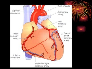
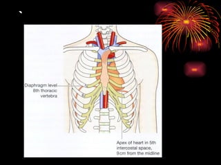
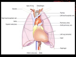
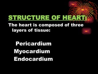
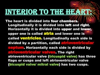
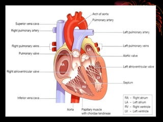
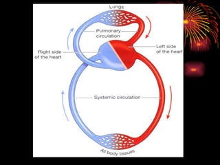
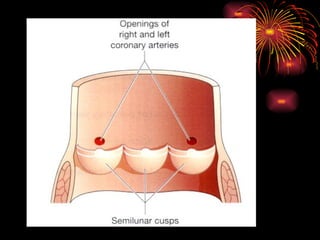
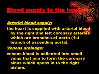
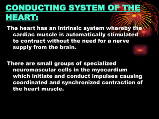
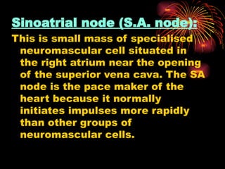
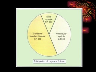
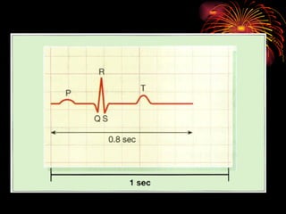
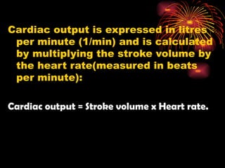
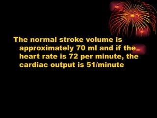
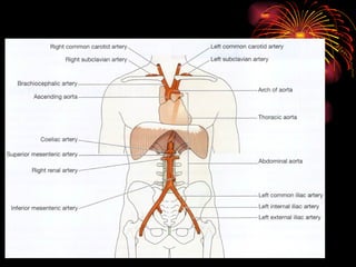
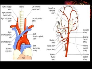
CARDIOVASCULAR
SYSTEM
Prepared by:
Mr. Sajjadhusen.S.S.
HEART
The heart is a roughly cone-
shaped hollow muscular
organ. It is situated in the
thoracic cavity in between
two lungs, the area is known
as mediastenum.
It is about 10 cm long and is
the size of the owner’s fist.
It weighs about 225 gm in
women and is heavier in
men (about 300-350 gm).
POSITION:
The heart lies in the thoracic cavity in
the mediastenum between to lungs. It
lies obliquely, a little more to the left
than the right, and presents base
above, and apex below. The base
extends at the level of 2nd
intercostals
space and apex rests at 5th
intecostal
space. The apex of heart is about 9 cm
away from the midline.
Organs associated with the
heart:
Inferiorly: diaphragm (apex rests)
Superiorly: the great blood vessels, i.e. the
aorta, superior venacava, pulmonary artery
and pulmonary vein.
Posteriorly: the esophagus, trachea, left and
right bronchus, descending aorta, inferior
venacava and thoracic vertebrae.
Laterally: the lungs (right and left)
Anteriorly: the sternum, ribs and intercostals
muscles.
`
STRUCTURE OF HEART:
The heart is composed of three
layers of tissue:
Pericardium
1.Myocardium
2.Endocardium
Pericardium:
The pericardium is made up of
two sacs. The outer sac
consists of fibrous tissue and
the inner of a continuous double
layer of serous membrane.
Myocardium:
The myocardium is composed of
specialized cardiac muscle
found only in the heart. It is not
under voluntary control .
Endocardium:
This forms the lining of the
myocardium and the heart
valves. It is a smooth, glistening
membrane which permits
smooth flow of blood inside the
heart. It consists of flattened
epithelial cells.
INTERIOR TO THE HEART:
The heart is divided into four chambers.
Longitudinally it is divided into left and right.
Horizontally it is divided into upper and lower,
upper one is called atria and lower one is
called ventricles. Longitudinally each side is
divided by a partition, called atrioventricular
septum. Horizontally each side is divided by
atrioventricular valves. The right
atrioventricular valve (tricuspid valve) has three
flaps or cusps and left atrioventricular valve
(bicuspid valve/ mitral valve) has two cusps.
FLOW OF BLOOD THROUGH THE
HEART:
The two largest veins of the body, the superior and
inferior vena cavae, empty their content into the right
atrium. This blood passes via the right atrioventricular
valve in to the right ventricle, and from there it is
pumped into the pulmonary artery or trunk (the only
one artery in the body which carries deoxygenated
blood). The opening of pulmonary artery is guarded by
pulmonary valve, formed by semilunar cusps. This
valve prevents backflow of blood into the right
ventricle. After leaving the heart pulmonary artery
divides into left and right pulmonary arteries, which
carry the venous blood to the lungs where exchange of
gases takes place, i.e. carbon dioxide is excreted and
oxygen is absorbed.
Blood supply to the heart:
Arterial blood supply:
the heart is supplied with arterial blood
by the right and left coronary arteries
which are branches of aorta (1st
branch of ascending aorta).
Venous drainage:
venous blood is collected into small
veins that join to form the coronary
sinus which opens in to the right
atrium.
CONDUCTING SYSTEM OF THE
HEART:
The heart has an intrinsic system whereby the
cardiac muscle is automatically stimulated
to contract without the need for a nerve
supply from the brain.
There are small groups of specialized
neuromascular cells in the myocardium
which initiate and conduct impulses causing
coordinated and synchronized contraction of
the heart muscle.
Sinoatrial node (S.A. node):
This is small mass of specialised
neuromascular cell situated in
the right atrium near the opening
of the superior vena cava. The SA
node is the pace maker of the
heart because it normally
initiates impulses more rapidly
than other groups of
neuromascular cells.
Atrioventricular node (A.V. node):
This is small mass of neuromascular
tissue situated in the wall of the atrial
septum near the atrioventricular
valves. Normally the AV node is
stimulated by impulses that sweep
over the atrial myocardium. However,
it too is capable of initiating impulses
that cause contraction but at a slower
rate than the SA node.
Atrioventricular bundle (AV bundle or
bundle of His):
This is a mass of specialized fibres
that originate from the AV node.
The AV bundle crosses the fibrous
ring that seprarates atria and
ventricles then, at the upper end of
the ventricular septum, it divides
into right and left bundle branches.
Within the ventricular myocardium
the branches break up into fine
fibres, called the purkinje fibres.
The AV bundle, bundle branches
and purkinje fibres convey
electrical impulses from the AV
node to the apex of the
myocardium where the wave of
ventricular contraction begins,
then sweeps upwards and
outwards, pumping blood into the
pulmonary artery and the aorta.
Factors affecting heart rate
Autonomic nervous system:
The rate at which the heart beats is a balance of sympathetic
and parasympathetic activity and this is the most important
factor in determining heart rate.
Circulating chemicals:
The hormones adrenaline and noradrenaline, secreted by the
adrenal medulla, have the same effect as sympathetic
stimulation, i.e. they increase the heart rate Other hormones
including thyroxine increase heart rate by their metabolic
effect. Some drugs, dissolved gases and electrolytes in the
blood may either increase or decrease the heart rate.
Position.:
When the person is upright, the heart rate is usually faster than
when lying down.
Exercise.
Active muscles need more blood than resting
muscles and this is achieved by an increased heart rate
and selective vasodilatation.
Emotional states.
During excitement, fear or anxiety the heart rate is increased.
Other effects mediated by the sympathetic nervous system
may be present.
Gender.
The heart rate is faster in women than men.
Age.
In babies and small children the heart rate is more rapid than in
older children and adults.
THE CARDIAC CYCLE:
The function of the heart is to maintain
a constant circulation of blood
throughout the body. The heart acts
as a pump and its action consists of a
series of events known as the cardiac
cycle.
During each heartbeat, or cardiac cycle,
the heart contacts and then relaxes.
The period of contraction is called
systole and the period of relaxation is
called diastole.
Stages of the cardiac cycle:
The normal number of cardiac cycles per
minute ranges from 60-80. Taking 74 as an
example each cycle lasts about 0.8 of a
second and consists of:
Atrial systole: contraction of the atria
Ventricular systole: contraction of the
Complete cardiac diastole: relaxation
of the atria and ventricles.
Heart sounds
. Two sounds, separated by a short pause,
can be clearly distinguished. They are
described in words as 'lub dup'. The first
sound, 'lub', is fairly loud and is due to the
closure of the atrioventricular valves. This
corresponds with ventricular systole. The
second sound, 'dup', is softer and is due to
the closure of the aortic and pulmonary
valves. This corresponds with atrial
systole.
Elecrocardiogram (ECG)
The pattern of electrical activity
may be displayed on an
oscilloscope screen or traced
on paper. The apparatus used is
an electrocardiograph and the
tracing is an electrocardiogram
(ECG).
The normal ECG tracing
shows five waves which,
by convention, have been
named P, Q, R, S and T.
The P wave arises when the impulse
from the SA node sweeps over the
atria.
The QRS complex represents the very
rapid spread of the impulse from the
AV node through the AV bundle and the
Purkinje fibres and the electrical
activity of the ventricular muscle.
The T wave represents the relaxation of
the ventricular muscle.
The ECG described above
originates from the SA node and
is known as sinus rhythm. The
rate of sinus rhythm is 60 to 100
beats per minute. A faster heart
rate is called tachycardia and a
slower heart rate, bradycardia.
Cardiac output
The cardiac output is the amount of
blood ejected from the heart. The
amount expelled by each
contraction of the ventricles is
called stroke volume.
Cardiac output is expressed in litres
per minute (1/min) and is calculated
by multiplying the stroke volume by
the heart rate(measured in beats
per minute):
Cardiac output = Stroke volume x Heart rate.
The normal stroke volume is
approximately 70 ml and if the
heart rate is 72 per minute, the
cardiac output is 51/minute
Heart n Circulation.pptx for GnM students

Heart n Circulation.pptx for GnM students

  • 1.
  • 2.
    HEART The heart isa roughly cone- shaped hollow muscular organ. It is situated in the thoracic cavity in between two lungs, the area is known as mediastenum.
  • 3.
    It is about10 cm long and is the size of the owner’s fist. It weighs about 225 gm in women and is heavier in men (about 300-350 gm).
  • 4.
    POSITION: The heart liesin the thoracic cavity in the mediastenum between to lungs. It lies obliquely, a little more to the left than the right, and presents base above, and apex below. The base extends at the level of 2nd intercostals space and apex rests at 5th intecostal space. The apex of heart is about 9 cm away from the midline.
  • 5.
    Organs associated withthe heart: Inferiorly: diaphragm (apex rests) Superiorly: the great blood vessels, i.e. the aorta, superior venacava, pulmonary artery and pulmonary vein. Posteriorly: the esophagus, trachea, left and right bronchus, descending aorta, inferior venacava and thoracic vertebrae. Laterally: the lungs (right and left) Anteriorly: the sternum, ribs and intercostals muscles.
  • 7.
  • 9.
    STRUCTURE OF HEART: Theheart is composed of three layers of tissue: Pericardium 1.Myocardium 2.Endocardium
  • 10.
    Pericardium: The pericardium ismade up of two sacs. The outer sac consists of fibrous tissue and the inner of a continuous double layer of serous membrane.
  • 11.
    Myocardium: The myocardium iscomposed of specialized cardiac muscle found only in the heart. It is not under voluntary control .
  • 12.
    Endocardium: This forms thelining of the myocardium and the heart valves. It is a smooth, glistening membrane which permits smooth flow of blood inside the heart. It consists of flattened epithelial cells.
  • 13.
    INTERIOR TO THEHEART: The heart is divided into four chambers. Longitudinally it is divided into left and right. Horizontally it is divided into upper and lower, upper one is called atria and lower one is called ventricles. Longitudinally each side is divided by a partition, called atrioventricular septum. Horizontally each side is divided by atrioventricular valves. The right atrioventricular valve (tricuspid valve) has three flaps or cusps and left atrioventricular valve (bicuspid valve/ mitral valve) has two cusps.
  • 15.
    FLOW OF BLOODTHROUGH THE HEART: The two largest veins of the body, the superior and inferior vena cavae, empty their content into the right atrium. This blood passes via the right atrioventricular valve in to the right ventricle, and from there it is pumped into the pulmonary artery or trunk (the only one artery in the body which carries deoxygenated blood). The opening of pulmonary artery is guarded by pulmonary valve, formed by semilunar cusps. This valve prevents backflow of blood into the right ventricle. After leaving the heart pulmonary artery divides into left and right pulmonary arteries, which carry the venous blood to the lungs where exchange of gases takes place, i.e. carbon dioxide is excreted and oxygen is absorbed.
  • 19.
    Blood supply tothe heart: Arterial blood supply: the heart is supplied with arterial blood by the right and left coronary arteries which are branches of aorta (1st branch of ascending aorta). Venous drainage: venous blood is collected into small veins that join to form the coronary sinus which opens in to the right atrium.
  • 20.
    CONDUCTING SYSTEM OFTHE HEART: The heart has an intrinsic system whereby the cardiac muscle is automatically stimulated to contract without the need for a nerve supply from the brain. There are small groups of specialized neuromascular cells in the myocardium which initiate and conduct impulses causing coordinated and synchronized contraction of the heart muscle.
  • 21.
    Sinoatrial node (S.A.node): This is small mass of specialised neuromascular cell situated in the right atrium near the opening of the superior vena cava. The SA node is the pace maker of the heart because it normally initiates impulses more rapidly than other groups of neuromascular cells.
  • 22.
    Atrioventricular node (A.V.node): This is small mass of neuromascular tissue situated in the wall of the atrial septum near the atrioventricular valves. Normally the AV node is stimulated by impulses that sweep over the atrial myocardium. However, it too is capable of initiating impulses that cause contraction but at a slower rate than the SA node.
  • 23.
    Atrioventricular bundle (AVbundle or bundle of His): This is a mass of specialized fibres that originate from the AV node. The AV bundle crosses the fibrous ring that seprarates atria and ventricles then, at the upper end of the ventricular septum, it divides into right and left bundle branches.
  • 24.
    Within the ventricularmyocardium the branches break up into fine fibres, called the purkinje fibres. The AV bundle, bundle branches and purkinje fibres convey electrical impulses from the AV node to the apex of the myocardium where the wave of ventricular contraction begins, then sweeps upwards and outwards, pumping blood into the pulmonary artery and the aorta.
  • 26.
    Factors affecting heartrate Autonomic nervous system: The rate at which the heart beats is a balance of sympathetic and parasympathetic activity and this is the most important factor in determining heart rate. Circulating chemicals: The hormones adrenaline and noradrenaline, secreted by the adrenal medulla, have the same effect as sympathetic stimulation, i.e. they increase the heart rate Other hormones including thyroxine increase heart rate by their metabolic effect. Some drugs, dissolved gases and electrolytes in the blood may either increase or decrease the heart rate. Position.: When the person is upright, the heart rate is usually faster than when lying down.
  • 27.
    Exercise. Active muscles needmore blood than resting muscles and this is achieved by an increased heart rate and selective vasodilatation. Emotional states. During excitement, fear or anxiety the heart rate is increased. Other effects mediated by the sympathetic nervous system may be present. Gender. The heart rate is faster in women than men. Age. In babies and small children the heart rate is more rapid than in older children and adults.
  • 28.
    THE CARDIAC CYCLE: Thefunction of the heart is to maintain a constant circulation of blood throughout the body. The heart acts as a pump and its action consists of a series of events known as the cardiac cycle. During each heartbeat, or cardiac cycle, the heart contacts and then relaxes. The period of contraction is called systole and the period of relaxation is called diastole.
  • 29.
    Stages of thecardiac cycle: The normal number of cardiac cycles per minute ranges from 60-80. Taking 74 as an example each cycle lasts about 0.8 of a second and consists of: Atrial systole: contraction of the atria Ventricular systole: contraction of the Complete cardiac diastole: relaxation of the atria and ventricles.
  • 31.
    Heart sounds . Twosounds, separated by a short pause, can be clearly distinguished. They are described in words as 'lub dup'. The first sound, 'lub', is fairly loud and is due to the closure of the atrioventricular valves. This corresponds with ventricular systole. The second sound, 'dup', is softer and is due to the closure of the aortic and pulmonary valves. This corresponds with atrial systole.
  • 32.
    Elecrocardiogram (ECG) The patternof electrical activity may be displayed on an oscilloscope screen or traced on paper. The apparatus used is an electrocardiograph and the tracing is an electrocardiogram (ECG).
  • 33.
    The normal ECGtracing shows five waves which, by convention, have been named P, Q, R, S and T.
  • 34.
    The P wavearises when the impulse from the SA node sweeps over the atria. The QRS complex represents the very rapid spread of the impulse from the AV node through the AV bundle and the Purkinje fibres and the electrical activity of the ventricular muscle. The T wave represents the relaxation of the ventricular muscle.
  • 35.
    The ECG describedabove originates from the SA node and is known as sinus rhythm. The rate of sinus rhythm is 60 to 100 beats per minute. A faster heart rate is called tachycardia and a slower heart rate, bradycardia.
  • 37.
    Cardiac output The cardiacoutput is the amount of blood ejected from the heart. The amount expelled by each contraction of the ventricles is called stroke volume.
  • 38.
    Cardiac output isexpressed in litres per minute (1/min) and is calculated by multiplying the stroke volume by the heart rate(measured in beats per minute): Cardiac output = Stroke volume x Heart rate.
  • 39.
    The normal strokevolume is approximately 70 ml and if the heart rate is 72 per minute, the cardiac output is 51/minute