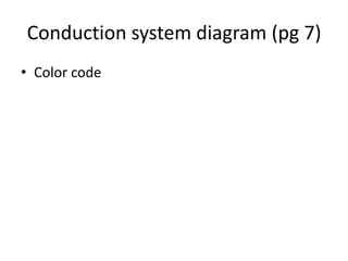
Heart Physiology (pg 6)
Conduction System
• To pump blood efficiently, both atria need to
  contract at the same time, followed by
  contraction of both ventricles (the cardiac
  cycle)
Conduction System
• To pump blood efficiently, both atria need to
  contract at the same time, followed by
  contraction of both ventricles (the cardiac
  cycle)
• Contraction is coordinated by the conduction
  system, which carries the signal to contract
  throughout the heart
Conduction System
• To pump blood efficiently, both atria need to
  contract at the same time, followed by
  contraction of both ventricles (the cardiac
  cycle)
• Contraction is coordinated by the conduction
  system, which carries the signal to contract
  throughout the heart
• Intercalated disks allow signal to travel rapidly
  between adjacent cells
Components of the Conduction System
1. Sinoatrial node: located on the posterior wall
   of the right atrium, near the entrance of the
   superior vena cava. Initiates impulses 70-80
   times per minute. Pacemaker of the heart.
   Impulses travel rapidly throughout the atrial
   myocardium and cause the two atria to
   contract simultaneously
2. Atrioventricular (AV) node: Located on the
   floor of the right atrium. Cells in the AV node
   conduct impulses slowly. This allows time for
   the atria to finish contracting before the
   ventricles begin contracting
3. AV bundle (bundle of His): Conducts impulses
  from atria to ventricles
3. AV bundle (bundle of His): Conducts impulses
  from atria to ventricles
4. Right and left bundle branches: extend down
  the sides of the interventricular septum.
  Branch profusely to form the conduction
  myofibers
3. AV bundle (bundle of His): Conducts impulses
  from atria to ventricles
4. Right and left bundle branches: extend down
  the sides of the interventricular septum.
  Branch profusely to form the conduction
  myofibers
5. Conduction myofibers (Purkinje fibers):
  Transmit impulses throughout the ventricular
  myocardium so that both ventricles contract
  at the same time
Conduction system diagram (pg 7)
• Color code
Electrocardiograms (ECG or EKG)
• Impulses conducted through the heart during
  a cardiac cycle produce electric currents that
  can be measured on the surface of the body
  to produce an electrocardiogram
Electrocardiograms (ECG or EKG)
• Impulses conducted through the heart during
  a cardiac cycle produce electric currents that
  can be measured on the surface of the body
  to produce an electrocardiogram
• Diagram
Electrocardiograms (ECG or EKG)
• Impulses conducted through the heart during
  a cardiac cycle produce electric currents that
  can be measured on the surface of the body
  to produce an electrocardiogram
• Diagram
• Useful in evaluating heart function. For
  example, a heightened p wave indicates an
  enlarged atrium
Cardiac cycle
• Systole = contraction phase
• Diastole = relaxation phase
Cardiac Output
• Volume of blood pumped by a ventricle in 1
  minute
Cardiac Output
• Volume of blood pumped by a ventricle in 1
  minute
• = Stroke volume (ml/cycle) x Heart Rate
  (cycles/minute)
Cardiac Output
• Volume of blood pumped by a ventricle in 1
  minute
• = Stroke volume (ml/cycle) x Heart Rate
  (cycles/minute)
• Heart rate: SA node sets basic rate, but it can be
  changed by the cardiac center in the medulla
  oblongata. Rate increases in response to
  epinephrine, low blood pressure, high CO2, low
  pH, high temperature, emotions (fear, anxiety,
  excitement, anger)

Heart physiology

  • 1.
  • 2.
    Conduction System • Topump blood efficiently, both atria need to contract at the same time, followed by contraction of both ventricles (the cardiac cycle)
  • 3.
    Conduction System • Topump blood efficiently, both atria need to contract at the same time, followed by contraction of both ventricles (the cardiac cycle) • Contraction is coordinated by the conduction system, which carries the signal to contract throughout the heart
  • 4.
    Conduction System • Topump blood efficiently, both atria need to contract at the same time, followed by contraction of both ventricles (the cardiac cycle) • Contraction is coordinated by the conduction system, which carries the signal to contract throughout the heart • Intercalated disks allow signal to travel rapidly between adjacent cells
  • 5.
    Components of theConduction System 1. Sinoatrial node: located on the posterior wall of the right atrium, near the entrance of the superior vena cava. Initiates impulses 70-80 times per minute. Pacemaker of the heart. Impulses travel rapidly throughout the atrial myocardium and cause the two atria to contract simultaneously
  • 6.
    Components of theConduction System 1. Sinoatrial node: located on the posterior wall of the right atrium, near the entrance of the superior vena cava. Initiates impulses 70-80 times per minute. Pacemaker of the heart. Impulses travel rapidly throughout the atrial myocardium and cause the two atria to contract simultaneously 2. Atrioventricular (AV) node: Located on the floor of the right atrium. Cells in the AV node conduct impulses slowly. This allows time for the atria to finish contracting before the ventricles begin contracting
  • 7.
    3. AV bundle(bundle of His): Conducts impulses from atria to ventricles
  • 8.
    3. AV bundle(bundle of His): Conducts impulses from atria to ventricles 4. Right and left bundle branches: extend down the sides of the interventricular septum. Branch profusely to form the conduction myofibers
  • 9.
    3. AV bundle(bundle of His): Conducts impulses from atria to ventricles 4. Right and left bundle branches: extend down the sides of the interventricular septum. Branch profusely to form the conduction myofibers 5. Conduction myofibers (Purkinje fibers): Transmit impulses throughout the ventricular myocardium so that both ventricles contract at the same time
  • 10.
    Conduction system diagram(pg 7) • Color code
  • 11.
    Electrocardiograms (ECG orEKG) • Impulses conducted through the heart during a cardiac cycle produce electric currents that can be measured on the surface of the body to produce an electrocardiogram
  • 12.
    Electrocardiograms (ECG orEKG) • Impulses conducted through the heart during a cardiac cycle produce electric currents that can be measured on the surface of the body to produce an electrocardiogram • Diagram
  • 13.
    Electrocardiograms (ECG orEKG) • Impulses conducted through the heart during a cardiac cycle produce electric currents that can be measured on the surface of the body to produce an electrocardiogram • Diagram • Useful in evaluating heart function. For example, a heightened p wave indicates an enlarged atrium
  • 14.
    Cardiac cycle • Systole= contraction phase • Diastole = relaxation phase
  • 15.
    Cardiac Output • Volumeof blood pumped by a ventricle in 1 minute
  • 16.
    Cardiac Output • Volumeof blood pumped by a ventricle in 1 minute • = Stroke volume (ml/cycle) x Heart Rate (cycles/minute)
  • 17.
    Cardiac Output • Volumeof blood pumped by a ventricle in 1 minute • = Stroke volume (ml/cycle) x Heart Rate (cycles/minute) • Heart rate: SA node sets basic rate, but it can be changed by the cardiac center in the medulla oblongata. Rate increases in response to epinephrine, low blood pressure, high CO2, low pH, high temperature, emotions (fear, anxiety, excitement, anger)