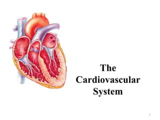
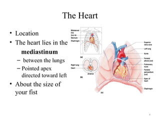
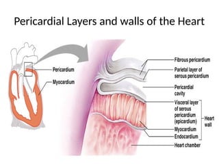
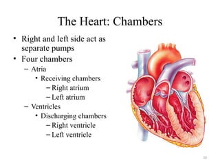
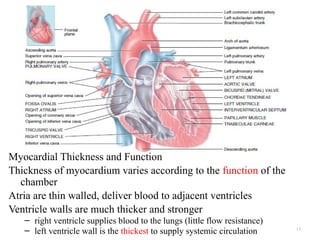
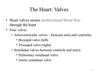
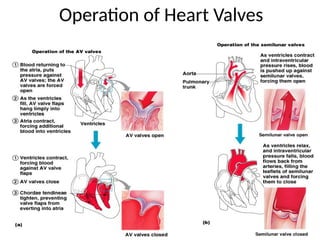
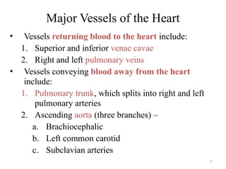
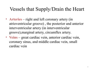
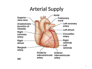
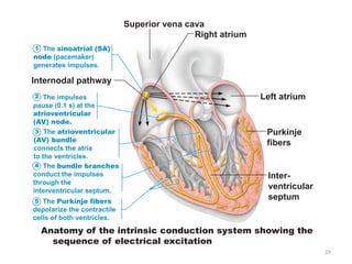
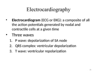
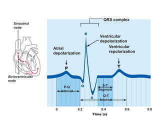
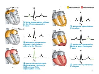
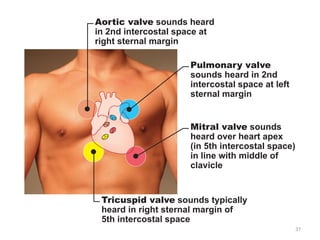
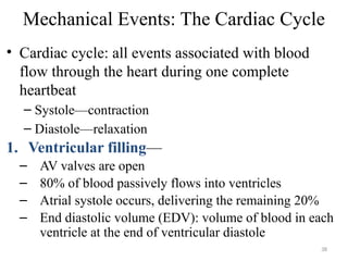
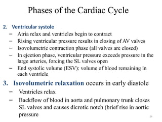
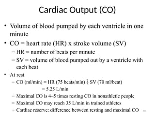
1
Hawassa University
CMHS
School of Medicine
Physiology unit
2022G.C.
BY Nibretie M.
2
The
Cardiovascular
System
3
The Cardiovascular System
• A closed system of the heart and blood vessels
– The heart pumps blood
– Blood vessels allow blood to circulate to all parts
of the body
• The function of the cardiovascular system is to
deliver oxygen and nutrients and to remove
carbon dioxide and other waste products
4
The Heart
• Location
• The heart lies in the
mediastinum
– between the lungs
– Pointed apex
directed toward left
• About the size of
your fist
5
6
Coverings of the Heart:
• Pericardium – a double-walled sac around the
heart composed of:
1. A superficial fibrous pericardium: is composed of
tough, inelastic, dense irregular connective tissue.
2. A deep two-layer serous pericardium
a. The parietal layer lines the internal surface of the
fibrous pericardium
b. The visceral layer or epicardium lines the surface
of the heart
They are separated by the fluid-filled pericardial
cavity
7
Coverings of the Heart:
Between the parietal and visceral layers of the serous
pericardium is a thin film of lubricating serous fluid.
This slippery secretion of the pericardial cells, known
as pericardial fluid,
The Function of the Pericardium:
– Protects and anchors the heart
– Prevents overfilling of the heart with blood
– Allows for the heart to work in a relatively friction-
free environment
8
The Heart Wall: 3 layers
• Epicardium
• Outside layer
• This layer is the visceral pericardium
• Connective tissue layer
• Myocardium
• Middle layer
• Mostly cardiac muscle is responsible for the pumping action
of the heart and is composed of cardiac muscle tissue.
• It makes up approximately 95% of the heart wall.
• Endocardium
• Inner layer
It provides a smooth lining for the chambers of the heart
and covers the valves of the heart.
9
Pericardial Layers and walls of the Heart
10
The Heart: Chambers
• Right and left side act as
separate pumps
• Four chambers
– Atria
• Receiving chambers
– Right atrium
– Left atrium
– Ventricles
• Discharging chambers
– Right ventricle
– Left ventricle
11
Atria of the Heart
• Atria are the receiving chambers of the heart
• Each atrium has a protruding auricle
• Pectinate muscles mark atrial walls
• Blood enters right atria from superior and
inferior venae cavae and coronary sinus
• Blood enters left atria from pulmonary veins
12
Ventricles of the Heart
• Ventricles are the discharging chambers of the
heart
• Papillary muscles and trabeculae carneae
muscles mark ventricular walls
• Right ventricle pumps blood into the pulmonary
trunk
• Left ventricle pumps blood into the aorta
13
Myocardial Thickness and Function
Thickness of myocardium varies according to the function of the
chamber
Atria are thin walled, deliver blood to adjacent ventricles
Ventricle walls are much thicker and stronger
– right ventricle supplies blood to the lungs (little flow resistance)
– left ventricle wall is the thickest to supply systemic circulation
14
The Heart: Valves
• Heart valves ensure unidirectional blood flow
through the heart
• Four valves
– Atrioventricular valves – between atria and ventricles
• Bicuspid valve (left)
• Tricuspid valve (right)
– Semilunar valves between ventricle and artery
• Pulmonary semilunar valve
• Aortic semilunar valve
15
The Heart: Valves
• Valves open as blood is pumped through
• Held in place by chordae tendineae (“heart
strings”)
• Close to prevent backflow
• Atrioventricular (AV) valves lie between the
atria and the ventricles
– AV valves prevent backflow into the atria when
ventricles contract
16
Heart Valves
• Semilunar valves prevent backflow of blood
into the ventricles
• Aortic semilunar valve lies between the left
ventricle and the aorta
• Pulmonary semilunar valve lies between the
right ventricle and pulmonary trunk
17
Operation of Heart Valves
18
• Vessels returning blood to the heart include:
1. Superior and inferior venae cavae
2. Right and left pulmonary veins
• Vessels conveying blood away from the heart
include:
1. Pulmonary trunk, which splits into right and left
pulmonary arteries
2. Ascending aorta (three branches) –
a. Brachiocephalic
b. Left common carotid
c. Subclavian arteries
Major Vessels of the Heart
19
• Arteries – right and left coronary artery (in
atrioventricular groove) , the posterior and anterior
interventricular artery (in interventricular
groove),marginal artery, circumflex artery.
• Veins – great cardiac vein, anterior cardiac vein,
coronary sinus, and middle cardiac vein, small
cardiac vein
Vessels that Supply/Drain the Heart
20
Arterial Supply
21
Venous Supply to Heart
22
Peripheral circulation
Pathway of Blood Through the Heart and Lungs
• Right atrium  tricuspid valve  right ventricle
Right ventricle  pulmonary semilunar valve 
pulmonary arteries  lungs Pulmonary
circulation
• Lungs  pulmonary veins  left atrium
Left atrium  bicuspid valve  left ventricle
Left ventricle  aortic semilunar valve  aorta
Aorta  systemic circulation
23
Blood
Circulation
24
Coronary Circulation
• Blood in the heart chambers does not nourish
the myocardium
• The heart has its own nourishing circulatory
system
– Coronary arteries
– Cardiac veins
– Blood empties into the right atrium via the
coronary sinus
25
Cardiac Muscle Contraction
• Heart muscle:
– Is stimulated by nerves and is self-excitable
(automaticity)
• Cardiac muscle contraction is similar to
skeletal muscle contraction
26
Conduction System
Intrinsic cardiac conduction system
A network of noncontractile (autorhythmic) cells that
initiate and distribute impulses to coordinate the
depolarization and contraction of the heart.
Have unstable resting potentials(pacemaker
potentials or prepotentials) due to open slow Na+
channels
• At threshold, Ca2+
channels open
• Explosive Ca2+
influx produces the rising phase of the
action potential
• Repolarization results from inactivation of Ca2+
channels and opening of voltage-gated K+
channels
27
Sequence of Excitation
1. Sinoatrial (SA) node (pacemaker)
– Generates impulses about 75 times/minute (sinus
rhythm)
– Depolarizes faster than any other part of the
myocardium
2. Atrioventricular (AV) node
– Smaller diameter fibers; fewer gap junctions
– Delays impulses approximately 0.1 second
– Depolarizes 50 times per minute in absence of
SA node input
28
Sequence of Excitation
3. Atrioventricular (AV) bundle (bundle of His)
– Only electrical connection between the atria and
ventricles
4. Right and left bundle branches
– Two pathways in the interventricular septum that
carry the impulses toward the apex of the heart
5. Purkinje fibers
– Complete the pathway into the apex and ventricular
walls
– AV bundle and Purkinje fibers depolarize only
30 times per minute in absence of AV node input
29
Anatomy of the intrinsic conduction system showing the
sequence of electrical excitation
Internodal pathway
Superior vena cava
Right atrium
Left atrium
Purkinje
fibers
Inter-
ventricular
septum
1 The sinoatrial (SA)
node (pacemaker)
generates impulses.
2 The impulses
pause (0.1 s) at the
atrioventricular
(AV) node.
The atrioventricular
(AV) bundle
connects the atria
to the ventricles.
4 The bundle branches
conduct the impulses
through the
interventricular septum.
3
The Purkinje fibers
depolarize the contractile
cells of both ventricles.
5
30
Electrocardiography
• Electrocardiogram (ECG or EKG): a composite of all
the action potentials generated by nodal and
contractile cells at a given time
• Three waves
1. P wave: depolarization of SA node
2. QRS complex: ventricular depolarization
3. T wave: ventricular repolarization
31
Sinoatrial
node
Atrioventricular
node
Atrial
depolarization
QRS complex
Ventricular
depolarization
Ventricular
repolarization
P-Q
Interval
S-T
Segment
Q-T
Interval
32
Atrial depolarization, initiated
by the SA node, causes the
P wave.
P
R
T
Q
S
SA node
AV node
With atrial depolarization
complete, the impulse is
delayed at the AV node.
Ventricular depolarization
begins at apex, causing the
QRS complex. Atrial
repolarization occurs.
P
R
T
Q
S
P
R
T
Q
S
Ventricular depolarization
is complete.
Ventricular repolarization
begins at apex, causing the
T wave.
Ventricular repolarization
is complete.
P
R
T
Q
S
P
R
T
Q
S
P
R
T
Q
S
Depolarization Repolarization
1
2
3
4
5
6
33
Control of Heart Rhythmicity
 Control of Heart Rhythmicity
Both sympathetic and parasympathetic nerves
Parasympathetic nerves (vagi) are distributed mainly
to the S-A and A-V nodes
Sympathetic nerves are distributed to all parts of the
heart
 Effect of Parasympathetic (Vagal) Stimulation
‹ Decreases the rate of rhythm of the sinus node
‹ Decreases excitability of the A-V junctional fibers
between the atrial musculature and the A-V node
slowing transmission of the cardiac impulse into the
ventricles
34
35
(a) Normal sinus rhythm.
(c) Second-degree heart block.
Some P waves are not conducted
through the AV node; hence more
P than QRS waves are seen. In
this tracing, the ratio of P waves
to QRS waves is mostly 2:1.
(d) Ventricular fibrillation. These
chaotic, grossly irregular ECG
deflections are seen in acute
heart attack and electrical shock.
(b) Junctional rhythm. The SA
node is nonfunctional, P waves
are absent, and heart is paced by
the AV node at 40 - 60 beats/min.
36
Heart Sounds
• Two sounds (lub-dup) associated with closing
of heart valves
– First sound occurs as AV valves close and signifies
beginning of systole
– Second sound occurs when SL valves close at the
beginning of ventricular diastole
• Heart murmurs: abnormal heart sounds most
often indicative of valve problems
37
Tricuspid valve sounds typically
heard in right sternal margin of
5th intercostal space
Aortic valve sounds heard
in 2nd intercostal space at
right sternal margin
Pulmonary valve
sounds heard in 2nd
intercostal space at left
sternal margin
Mitral valve sounds
heard over heart apex
(in 5th intercostal space)
in line with middle of
clavicle
38
Mechanical Events: The Cardiac Cycle
• Cardiac cycle: all events associated with blood
flow through the heart during one complete
heartbeat
– Systole—contraction
– Diastole—relaxation
1. Ventricular filling—
– AV valves are open
– 80% of blood passively flows into ventricles
– Atrial systole occurs, delivering the remaining 20%
– End diastolic volume (EDV): volume of blood in each
ventricle at the end of ventricular diastole
39
Phases of the Cardiac Cycle
2. Ventricular systole
– Atria relax and ventricles begin to contract
– Rising ventricular pressure results in closing of AV valves
– Isovolumetric contraction phase (all valves are closed)
– In ejection phase, ventricular pressure exceeds pressure in the
large arteries, forcing the SL valves open
– End systolic volume (ESV): volume of blood remaining in
each ventricle
3. Isovolumetric relaxation occurs in early diastole
– Ventricles relax
– Backflow of blood in aorta and pulmonary trunk closes
SL valves and causes dicrotic notch (brief rise in aortic
pressure
40
Cardiac Output (CO)
• Volume of blood pumped by each ventricle in one
minute
• CO = heart rate (HR) x stroke volume (SV)
– HR = number of beats per minute
– SV = volume of blood pumped out by a ventricle with
each beat
• At rest
– CO (ml/min) = HR (75 beats/min)  SV (70 ml/beat)
= 5.25 L/min
– Maximal CO is 4–5 times resting CO in nonathletic people
– Maximal CO may reach 35 L/min in trained athletes
– Cardiac reserve: difference between resting and maximal CO
41
Regulation of Stroke Volume
• SV = EDV – ESV
• Three main factors affect SV
– Preload
– Contractility
– Afterload
• Preload: degree of stretch of cardiac muscle cells before
they contract (Frank-Starling law of the heart)
– Cardiac muscle exhibits a length-tension relationship
– At rest, cardiac muscle cells are shorter than optimal length
– Slow heartbeat and exercise increase venous return
– Increased venous return distends (stretches) the ventricles and
increases contraction force
42
Regulation of Stroke Volume
• Contractility: contractile strength at a given muscle length,
independent of muscle stretch and EDV
• Positive inotropic agents increase contractility
– Increased Ca2+
influx due to sympathetic stimulation
– Hormones ( epinephrine)
• Negative inotropic agents decrease contractility
_ Increased extracellular K+
– Calcium channel blockers
• Afterload:Is back pressure exerted by blood in the large
arteries leaving the heart
• pressure that must be overcome for ventricles to eject blood
43
Regulation of Stroke Volume
• Hypertension increases afterload, resulting in increased
ESV and reduced SV
• Regulation of Heart Rate
• Positive chronotropic factors increase heart rate
– Example: Exercise ,decreased blood
volume ,sympathetic nervous system and hormones
• Negative chronotropic factors decrease heart rate
Decreased heart rate: Example
– Parasympathetic nervous system
– High blood pressure or blood volume
– Decreased venous return
44
Venous
return
Contractility
Sympathetic
activity
Parasympathetic
activity
EDV
(preload)
Stroke
volume
Heart
rate
Cardiac
output
ESV
Exercise (by
skeletal muscle and
respiratory pumps;
Heart rate
(allows more
time for
ventricular
filling)
Bloodborne
epinephrine,
thyroxine,
excess Ca2+
Exercise,
fright, anxiety
Initial stimulus
Result
Physiological response
45
Blood Pressure
• the hydrostatic pressure exerted by blood on the
walls of a blood vessel.
• Measurements by health professionals are made
on the pressure in large arteries
– Systolic – pressure at the peak of ventricular
contraction
– Diastolic – pressure when ventricles relax
• Pressure in blood vessels decreases as the
distance away from the heart increases
46
Measuring Arterial Blood Pressure
47
Variations in Blood Pressure
• Human normal range is variable
– Normal
• 140–110 mm Hg systolic
• 80–75 mm Hg diastolic
• The average BP 120/80 mm HG
Homeostatic Imbalances
Tachycardia: abnormally fast heart rate (>100 bpm)
– If persistent, may lead to fibrillation
 Bradycardia: heart rate slower than 60 bpm
– May result in grossly inadequate blood circulation
--May be desirable result of endurance training
48
Homeostatic Imbalances
Hypotension
• Low systolic (below 110 mm HG)
• Low diastolic (below 70 mm HG)
• Often associated with illness
Hypertension
• High systolic (above 140 mm HG)
• High diastolic (above 90 mm HG)
• Can be dangerous if it is chronic
Classification

cardiac physiology.pptx cardiac physiology

  • 1.
    1 Hawassa University CMHS School ofMedicine Physiology unit 2022G.C. BY Nibretie M.
  • 2.
  • 3.
    3 The Cardiovascular System •A closed system of the heart and blood vessels – The heart pumps blood – Blood vessels allow blood to circulate to all parts of the body • The function of the cardiovascular system is to deliver oxygen and nutrients and to remove carbon dioxide and other waste products
  • 4.
    4 The Heart • Location •The heart lies in the mediastinum – between the lungs – Pointed apex directed toward left • About the size of your fist
  • 5.
  • 6.
    6 Coverings of theHeart: • Pericardium – a double-walled sac around the heart composed of: 1. A superficial fibrous pericardium: is composed of tough, inelastic, dense irregular connective tissue. 2. A deep two-layer serous pericardium a. The parietal layer lines the internal surface of the fibrous pericardium b. The visceral layer or epicardium lines the surface of the heart They are separated by the fluid-filled pericardial cavity
  • 7.
    7 Coverings of theHeart: Between the parietal and visceral layers of the serous pericardium is a thin film of lubricating serous fluid. This slippery secretion of the pericardial cells, known as pericardial fluid, The Function of the Pericardium: – Protects and anchors the heart – Prevents overfilling of the heart with blood – Allows for the heart to work in a relatively friction- free environment
  • 8.
    8 The Heart Wall:3 layers • Epicardium • Outside layer • This layer is the visceral pericardium • Connective tissue layer • Myocardium • Middle layer • Mostly cardiac muscle is responsible for the pumping action of the heart and is composed of cardiac muscle tissue. • It makes up approximately 95% of the heart wall. • Endocardium • Inner layer It provides a smooth lining for the chambers of the heart and covers the valves of the heart.
  • 9.
    9 Pericardial Layers andwalls of the Heart
  • 10.
    10 The Heart: Chambers •Right and left side act as separate pumps • Four chambers – Atria • Receiving chambers – Right atrium – Left atrium – Ventricles • Discharging chambers – Right ventricle – Left ventricle
  • 11.
    11 Atria of theHeart • Atria are the receiving chambers of the heart • Each atrium has a protruding auricle • Pectinate muscles mark atrial walls • Blood enters right atria from superior and inferior venae cavae and coronary sinus • Blood enters left atria from pulmonary veins
  • 12.
    12 Ventricles of theHeart • Ventricles are the discharging chambers of the heart • Papillary muscles and trabeculae carneae muscles mark ventricular walls • Right ventricle pumps blood into the pulmonary trunk • Left ventricle pumps blood into the aorta
  • 13.
    13 Myocardial Thickness andFunction Thickness of myocardium varies according to the function of the chamber Atria are thin walled, deliver blood to adjacent ventricles Ventricle walls are much thicker and stronger – right ventricle supplies blood to the lungs (little flow resistance) – left ventricle wall is the thickest to supply systemic circulation
  • 14.
    14 The Heart: Valves •Heart valves ensure unidirectional blood flow through the heart • Four valves – Atrioventricular valves – between atria and ventricles • Bicuspid valve (left) • Tricuspid valve (right) – Semilunar valves between ventricle and artery • Pulmonary semilunar valve • Aortic semilunar valve
  • 15.
    15 The Heart: Valves •Valves open as blood is pumped through • Held in place by chordae tendineae (“heart strings”) • Close to prevent backflow • Atrioventricular (AV) valves lie between the atria and the ventricles – AV valves prevent backflow into the atria when ventricles contract
  • 16.
    16 Heart Valves • Semilunarvalves prevent backflow of blood into the ventricles • Aortic semilunar valve lies between the left ventricle and the aorta • Pulmonary semilunar valve lies between the right ventricle and pulmonary trunk
  • 17.
  • 18.
    18 • Vessels returningblood to the heart include: 1. Superior and inferior venae cavae 2. Right and left pulmonary veins • Vessels conveying blood away from the heart include: 1. Pulmonary trunk, which splits into right and left pulmonary arteries 2. Ascending aorta (three branches) – a. Brachiocephalic b. Left common carotid c. Subclavian arteries Major Vessels of the Heart
  • 19.
    19 • Arteries –right and left coronary artery (in atrioventricular groove) , the posterior and anterior interventricular artery (in interventricular groove),marginal artery, circumflex artery. • Veins – great cardiac vein, anterior cardiac vein, coronary sinus, and middle cardiac vein, small cardiac vein Vessels that Supply/Drain the Heart
  • 20.
  • 21.
  • 22.
    22 Peripheral circulation Pathway ofBlood Through the Heart and Lungs • Right atrium  tricuspid valve  right ventricle Right ventricle  pulmonary semilunar valve  pulmonary arteries  lungs Pulmonary circulation • Lungs  pulmonary veins  left atrium Left atrium  bicuspid valve  left ventricle Left ventricle  aortic semilunar valve  aorta Aorta  systemic circulation
  • 23.
  • 24.
    24 Coronary Circulation • Bloodin the heart chambers does not nourish the myocardium • The heart has its own nourishing circulatory system – Coronary arteries – Cardiac veins – Blood empties into the right atrium via the coronary sinus
  • 25.
    25 Cardiac Muscle Contraction •Heart muscle: – Is stimulated by nerves and is self-excitable (automaticity) • Cardiac muscle contraction is similar to skeletal muscle contraction
  • 26.
    26 Conduction System Intrinsic cardiacconduction system A network of noncontractile (autorhythmic) cells that initiate and distribute impulses to coordinate the depolarization and contraction of the heart. Have unstable resting potentials(pacemaker potentials or prepotentials) due to open slow Na+ channels • At threshold, Ca2+ channels open • Explosive Ca2+ influx produces the rising phase of the action potential • Repolarization results from inactivation of Ca2+ channels and opening of voltage-gated K+ channels
  • 27.
    27 Sequence of Excitation 1.Sinoatrial (SA) node (pacemaker) – Generates impulses about 75 times/minute (sinus rhythm) – Depolarizes faster than any other part of the myocardium 2. Atrioventricular (AV) node – Smaller diameter fibers; fewer gap junctions – Delays impulses approximately 0.1 second – Depolarizes 50 times per minute in absence of SA node input
  • 28.
    28 Sequence of Excitation 3.Atrioventricular (AV) bundle (bundle of His) – Only electrical connection between the atria and ventricles 4. Right and left bundle branches – Two pathways in the interventricular septum that carry the impulses toward the apex of the heart 5. Purkinje fibers – Complete the pathway into the apex and ventricular walls – AV bundle and Purkinje fibers depolarize only 30 times per minute in absence of AV node input
  • 29.
    29 Anatomy of theintrinsic conduction system showing the sequence of electrical excitation Internodal pathway Superior vena cava Right atrium Left atrium Purkinje fibers Inter- ventricular septum 1 The sinoatrial (SA) node (pacemaker) generates impulses. 2 The impulses pause (0.1 s) at the atrioventricular (AV) node. The atrioventricular (AV) bundle connects the atria to the ventricles. 4 The bundle branches conduct the impulses through the interventricular septum. 3 The Purkinje fibers depolarize the contractile cells of both ventricles. 5
  • 30.
    30 Electrocardiography • Electrocardiogram (ECGor EKG): a composite of all the action potentials generated by nodal and contractile cells at a given time • Three waves 1. P wave: depolarization of SA node 2. QRS complex: ventricular depolarization 3. T wave: ventricular repolarization
  • 31.
  • 32.
    32 Atrial depolarization, initiated bythe SA node, causes the P wave. P R T Q S SA node AV node With atrial depolarization complete, the impulse is delayed at the AV node. Ventricular depolarization begins at apex, causing the QRS complex. Atrial repolarization occurs. P R T Q S P R T Q S Ventricular depolarization is complete. Ventricular repolarization begins at apex, causing the T wave. Ventricular repolarization is complete. P R T Q S P R T Q S P R T Q S Depolarization Repolarization 1 2 3 4 5 6
  • 33.
    33 Control of HeartRhythmicity  Control of Heart Rhythmicity Both sympathetic and parasympathetic nerves Parasympathetic nerves (vagi) are distributed mainly to the S-A and A-V nodes Sympathetic nerves are distributed to all parts of the heart  Effect of Parasympathetic (Vagal) Stimulation ‹ Decreases the rate of rhythm of the sinus node ‹ Decreases excitability of the A-V junctional fibers between the atrial musculature and the A-V node slowing transmission of the cardiac impulse into the ventricles
  • 34.
  • 35.
    35 (a) Normal sinusrhythm. (c) Second-degree heart block. Some P waves are not conducted through the AV node; hence more P than QRS waves are seen. In this tracing, the ratio of P waves to QRS waves is mostly 2:1. (d) Ventricular fibrillation. These chaotic, grossly irregular ECG deflections are seen in acute heart attack and electrical shock. (b) Junctional rhythm. The SA node is nonfunctional, P waves are absent, and heart is paced by the AV node at 40 - 60 beats/min.
  • 36.
    36 Heart Sounds • Twosounds (lub-dup) associated with closing of heart valves – First sound occurs as AV valves close and signifies beginning of systole – Second sound occurs when SL valves close at the beginning of ventricular diastole • Heart murmurs: abnormal heart sounds most often indicative of valve problems
  • 37.
    37 Tricuspid valve soundstypically heard in right sternal margin of 5th intercostal space Aortic valve sounds heard in 2nd intercostal space at right sternal margin Pulmonary valve sounds heard in 2nd intercostal space at left sternal margin Mitral valve sounds heard over heart apex (in 5th intercostal space) in line with middle of clavicle
  • 38.
    38 Mechanical Events: TheCardiac Cycle • Cardiac cycle: all events associated with blood flow through the heart during one complete heartbeat – Systole—contraction – Diastole—relaxation 1. Ventricular filling— – AV valves are open – 80% of blood passively flows into ventricles – Atrial systole occurs, delivering the remaining 20% – End diastolic volume (EDV): volume of blood in each ventricle at the end of ventricular diastole
  • 39.
    39 Phases of theCardiac Cycle 2. Ventricular systole – Atria relax and ventricles begin to contract – Rising ventricular pressure results in closing of AV valves – Isovolumetric contraction phase (all valves are closed) – In ejection phase, ventricular pressure exceeds pressure in the large arteries, forcing the SL valves open – End systolic volume (ESV): volume of blood remaining in each ventricle 3. Isovolumetric relaxation occurs in early diastole – Ventricles relax – Backflow of blood in aorta and pulmonary trunk closes SL valves and causes dicrotic notch (brief rise in aortic pressure
  • 40.
    40 Cardiac Output (CO) •Volume of blood pumped by each ventricle in one minute • CO = heart rate (HR) x stroke volume (SV) – HR = number of beats per minute – SV = volume of blood pumped out by a ventricle with each beat • At rest – CO (ml/min) = HR (75 beats/min)  SV (70 ml/beat) = 5.25 L/min – Maximal CO is 4–5 times resting CO in nonathletic people – Maximal CO may reach 35 L/min in trained athletes – Cardiac reserve: difference between resting and maximal CO
  • 41.
    41 Regulation of StrokeVolume • SV = EDV – ESV • Three main factors affect SV – Preload – Contractility – Afterload • Preload: degree of stretch of cardiac muscle cells before they contract (Frank-Starling law of the heart) – Cardiac muscle exhibits a length-tension relationship – At rest, cardiac muscle cells are shorter than optimal length – Slow heartbeat and exercise increase venous return – Increased venous return distends (stretches) the ventricles and increases contraction force
  • 42.
    42 Regulation of StrokeVolume • Contractility: contractile strength at a given muscle length, independent of muscle stretch and EDV • Positive inotropic agents increase contractility – Increased Ca2+ influx due to sympathetic stimulation – Hormones ( epinephrine) • Negative inotropic agents decrease contractility _ Increased extracellular K+ – Calcium channel blockers • Afterload:Is back pressure exerted by blood in the large arteries leaving the heart • pressure that must be overcome for ventricles to eject blood
  • 43.
    43 Regulation of StrokeVolume • Hypertension increases afterload, resulting in increased ESV and reduced SV • Regulation of Heart Rate • Positive chronotropic factors increase heart rate – Example: Exercise ,decreased blood volume ,sympathetic nervous system and hormones • Negative chronotropic factors decrease heart rate Decreased heart rate: Example – Parasympathetic nervous system – High blood pressure or blood volume – Decreased venous return
  • 44.
    44 Venous return Contractility Sympathetic activity Parasympathetic activity EDV (preload) Stroke volume Heart rate Cardiac output ESV Exercise (by skeletal muscleand respiratory pumps; Heart rate (allows more time for ventricular filling) Bloodborne epinephrine, thyroxine, excess Ca2+ Exercise, fright, anxiety Initial stimulus Result Physiological response
  • 45.
    45 Blood Pressure • thehydrostatic pressure exerted by blood on the walls of a blood vessel. • Measurements by health professionals are made on the pressure in large arteries – Systolic – pressure at the peak of ventricular contraction – Diastolic – pressure when ventricles relax • Pressure in blood vessels decreases as the distance away from the heart increases
  • 46.
  • 47.
    47 Variations in BloodPressure • Human normal range is variable – Normal • 140–110 mm Hg systolic • 80–75 mm Hg diastolic • The average BP 120/80 mm HG Homeostatic Imbalances Tachycardia: abnormally fast heart rate (>100 bpm) – If persistent, may lead to fibrillation  Bradycardia: heart rate slower than 60 bpm – May result in grossly inadequate blood circulation --May be desirable result of endurance training
  • 48.
    48 Homeostatic Imbalances Hypotension • Lowsystolic (below 110 mm HG) • Low diastolic (below 70 mm HG) • Often associated with illness Hypertension • High systolic (above 140 mm HG) • High diastolic (above 90 mm HG) • Can be dangerous if it is chronic
  • 49.