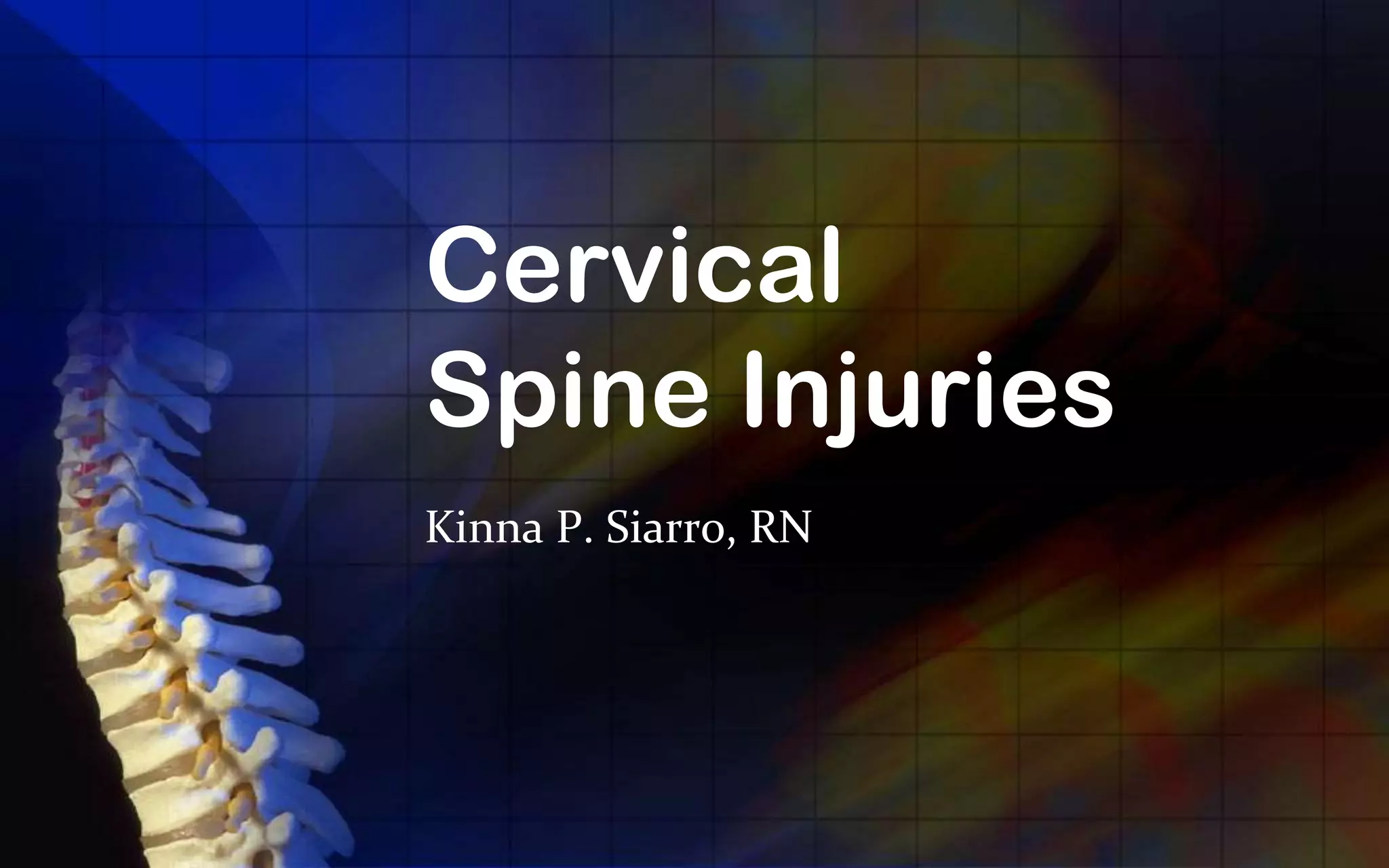
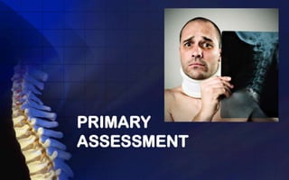
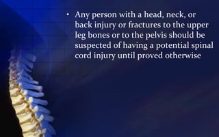
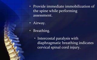
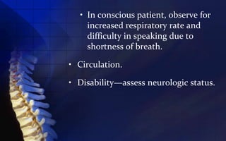
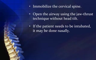
Cervical
Spine Injuries
Kinna P. Siarro, RN
• Injuries to the cervical spine are
  serious because the crushing,
  stretching, and rotational shear
  forces exerted on the cord at the time
  of trauma can produce severe
  neurologic deficits

• Edema and cord swelling contribute
  further to the loss of spinal cord
  function.
PRIMARY
ASSESSMENT
• Any person with a head, neck, or
  back injury or fractures to the upper
  leg bones or to the pelvis should be
  suspected of having a potential spinal
  cord injury until proved otherwise
• Provide immediate immobilization of
  the spine while performing
  assessment.

• Airway.

• Breathing.

  • Intercostal paralysis with
    diaphragmatic breathing indicates
    cervical spinal cord injury.
• In conscious patient, observe for
    increased respiratory rate and
    difficulty in speaking due to
    shortness of breath.

• Circulation.

• Disability—assess neurologic status.
PRIMARY
INTERVENTIONS
• Immobilize the cervical spine.

• Open the airway using the jaw-thrust
  technique without head tilt.

• If the patient needs to be intubated,
  it may be done nasally.
• If respirations are shallow, assist with
  a bag-valve mask.

• Administer high-flow oxygen to
  minimize potential hypoxic spinal
  cord damage.
SUBSEQUENT
ASSESSMENT
• Assess the position of the patient
  when found; this may indicate the
  type of injury incurred.
• Hypotension and bradycardia
  accompanied by warm, dry skin—
  suggests spinal shock.
• Neck and back pain/extremity pain
  or burning sensation to the skin.
• History of unconsciousness.
• Total sensory loss and motor
  paralysis below level of injury.
• Loss of bowel and bladder control;
  usually urinary retention and bladder
  distention.
• Loss of sweating and vasomotor tone
  below level of cord lesion.
• Priapism—persistent erection of
  penis.
• Hypothermia—due to the inability to
  constrict peripheral blood vessels
  and conserve body heat.
• Loss of rectal tone.
GENERAL
INTERVENTIONS
NURSING ALERT

• A spinal cord injury can be made
  worse during the acute phase of
  injury, resulting in permanent
  neurologic damage.
• Proper handling is an immediate
  priority.
• Insert an NG tube.
• Keep the patient warm.
• Initiate I.V. access.
• Insert an indwelling urinary catheter to
  avoid bladder distention.
• Monitor for hypotension, hypothermia, and
  bradycardia.
• Continue with repeated neurologic
  examinations to determine if there is
  deterioration of the spinal cord injury.
• Be prepared to manage seizures.
• Pharmacologic interventions: high-dose
  steroids (methylprednisolone).
   • The standard regimen is 30 mg/kg I.V.
     loading dose over 15 minutes, followed
     by a 5.4 mg/kg/hour infusion to be
     initiated 45 minutes later.
   • Continue the infusion for 23 hours.
Cervical spine injuries

Cervical spine injuries

  • 1.
  • 2.
    • Injuries tothe cervical spine are serious because the crushing, stretching, and rotational shear forces exerted on the cord at the time of trauma can produce severe neurologic deficits • Edema and cord swelling contribute further to the loss of spinal cord function.
  • 3.
  • 4.
    • Any personwith a head, neck, or back injury or fractures to the upper leg bones or to the pelvis should be suspected of having a potential spinal cord injury until proved otherwise
  • 5.
    • Provide immediateimmobilization of the spine while performing assessment. • Airway. • Breathing. • Intercostal paralysis with diaphragmatic breathing indicates cervical spinal cord injury.
  • 6.
    • In consciouspatient, observe for increased respiratory rate and difficulty in speaking due to shortness of breath. • Circulation. • Disability—assess neurologic status.
  • 7.
  • 8.
    • Immobilize thecervical spine. • Open the airway using the jaw-thrust technique without head tilt. • If the patient needs to be intubated, it may be done nasally.
  • 9.
    • If respirationsare shallow, assist with a bag-valve mask. • Administer high-flow oxygen to minimize potential hypoxic spinal cord damage.
  • 10.
  • 11.
    • Assess theposition of the patient when found; this may indicate the type of injury incurred. • Hypotension and bradycardia accompanied by warm, dry skin— suggests spinal shock. • Neck and back pain/extremity pain or burning sensation to the skin. • History of unconsciousness.
  • 12.
    • Total sensoryloss and motor paralysis below level of injury. • Loss of bowel and bladder control; usually urinary retention and bladder distention. • Loss of sweating and vasomotor tone below level of cord lesion. • Priapism—persistent erection of penis.
  • 13.
    • Hypothermia—due tothe inability to constrict peripheral blood vessels and conserve body heat. • Loss of rectal tone.
  • 14.
  • 15.
    NURSING ALERT • Aspinal cord injury can be made worse during the acute phase of injury, resulting in permanent neurologic damage. • Proper handling is an immediate priority.
  • 16.
    • Insert anNG tube. • Keep the patient warm. • Initiate I.V. access. • Insert an indwelling urinary catheter to avoid bladder distention. • Monitor for hypotension, hypothermia, and bradycardia. • Continue with repeated neurologic examinations to determine if there is deterioration of the spinal cord injury.
  • 17.
    • Be preparedto manage seizures. • Pharmacologic interventions: high-dose steroids (methylprednisolone). • The standard regimen is 30 mg/kg I.V. loading dose over 15 minutes, followed by a 5.4 mg/kg/hour infusion to be initiated 45 minutes later. • Continue the infusion for 23 hours.