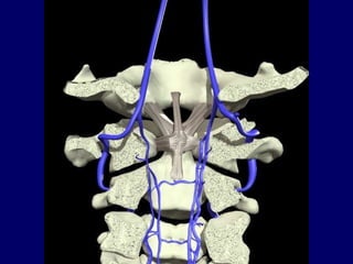
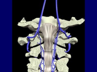
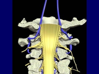
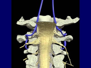
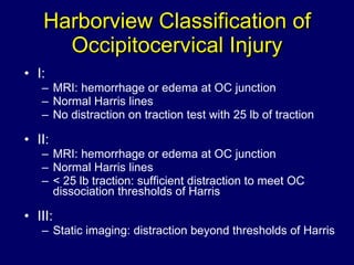
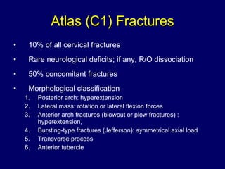
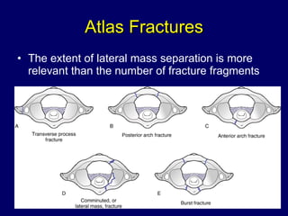
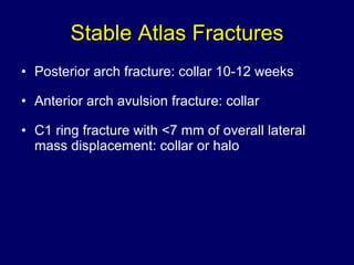
Upper Cervical Trauma Sohail Bajammal, MBChB, MSc November 14, 2006 St. Joseph’s Healthcare, Hamilton  Weekly Orthopaedic Teaching Rounds
Upper Cervical Trauma a.k.a. Cranio-cervical Trauma Occipito-cervical Trauma Occipito-atlanto-axial Trauma
Outline The Problem Anatomy X-rays Fractures of O, C1, C2
The Problem Better recognition Better pre-hospital care (ATLS, Orthopod) Radiographic finding Improved cars safety: Less mortality at scene, more of OC injury
Evaluation History: mechanism of injury Physical: ATLS 2° survey: thorough neurological exam Radiology: 3-views C-spine, CT, MRI
Anatomy
Anatomy Unique anatomy of O-C1-C2 C1: no body, 2 articular pillars connected by 2 arches C2: dens, flat C1-2 Ligaments: Intrinsic (within spinal canal): Odontoid: alar, apical Cruciate: transverse lig, vertical bands Tectorial membrane: thickening of PLL Extrinsic: Ligamentum nuchae Anterior and posterior atlanto-occipital membrane Anterior and posterior atlanto-axial membrane Joint capsules Vertebral artery
Courtesy of AnatomyTV


Ponticulus Posticus Latin , little posterior bridge Young et al., 2005 JBJS(A) 15.5% prevalence of arcuate foramen in 464 lateral c-spine x-rays
Ponticulus Posticus
Ponticulus Posticus
X-rays
Cervical X-rays ABCDEs A: adequacy, alignment B: bones C: cartilage D: disc space E: else (skull, clavicle) S: soft tissue
Lateral C-spine
Harris Lines SAC: > 13 mm
BC/OA >1 considered abnormal Limited Usefulness Positive only in Anterior Translational injuries False Negative with pure distraction Powers’ Ratio
Open-mouth View
Occipital Condyle  Fractures CT,  R/O OC dissociation I: comminuted, axial impaction Stable    Collar 6-8 weeks II: extension of basilar skull fracture into condyle Potentially unstable    Collar 6-8 weeks III: avulsion of alar lig Minimal displaced    Halo vest, 8-12 weeks Displaced    O-C2 fusion Consider surgery if OC dissociation
Occipito-cervical Dissociation Rare and usually fatal Often assoc. with facial injuries, chest trauma Deceleration AVOID traction!! Halo until surgery 1 º  treatment:  Oc-C2 fusion if good screw purchase Oc-C3 fusion otherwise Biomechanically: plate & screw > screws > wires
Traynelis Classification of Occipito-cervical Dissociation
Harborview Classification of Occipitocervical Injury I:  MRI: hemorrhage or edema at OC junction Normal Harris lines No distraction on traction test with 25 lb of traction  II: MRI: hemorrhage or edema at OC junction Normal Harris lines < 25 lb traction: sufficient distraction to meet OC dissociation thresholds of Harris III: Static imaging: distraction beyond thresholds of Harris
Atlas (C1) Fractures 10% of all cervical fractures Rare neurological deficits; if any, R/O dissociation 50% concomitant fractures Morphological classification Posterior arch: hyperextension  Lateral mass: rotation or lateral flexion forces  Anterior arch fractures (blowout or plow fractures) : hyperextension,  Bursting-type fractures (Jefferson): symmetrical axial load Transverse process Anterior tubercle
Atlas Fractures The extent of lateral mass separation is more relevant than the number of fracture fragments
Stable Atlas Fractures Posterior arch fracture: collar 10-12 weeks Anterior arch avulsion fracture: collar C1 ring fracture with <7 mm of overall lateral mass displacement: collar or halo
Unstable Atlas Fractures C1 ring fracture with ≥7 mm of overall C1 lateral mass displacement: prolonged halo or fusion (C1-C2, or Occiput-C2)  Plough fracture: reduction with halo in slight flexion or C1-C2 fusion or occiput-C2
Plough Fracture
Rupture of Transverse Ligament Flexion force Dickman Classification: Mid-substance tear Avulsion of lateral mass of C1 As force increases, alar and apical lig tear (ADI > 7mm) Treatment: If ADI ≤5mm    collar If ADI >5mm and type I    C1-C2 fusion If ADI >5mm and type II    halo
Atlanto-Axial Instability A: Rotational Around the dens Treated with closed reduction and immobilization.  Beware of associated fractures B: Translational Translation between C1–C2, where transverse lig is disrupted Mid-substance transverse ligament tears (type I) are treated with C1–C2 arthrodesis C: Distraction: Indicating craniocervical dissociation Bony avulsions (type II) may be treated with halo or C1–C2 arthrodesis
Rotatory Atlanto-Axial Instability Collar or Halo C1-C2 Fusion
Axis (C2) Fractures Odontoid fractures Traumatic spondylolisthesis of the axis (hangman's fracture)
Odontoid Fractures 60% of C2 fractures 10-20% of all c-spine fractures Neurological deficits in 10-20% Bimodal:  young (high energy), elderly (falls) Anderson and D'Alonzo Classification
 
Type I Odontoid Occurs at tip, cephalad to the transverse Least common  Represent an avulsion of the alar ligament Treated with collar or halo 6-8 weeks Surgery (occiput-C2 fusion) if associated with occipitocervical dissociation
Type III Odontoid Extends into the body of the axis More stable than type II fractures  Higher union rate with non-surgical Treated with a halo or brace 8-12 weeks after reduction if displaced
Type II Odontoid At the junction of the base of the odontoid and body of the axis  The most common fracture type The least likely to heal with non-surgical (10-77% non-union) IIA: new addition, comminution at base Treatment: controversial
Type II Odontoid Higher risk of non-union: Initial displacement > 5mm Posterior displacement Angulation > 10 0 Age > 50 Smoking Delay in diagnosis > 3 weeks Inability to achieve or maintain reduction
Options for High Risk type II Collar: very high risk of non-union Reduction and Halo: risk of complications in elderly Anterior Odontoid Screw(s) Pros: High union rate, preserves atlanto-axial motion Cons: Poor fixation in osteoporotic, difficult in large chest or posteriorly displaced C/I: reverse obliquity Posterior C1-C2 arthrodesis: C1-2 transarticular screw > segmental C1-2 fixation > wires techniques
Anterior Odontoid Screw
Traumatic spondylolisthesis of the Axis (Hangman's fracture) 2 nd  most common fracture of C2 15% of all cervical spine fractures Higher energy injury, associated spinal #: 30%  Younger age group, MVC  MOI: hyperextension + axial compression; additional flexion moment leads to very unstable injury Rare neurological involvement
Hangman's Fracture   Effendi    Levine & Edwards Classification
Type I Hangman’s Most common Bilateral pars fractures with translation <3 mm and no angulation Treated with collar, occasionally halo
Type IA Hangman’s Atypical fracture, recently recognized Minimal translation and little or no angulation Elongation of the C2 body  CT: extension of fracture line into the body and often through the foramen transversarium (vertebral artery injury may occur) May have canal compromise Usually halo, surgery if neuro deficits Surgical options: anterior C2–C3 arthrodesis, posterior C1–C3 vs C2–C3 arthrodesis, or combined approach
Type IA Hangman’s
Type II Hangman’s C2-3 disc and PLL are disrupted, resulting in translation >3 mm and marked angulation ALL  generally remains intact but is stripped from its bony attachment Halo: after reduction in slight extension
Type IIA Hangman’s Less common; MOI: hyperext, axial then flex Fracture line is more oblique than vertical (vs II) Little or no translation, but significant angulation. Avoid traction Halo, and if markedly displaced, possibly direct fixation of fractured arch through a posterior approach C1-C3, or by C2–C3 anterior discectomy and arthrodesis
Type III Hangman’s A combination of pars fracture with dislocation of the C2-3 facet joints Very unstable, with free-floating inferior articular processes The most common injury to be associated with neurological deficit Requires surgery; it is irreducible by closed means Options: Anterior C2-3 discectomy and fusion, or posterior open reduction and C1-3 fusion
Posterior C1-2 Fusion Gallie Technique
Posterior C1-2 Fusion Brooks-Jenkins technique
C1–C2 Transarticular Screw Fixation Magerl technique
Finally.. It’s over!
Halo Crown first then the vest Prep areas in infiltrate with local Normally put scoop under head (unless contraindicated) Put halo crown around head Landmarks: for the anterolateral pins, 1cm above the lateral 1/3 of the eyebrow and the same distance above the top of the ear  Place the anterior pins in bare skin NOT in the hairline
Halo Have patient close eyes before insertion of ant pins Place 4 pins in and tighten all 4 to engage skin and bone Tighten to 8 in-lb with the torque wrench Place the vest on X-ray!
Halo Care Routinely retighten after 24-48 hours If a pin is to be replaced, a new pin should be inserted satisfactorily before the loose pin is removed Pin sites should be cleaned daily Most commonly injured nerves are the supraorbital and supratrochlear
Halo Care Inserting anterolateral pins behind the hairline in hopes of obtaining a more cosmetically acceptable scar should be avoided - this location places the pin within the temporal fossa where the skull is the thinnest Pins located in the temporal fossa also pierce the temporalis muscle and often lead to painful mastication


Bajammal 2006 Upper Cervical Trauma

  • 1.
    Upper Cervical TraumaSohail Bajammal, MBChB, MSc November 14, 2006 St. Joseph’s Healthcare, Hamilton Weekly Orthopaedic Teaching Rounds
  • 2.
    Upper Cervical Traumaa.k.a. Cranio-cervical Trauma Occipito-cervical Trauma Occipito-atlanto-axial Trauma
  • 3.
    Outline The ProblemAnatomy X-rays Fractures of O, C1, C2
  • 4.
    The Problem Betterrecognition Better pre-hospital care (ATLS, Orthopod) Radiographic finding Improved cars safety: Less mortality at scene, more of OC injury
  • 5.
    Evaluation History: mechanismof injury Physical: ATLS 2° survey: thorough neurological exam Radiology: 3-views C-spine, CT, MRI
  • 6.
  • 7.
    Anatomy Unique anatomyof O-C1-C2 C1: no body, 2 articular pillars connected by 2 arches C2: dens, flat C1-2 Ligaments: Intrinsic (within spinal canal): Odontoid: alar, apical Cruciate: transverse lig, vertical bands Tectorial membrane: thickening of PLL Extrinsic: Ligamentum nuchae Anterior and posterior atlanto-occipital membrane Anterior and posterior atlanto-axial membrane Joint capsules Vertebral artery
  • 8.
  • 9.
  • 10.
  • 11.
  • 12.
  • 13.
  • 14.
  • 15.
    Ponticulus Posticus Latin, little posterior bridge Young et al., 2005 JBJS(A) 15.5% prevalence of arcuate foramen in 464 lateral c-spine x-rays
  • 16.
  • 17.
  • 18.
  • 19.
    Cervical X-rays ABCDEsA: adequacy, alignment B: bones C: cartilage D: disc space E: else (skull, clavicle) S: soft tissue
  • 20.
  • 21.
  • 22.
    BC/OA >1 consideredabnormal Limited Usefulness Positive only in Anterior Translational injuries False Negative with pure distraction Powers’ Ratio
  • 23.
  • 24.
    Occipital Condyle Fractures CT, R/O OC dissociation I: comminuted, axial impaction Stable  Collar 6-8 weeks II: extension of basilar skull fracture into condyle Potentially unstable  Collar 6-8 weeks III: avulsion of alar lig Minimal displaced  Halo vest, 8-12 weeks Displaced  O-C2 fusion Consider surgery if OC dissociation
  • 25.
    Occipito-cervical Dissociation Rareand usually fatal Often assoc. with facial injuries, chest trauma Deceleration AVOID traction!! Halo until surgery 1 º treatment: Oc-C2 fusion if good screw purchase Oc-C3 fusion otherwise Biomechanically: plate & screw > screws > wires
  • 26.
    Traynelis Classification ofOccipito-cervical Dissociation
  • 27.
    Harborview Classification ofOccipitocervical Injury I: MRI: hemorrhage or edema at OC junction Normal Harris lines No distraction on traction test with 25 lb of traction II: MRI: hemorrhage or edema at OC junction Normal Harris lines < 25 lb traction: sufficient distraction to meet OC dissociation thresholds of Harris III: Static imaging: distraction beyond thresholds of Harris
  • 28.
    Atlas (C1) Fractures10% of all cervical fractures Rare neurological deficits; if any, R/O dissociation 50% concomitant fractures Morphological classification Posterior arch: hyperextension Lateral mass: rotation or lateral flexion forces Anterior arch fractures (blowout or plow fractures) : hyperextension, Bursting-type fractures (Jefferson): symmetrical axial load Transverse process Anterior tubercle
  • 29.
    Atlas Fractures Theextent of lateral mass separation is more relevant than the number of fracture fragments
  • 30.
    Stable Atlas FracturesPosterior arch fracture: collar 10-12 weeks Anterior arch avulsion fracture: collar C1 ring fracture with <7 mm of overall lateral mass displacement: collar or halo
  • 31.
    Unstable Atlas FracturesC1 ring fracture with ≥7 mm of overall C1 lateral mass displacement: prolonged halo or fusion (C1-C2, or Occiput-C2) Plough fracture: reduction with halo in slight flexion or C1-C2 fusion or occiput-C2
  • 32.
  • 33.
    Rupture of TransverseLigament Flexion force Dickman Classification: Mid-substance tear Avulsion of lateral mass of C1 As force increases, alar and apical lig tear (ADI > 7mm) Treatment: If ADI ≤5mm  collar If ADI >5mm and type I  C1-C2 fusion If ADI >5mm and type II  halo
  • 34.
    Atlanto-Axial Instability A:Rotational Around the dens Treated with closed reduction and immobilization. Beware of associated fractures B: Translational Translation between C1–C2, where transverse lig is disrupted Mid-substance transverse ligament tears (type I) are treated with C1–C2 arthrodesis C: Distraction: Indicating craniocervical dissociation Bony avulsions (type II) may be treated with halo or C1–C2 arthrodesis
  • 35.
    Rotatory Atlanto-Axial InstabilityCollar or Halo C1-C2 Fusion
  • 36.
    Axis (C2) FracturesOdontoid fractures Traumatic spondylolisthesis of the axis (hangman's fracture)
  • 37.
    Odontoid Fractures 60%of C2 fractures 10-20% of all c-spine fractures Neurological deficits in 10-20% Bimodal: young (high energy), elderly (falls) Anderson and D'Alonzo Classification
  • 38.
  • 39.
    Type I OdontoidOccurs at tip, cephalad to the transverse Least common Represent an avulsion of the alar ligament Treated with collar or halo 6-8 weeks Surgery (occiput-C2 fusion) if associated with occipitocervical dissociation
  • 40.
    Type III OdontoidExtends into the body of the axis More stable than type II fractures Higher union rate with non-surgical Treated with a halo or brace 8-12 weeks after reduction if displaced
  • 41.
    Type II OdontoidAt the junction of the base of the odontoid and body of the axis The most common fracture type The least likely to heal with non-surgical (10-77% non-union) IIA: new addition, comminution at base Treatment: controversial
  • 42.
    Type II OdontoidHigher risk of non-union: Initial displacement > 5mm Posterior displacement Angulation > 10 0 Age > 50 Smoking Delay in diagnosis > 3 weeks Inability to achieve or maintain reduction
  • 43.
    Options for HighRisk type II Collar: very high risk of non-union Reduction and Halo: risk of complications in elderly Anterior Odontoid Screw(s) Pros: High union rate, preserves atlanto-axial motion Cons: Poor fixation in osteoporotic, difficult in large chest or posteriorly displaced C/I: reverse obliquity Posterior C1-C2 arthrodesis: C1-2 transarticular screw > segmental C1-2 fixation > wires techniques
  • 44.
  • 45.
    Traumatic spondylolisthesis ofthe Axis (Hangman's fracture) 2 nd most common fracture of C2 15% of all cervical spine fractures Higher energy injury, associated spinal #: 30% Younger age group, MVC MOI: hyperextension + axial compression; additional flexion moment leads to very unstable injury Rare neurological involvement
  • 46.
    Hangman's Fracture Effendi  Levine & Edwards Classification
  • 47.
    Type I Hangman’sMost common Bilateral pars fractures with translation <3 mm and no angulation Treated with collar, occasionally halo
  • 48.
    Type IA Hangman’sAtypical fracture, recently recognized Minimal translation and little or no angulation Elongation of the C2 body CT: extension of fracture line into the body and often through the foramen transversarium (vertebral artery injury may occur) May have canal compromise Usually halo, surgery if neuro deficits Surgical options: anterior C2–C3 arthrodesis, posterior C1–C3 vs C2–C3 arthrodesis, or combined approach
  • 49.
  • 50.
    Type II Hangman’sC2-3 disc and PLL are disrupted, resulting in translation >3 mm and marked angulation ALL generally remains intact but is stripped from its bony attachment Halo: after reduction in slight extension
  • 51.
    Type IIA Hangman’sLess common; MOI: hyperext, axial then flex Fracture line is more oblique than vertical (vs II) Little or no translation, but significant angulation. Avoid traction Halo, and if markedly displaced, possibly direct fixation of fractured arch through a posterior approach C1-C3, or by C2–C3 anterior discectomy and arthrodesis
  • 52.
    Type III Hangman’sA combination of pars fracture with dislocation of the C2-3 facet joints Very unstable, with free-floating inferior articular processes The most common injury to be associated with neurological deficit Requires surgery; it is irreducible by closed means Options: Anterior C2-3 discectomy and fusion, or posterior open reduction and C1-3 fusion
  • 53.
    Posterior C1-2 FusionGallie Technique
  • 54.
    Posterior C1-2 FusionBrooks-Jenkins technique
  • 55.
    C1–C2 Transarticular ScrewFixation Magerl technique
  • 56.
  • 57.
    Halo Crown firstthen the vest Prep areas in infiltrate with local Normally put scoop under head (unless contraindicated) Put halo crown around head Landmarks: for the anterolateral pins, 1cm above the lateral 1/3 of the eyebrow and the same distance above the top of the ear Place the anterior pins in bare skin NOT in the hairline
  • 58.
    Halo Have patientclose eyes before insertion of ant pins Place 4 pins in and tighten all 4 to engage skin and bone Tighten to 8 in-lb with the torque wrench Place the vest on X-ray!
  • 59.
    Halo Care Routinelyretighten after 24-48 hours If a pin is to be replaced, a new pin should be inserted satisfactorily before the loose pin is removed Pin sites should be cleaned daily Most commonly injured nerves are the supraorbital and supratrochlear
  • 60.
    Halo Care Insertinganterolateral pins behind the hairline in hopes of obtaining a more cosmetically acceptable scar should be avoided - this location places the pin within the temporal fossa where the skull is the thinnest Pins located in the temporal fossa also pierce the temporalis muscle and often lead to painful mastication
  • 61.
  • 62.