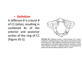
C1 Fracture
(Jefferson Fracture)
Dr. Dibyendunarayan Bid
• Definition
A Jefferson # is a burst #
of C1 (atlas), resulting in
combined #s of the
anterior and posterior
arches of the ring of C1
(Figure 35-1).
• Mechanism of Injury
• This # is caused by axial compression,
commonly from a fall on the head, such as
from diving into shallow water.
Radiographic Evaluation
• The standard trauma series for this cervical spine #
consists of anteroposterior, lateral, and open-mouth
(dens) views.
• Oblique radiographs help rule out associated #s in
the subaxial spine.
• The patient's head should not be passively moved.
• On the dens or odontoid view, lateral displacement
of the lateral articular masses of C1 relative to C2
suggests a Jefferson #.
• The # may not be evident on the lateral view, but soft
tissue swelling may be visualized anterior to the ring
of C1.
• The ring of the atlas fails in tension through both the
anterior and posterior arches.
• At least one # line is present through each arch.
• If the sum of displacement of the lateral masses, on
the odontoid view, is greater than 7 mm, this
strongly suggests a rupture of the transverse atlantal
ligament.
• Typically, the ligament fails by avulsion at its bony
insertion rather than as a mid-substance tear.
• A thin section computed tomography scan parallel to
the ring of the atlas best defines the # anatomy of a
C1 ring disruption as well as bony avulsion #s
associated with the transverse ligament.
Treatment Goals
• Orthopaedic Objectives
1. Obtain and maintain spinal alignment and
reduction of the #.
2. Provide spinal stability.
3. Prevent new neurologic deficits and attempt to
improve and prevent the exacerbation of existing
neurologic deficits.
4. Prevent future spinal deformity.
• Muscle Strength
– Restore and maintain the strength of paracervical
and cervical muscles, including the trapezius and the
upper extremity muscles.
– Restore the strength of the muscles of the lower
extremities that may atrophy from bed rest or
neurologic injury.
• Functional Goals
Develop cervical spinal flexibility for functional
independence.
• Expected Time of Bone Healing
Eight to 16 weeks before fusion is solid or # healing is
complete.
• Expected Duration of Rehabilitation
Three to 6 months.
Methods of Treatment
• Orthosis
– Hard collar (e.g., Philadelphia collar),
– Sternal-occiput-mandibular immobilization (SOMI) brace,
– cervicothoracic orthosis,
– four-poster brace, or halo vest (see Figures 8-7, 8-8, 8-9, 8-10, and 8-
11).
• Biomechanics: Stress-sharing device.
• Mode of bone healing: Secondary.
• Indication:
– A nondisplaced or minimally displaced # (less than 2
mm of combined displacement of the lateral masses
on the dens view) may be treated with a hard collar,
such as a Philadelphia collar, or one of the other rigid
nonhalo braces.
– A displaced # (2 to 7 mm of combined displacement
on the dens view) should first be reduced with halo
skeletal traction and then treated with a halo vest
once cervical muscle spasm has resolved.
– A # with a transverse ligament injury (greater than
7 mm of combined displacement of the lateral
masses on the dens view) should be treated with
4 weeks or more of halo skeletal traction followed
by halo vest.
– The length of skeletal traction before application
of the halo vest is still under debate.
Open Reduction and Posterior Spinal Fusion
• Biomechanics: Stress-sharing device until solid
arthrodesis occurs.
• Mode of bone healing: Secondary.
• Indications:
– Surgery is rarely necessary for isolated #s of the
ring of C1.
– Indications include failure to achieve or maintain
reduction in a halo vest, instability of the C1-2
motion segment, and failure of the # to reduce,
despite an adequate trial of skeletal traction.
– Operative treatment is also indicated if the
reduction is not maintained with halo vest
immobilization.
• Treatment generally consists of a posterior arthrodesis from
the occiput to C2 using various wiring techniques and
autogenous bone graft.
• Screw-plate fixations from the occiput to C2 may also be
used.
• An alternative technique that limits the fusion to the C1-2
joint complex and perhaps obviates the need for
postoperative halo immobilization is Magerl's C1-2
transarticular screw fixation.
Special Considerations of the Fracture
• Most Jefferson #s do not result in neurologic impairment
because the spinal canal is generally widened as a result of
the #, and there is sufficient space to accommodate the spinal
cord and associated swelling at the C1-2 level.
• The initial evaluation must include a thorough neurologic
examination and search for associated injuries.
• Halo traction should be used with the patient awake so that
the patient's neurologic status can be closely observed (see
page 557).
• Unstable injuries, in which greater than 7mm of
combined lateral mass widening has occurred, may
require a period of halo traction followed by halo
vest immobilization.
• The length of skeletal traction before application of
the halo vest is still under debate.
Associated Injuries
• Patients with Jefferson #s should be carefully
evaluated for head injuries as well as other
cervical spine #s, particularly #s of the upper
three cervical segments.
• In addition, injuries to the brachial plexus, thorax
and abdomen, and extremities may occur and
must be addressed in a timely fashion.
TREATMENT
LONG-TERM CONSIDERATIONS
• Residual pain may require temporary bracing and
anti-inflammatory medications.
• Residual neurologic deficits must be addressed and
may require extremity bracing, therapy, or surgery
for contracture releases and tendon transfers.
• If progressive cervical spine deformity occurs,
surgery may be necessary.
• There may be permanent loss of range of motion.
• The degree depends on the number of segments
arthrodesed and the level and extent of the fracture.
• Removal of the instrumentation may be necessary if
it is painful.
• If pseudoarthrosis occurs, reinstrumentation and
fusion may be necessary.
• Reflex sympathetic dystrophy may be a long-term
problem requiring cervical stellate ganglion blocks
and long-term physical therapy.
Reference
Cervical spine fractures dnbid 2020

Cervical spine fractures dnbid 2020

  • 1.
  • 2.
    • Definition A Jefferson# is a burst # of C1 (atlas), resulting in combined #s of the anterior and posterior arches of the ring of C1 (Figure 35-1).
  • 3.
    • Mechanism ofInjury • This # is caused by axial compression, commonly from a fall on the head, such as from diving into shallow water.
  • 4.
    Radiographic Evaluation • Thestandard trauma series for this cervical spine # consists of anteroposterior, lateral, and open-mouth (dens) views. • Oblique radiographs help rule out associated #s in the subaxial spine. • The patient's head should not be passively moved.
  • 5.
    • On thedens or odontoid view, lateral displacement of the lateral articular masses of C1 relative to C2 suggests a Jefferson #. • The # may not be evident on the lateral view, but soft tissue swelling may be visualized anterior to the ring of C1.
  • 6.
    • The ringof the atlas fails in tension through both the anterior and posterior arches. • At least one # line is present through each arch. • If the sum of displacement of the lateral masses, on the odontoid view, is greater than 7 mm, this strongly suggests a rupture of the transverse atlantal ligament.
  • 7.
    • Typically, theligament fails by avulsion at its bony insertion rather than as a mid-substance tear. • A thin section computed tomography scan parallel to the ring of the atlas best defines the # anatomy of a C1 ring disruption as well as bony avulsion #s associated with the transverse ligament.
  • 8.
    Treatment Goals • OrthopaedicObjectives 1. Obtain and maintain spinal alignment and reduction of the #. 2. Provide spinal stability. 3. Prevent new neurologic deficits and attempt to improve and prevent the exacerbation of existing neurologic deficits. 4. Prevent future spinal deformity.
  • 10.
    • Muscle Strength –Restore and maintain the strength of paracervical and cervical muscles, including the trapezius and the upper extremity muscles. – Restore the strength of the muscles of the lower extremities that may atrophy from bed rest or neurologic injury. • Functional Goals Develop cervical spinal flexibility for functional independence.
  • 11.
    • Expected Timeof Bone Healing Eight to 16 weeks before fusion is solid or # healing is complete. • Expected Duration of Rehabilitation Three to 6 months.
  • 12.
    Methods of Treatment •Orthosis – Hard collar (e.g., Philadelphia collar), – Sternal-occiput-mandibular immobilization (SOMI) brace, – cervicothoracic orthosis, – four-poster brace, or halo vest (see Figures 8-7, 8-8, 8-9, 8-10, and 8- 11). • Biomechanics: Stress-sharing device. • Mode of bone healing: Secondary.
  • 13.
    • Indication: – Anondisplaced or minimally displaced # (less than 2 mm of combined displacement of the lateral masses on the dens view) may be treated with a hard collar, such as a Philadelphia collar, or one of the other rigid nonhalo braces. – A displaced # (2 to 7 mm of combined displacement on the dens view) should first be reduced with halo skeletal traction and then treated with a halo vest once cervical muscle spasm has resolved.
  • 14.
    – A #with a transverse ligament injury (greater than 7 mm of combined displacement of the lateral masses on the dens view) should be treated with 4 weeks or more of halo skeletal traction followed by halo vest. – The length of skeletal traction before application of the halo vest is still under debate.
  • 15.
    Open Reduction andPosterior Spinal Fusion • Biomechanics: Stress-sharing device until solid arthrodesis occurs. • Mode of bone healing: Secondary.
  • 16.
    • Indications: – Surgeryis rarely necessary for isolated #s of the ring of C1. – Indications include failure to achieve or maintain reduction in a halo vest, instability of the C1-2 motion segment, and failure of the # to reduce, despite an adequate trial of skeletal traction. – Operative treatment is also indicated if the reduction is not maintained with halo vest immobilization.
  • 17.
    • Treatment generallyconsists of a posterior arthrodesis from the occiput to C2 using various wiring techniques and autogenous bone graft. • Screw-plate fixations from the occiput to C2 may also be used. • An alternative technique that limits the fusion to the C1-2 joint complex and perhaps obviates the need for postoperative halo immobilization is Magerl's C1-2 transarticular screw fixation.
  • 18.
    Special Considerations ofthe Fracture • Most Jefferson #s do not result in neurologic impairment because the spinal canal is generally widened as a result of the #, and there is sufficient space to accommodate the spinal cord and associated swelling at the C1-2 level. • The initial evaluation must include a thorough neurologic examination and search for associated injuries. • Halo traction should be used with the patient awake so that the patient's neurologic status can be closely observed (see page 557).
  • 19.
    • Unstable injuries,in which greater than 7mm of combined lateral mass widening has occurred, may require a period of halo traction followed by halo vest immobilization. • The length of skeletal traction before application of the halo vest is still under debate.
  • 20.
    Associated Injuries • Patientswith Jefferson #s should be carefully evaluated for head injuries as well as other cervical spine #s, particularly #s of the upper three cervical segments. • In addition, injuries to the brachial plexus, thorax and abdomen, and extremities may occur and must be addressed in a timely fashion.
  • 21.
  • 31.
    LONG-TERM CONSIDERATIONS • Residualpain may require temporary bracing and anti-inflammatory medications. • Residual neurologic deficits must be addressed and may require extremity bracing, therapy, or surgery for contracture releases and tendon transfers.
  • 32.
    • If progressivecervical spine deformity occurs, surgery may be necessary. • There may be permanent loss of range of motion. • The degree depends on the number of segments arthrodesed and the level and extent of the fracture.
  • 33.
    • Removal ofthe instrumentation may be necessary if it is painful. • If pseudoarthrosis occurs, reinstrumentation and fusion may be necessary. • Reflex sympathetic dystrophy may be a long-term problem requiring cervical stellate ganglion blocks and long-term physical therapy.
  • 34.