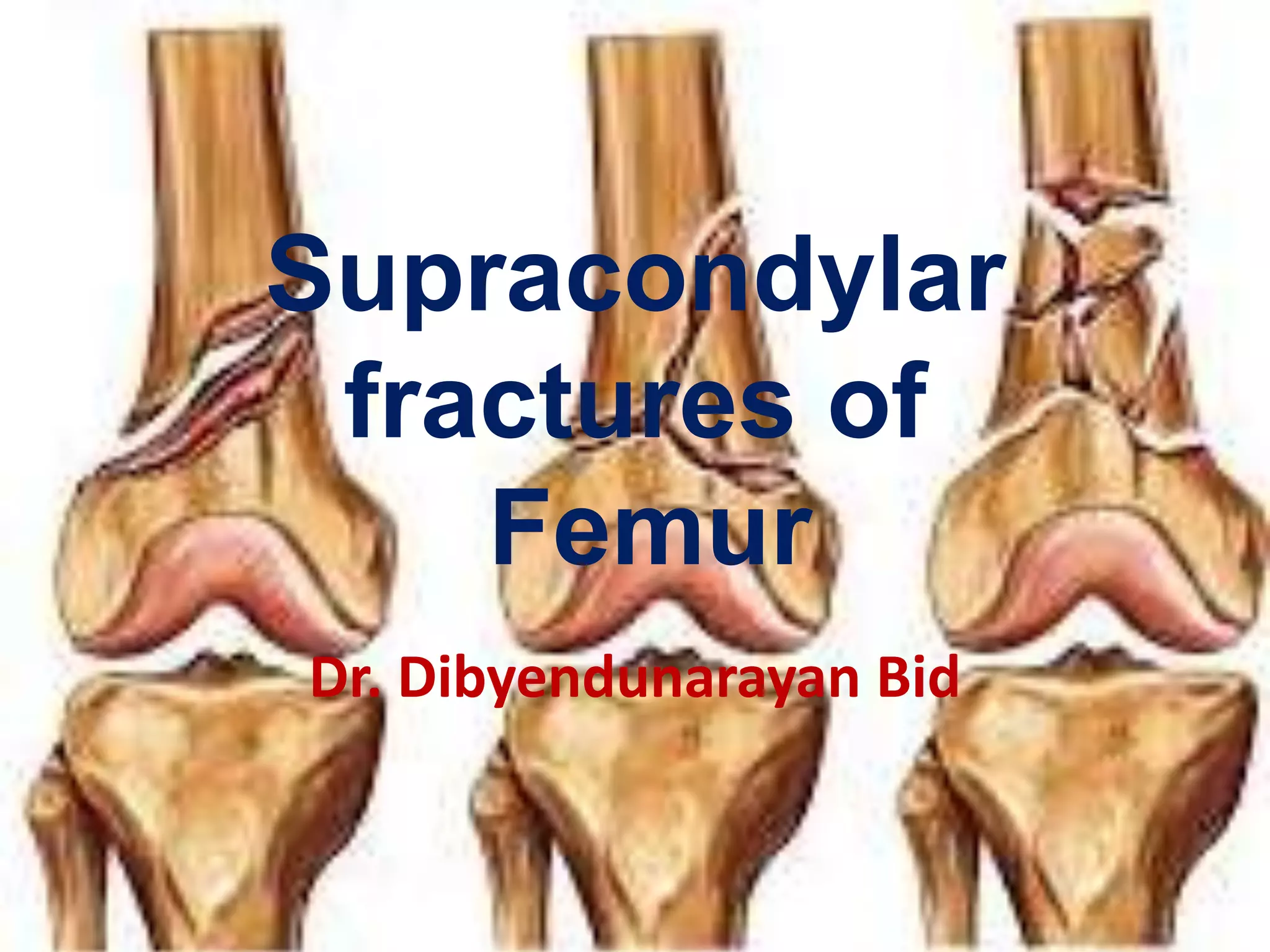
Supracondylar
fractures of
Femur
Dr. Dibyendunarayan Bid
Definition
• A supracondylar femur fracture involves the
distal aspect or metaphysis of the femur.
• This area includes the distal 8 to 15 cm of the
femur.
• The fracture frequently involves articular
surfaces.
12/4/2008 dnbid 2
• Complex classification systems have been
proposed for this fracture, all of which
attempt to define the amount of comminution
and the degree of displacement of the
fracture fragments.
12/4/2008 dnbid 3
• Muller's update of the AO classification system is
widely accepted.
• This involves dividing the fractures into extraarticular
(type A), unicondylar (type B), and bicondylar (type
C) fractures.
• These are then subdivided into types 1 through 3 in
each group.
• In progressing from type A to C, as well as subtypes 1 to 3, the
severity of the fracture increases and the prognosis for a good
result decreases (Figures 25-1, 25-2, 25-3, and 25-4).
•
12/4/2008 dnbid 4
12/4/2008 dnbid 5
12/4/2008 dnbid 6
Mechanism of Injury
• In younger patients, this fracture is usually secondary
to high-energy trauma, such as being struck by
an automobile.
• These cases commonly have other associated
injuries.
• In elderly patients, this fracture is usually secondary
to low-energy trauma, such as a simple fall.
• There are usually no other associated injuries in
these cases (Figure 25-5).
12/4/2008 dnbid 7
Treatment Goals
• Orthopaedic Objectives
Alignment
• Restore alignment by minimizing any residual
flexion/extension or varus/valgus angulation at the
fracture site.
• Any articular step-off must be less than 1 to 2 mm in
order to decrease or delay the risk of degenerative
changes and allow for functional range of motion
and normal gait.
12/4/2008 dnbid 8
Stability
• Stability is best achieved by restoring bony congruity
and using hardware to rigidly fix the fracture.
12/4/2008 dnbid 9
12/4/2008 dnbid 10
12/4/2008 dnbid 11
12/4/2008 dnbid 12
12/4/2008 dnbid 13
12/4/2008 dnbid 14
12/4/2008 dnbid 15
12/4/2008 dnbid 16
Casting or Traction
• Biomechanics: Stress-sharing device.
• Mode of Bone Healing: Secondary.
12/4/2008 dnbid 17
• Indication: Treatments involving traction or casting are
associated with a high risk of malunion, including varus,
valgus, and rotational deformities.
• Additionally, treatment with traction requires a prolonged
period of bed rest, with its associated risks of deep venous
thrombosis, bed sores, urinary tract infections, and
pulmonary compromise.
• This conservative treatment is indicated only for the
management of severely comminuted #s or for patients who
are at high medical risk for any operative management.
(Figures 25-10 and 25-11).
12/4/2008 dnbid 18
12/4/2008 dnbid 19
12/4/2008 dnbid 20
12/4/2008 dnbid 21
12/4/2008 dnbid 22
Gait Cycle
• Stance Phase
Stance phase, constituting 60% of the gait cycle, is most
affected by the fracture.
• Heel Strike
The quadriceps contracts concentrically to bring the
knee to full extension. However, a knee-flexion contracture
may exist. The patellar groove may be disrupted and the glide
mechanism of the patella in the groove may be affected,
causing pain. The extensor mechanism that surrounds the
knee joint may also be compromised. Occasionally the glide
mechanism of the quadriceps is also involved, thus limiting
terminal extension (see Figure 6-1).
12/4/2008 dnbid 23
• Foot-Flat
Foot-flat is usually not affected; however, the quadriceps
contracts eccentrically through elongation to keep the knee
under slight tension. Muscle contraction may cause some pain
(see Figure 6-2).
• Mid-Stance
Full weight passes through the fracture site at midstance,
because there is only single-limb support. Pain may be
present (see Figure 6-3).
• Push-Off
Push-off is usually not affected. The patella lies in the
trochlear groove at this phase and may cause pain (see Figure
6-4)12/4/2008 dnbid 24
• Swing Phase
• The swing phase constitutes 40% of the gait cycle.
• The quadriceps contract to cause acceleration of the
tibia on the femur.
• The quadriceps glide mechanism is necessary to
allow the knee to go to full extension before heel
strike.
• The quadriceps may not be able to contract enough
to achieve full extension (see Figures 6-6,6-7, and 6-8).
12/4/2008 dnbid 25
12/4/2008 dnbid 26
Orthopaedic and Rehabilitation
Considerations
• Physical Examination
• Initially, the most important aspect to check is the
patient's neurovascular status.
• Check pulses and capillary refill, as well as sensation
and active and passive range of motion of the
extremity.
• (Do not perform passive range of motion of the knee
unless absolutely rigid fixation has been achieved.)
12/4/2008 dnbid 27
• Check lower extremity compartments for softness
and monitor compartment pressures if there is any
suspicion of compartment syndrome.
• Additionally, check the wound for erythema or
discharge that might indicate infection.
• If edema is present, the patient should be instructed
to elevate the extremity properly.
12/4/2008 dnbid 28
• Dangers
Look for compartment syndrome, especially when
popliteal vessels are injured.
• Compartment syndrome is more common with high-
energy trauma and is usually noted in the first few
hours after injury.
12/4/2008 dnbid 29
• Radiography
• Check radiographs for varus, valgus, and rotational
alignment at the fracture site, as compared to the
unaffected knee.
• Also check for any displacement or loss of fixation.
• Because these fractures generally involve the
metaphyseal region, which has a good blood supply,
fracture healing usually occurs within the first 3
months after injury.
12/4/2008 dnbid 30
• Weight Bearing
The patient is non-weight bearing on the affected
extremity for 3 months.
12/4/2008 dnbid 31
• Range of Motion
• Gentle active ROM exercises are prescribed to the knee,
ankle, and hip.
• If fixation is not rigid, passive ROM exercises are avoided.
• If ankle edema is present, the ankle is elevated to decrease
swelling.
• Patients are encouraged to perform active ROM exercises at
the knee joint with the goal of full extension and 60 ° to 90 °
of knee flexion.
• Patients who are noncompliant or cannot follow orders are
placed in a hinged-knee brace or cast brace to allow for
protected ROM exercises.
12/4/2008 dnbid 32
• Muscle Strength
No strengthening exercises are prescribed at this
point to avoid risk of fracture displacement.
• Functional Activities
• The patient is instructed in stand/pivot transfers
using crutches or a walker, with no weight-bearing on
the affected extremity.
• The patient dons pants with the affected extremity first and
doffs them from the unaffected extremity first to decrease
stress on the fracture site.
12/4/2008 dnbid 33
• Gait
• The patient is instructed in a two-point gait using crutches or
a walker with no weight bearing on the affected extremity
(placing the crutches first and then hopping to the crutches on level
surfaces. See Figure 6-16).
• The patient ascends stairs with the uninvolved extremity first,
followed by the fractured extremity and the crutches, and
descends stairs with the crutches first, followed by the
fractured extremity first and then the uninvolved extremity
(see Figures 6-20,6-21,6-22, 6-23, 6-24, and 6-25).
12/4/2008 dnbid 34
12/4/2008 dnbid 35
12/4/2008 dnbid 36
12/4/2008 dnbid 37
12/4/2008 dnbid 38
12/4/2008 dnbid 39
12/4/2008 dnbid 40
12/4/2008 dnbid 41
12/4/2008 dnbid 42
12/4/2008 dnbid 43
12/4/2008 dnbid 44
12/4/2008 dnbid 45
12/4/2008 dnbid 46
12/4/2008 dnbid 47
Long-Term Considerations and Problems
• At every stage of treatment, radiographs should be
obtained to evaluate for loss of reduction, because a
malunion may lead to shortening or rotational
deformities and may hasten the onset of
degenerative joint disease of the knee.
12/4/2008 dnbid 48
• Additionally, a loss of motion of the knee is common.
• Therefore, every attempt should be made to obtain
rigid fixation, thus allowing the patient to begin
range-of-motion exercises early in treatment.
• Patients are not permitted to participate in sports,
repetitive pounding activities, jogging, or jumping for
6 months from the time of injury.
12/4/2008 dnbid 49
Reference
12/4/2008 dnbid 50
12/4/2008 dnbid 51
12/4/2008 dnbid 52

Femur supracondylar fractures

  • 1.
  • 2.
    Definition • A supracondylarfemur fracture involves the distal aspect or metaphysis of the femur. • This area includes the distal 8 to 15 cm of the femur. • The fracture frequently involves articular surfaces. 12/4/2008 dnbid 2
  • 3.
    • Complex classificationsystems have been proposed for this fracture, all of which attempt to define the amount of comminution and the degree of displacement of the fracture fragments. 12/4/2008 dnbid 3
  • 4.
    • Muller's updateof the AO classification system is widely accepted. • This involves dividing the fractures into extraarticular (type A), unicondylar (type B), and bicondylar (type C) fractures. • These are then subdivided into types 1 through 3 in each group. • In progressing from type A to C, as well as subtypes 1 to 3, the severity of the fracture increases and the prognosis for a good result decreases (Figures 25-1, 25-2, 25-3, and 25-4). • 12/4/2008 dnbid 4
  • 5.
  • 6.
  • 7.
    Mechanism of Injury •In younger patients, this fracture is usually secondary to high-energy trauma, such as being struck by an automobile. • These cases commonly have other associated injuries. • In elderly patients, this fracture is usually secondary to low-energy trauma, such as a simple fall. • There are usually no other associated injuries in these cases (Figure 25-5). 12/4/2008 dnbid 7
  • 8.
    Treatment Goals • OrthopaedicObjectives Alignment • Restore alignment by minimizing any residual flexion/extension or varus/valgus angulation at the fracture site. • Any articular step-off must be less than 1 to 2 mm in order to decrease or delay the risk of degenerative changes and allow for functional range of motion and normal gait. 12/4/2008 dnbid 8
  • 9.
    Stability • Stability isbest achieved by restoring bony congruity and using hardware to rigidly fix the fracture. 12/4/2008 dnbid 9
  • 10.
  • 11.
  • 12.
  • 13.
  • 14.
  • 15.
  • 16.
  • 17.
    Casting or Traction •Biomechanics: Stress-sharing device. • Mode of Bone Healing: Secondary. 12/4/2008 dnbid 17
  • 18.
    • Indication: Treatmentsinvolving traction or casting are associated with a high risk of malunion, including varus, valgus, and rotational deformities. • Additionally, treatment with traction requires a prolonged period of bed rest, with its associated risks of deep venous thrombosis, bed sores, urinary tract infections, and pulmonary compromise. • This conservative treatment is indicated only for the management of severely comminuted #s or for patients who are at high medical risk for any operative management. (Figures 25-10 and 25-11). 12/4/2008 dnbid 18
  • 19.
  • 20.
  • 21.
  • 22.
  • 23.
    Gait Cycle • StancePhase Stance phase, constituting 60% of the gait cycle, is most affected by the fracture. • Heel Strike The quadriceps contracts concentrically to bring the knee to full extension. However, a knee-flexion contracture may exist. The patellar groove may be disrupted and the glide mechanism of the patella in the groove may be affected, causing pain. The extensor mechanism that surrounds the knee joint may also be compromised. Occasionally the glide mechanism of the quadriceps is also involved, thus limiting terminal extension (see Figure 6-1). 12/4/2008 dnbid 23
  • 24.
    • Foot-Flat Foot-flat isusually not affected; however, the quadriceps contracts eccentrically through elongation to keep the knee under slight tension. Muscle contraction may cause some pain (see Figure 6-2). • Mid-Stance Full weight passes through the fracture site at midstance, because there is only single-limb support. Pain may be present (see Figure 6-3). • Push-Off Push-off is usually not affected. The patella lies in the trochlear groove at this phase and may cause pain (see Figure 6-4)12/4/2008 dnbid 24
  • 25.
    • Swing Phase •The swing phase constitutes 40% of the gait cycle. • The quadriceps contract to cause acceleration of the tibia on the femur. • The quadriceps glide mechanism is necessary to allow the knee to go to full extension before heel strike. • The quadriceps may not be able to contract enough to achieve full extension (see Figures 6-6,6-7, and 6-8). 12/4/2008 dnbid 25
  • 26.
  • 27.
    Orthopaedic and Rehabilitation Considerations •Physical Examination • Initially, the most important aspect to check is the patient's neurovascular status. • Check pulses and capillary refill, as well as sensation and active and passive range of motion of the extremity. • (Do not perform passive range of motion of the knee unless absolutely rigid fixation has been achieved.) 12/4/2008 dnbid 27
  • 28.
    • Check lowerextremity compartments for softness and monitor compartment pressures if there is any suspicion of compartment syndrome. • Additionally, check the wound for erythema or discharge that might indicate infection. • If edema is present, the patient should be instructed to elevate the extremity properly. 12/4/2008 dnbid 28
  • 29.
    • Dangers Look forcompartment syndrome, especially when popliteal vessels are injured. • Compartment syndrome is more common with high- energy trauma and is usually noted in the first few hours after injury. 12/4/2008 dnbid 29
  • 30.
    • Radiography • Checkradiographs for varus, valgus, and rotational alignment at the fracture site, as compared to the unaffected knee. • Also check for any displacement or loss of fixation. • Because these fractures generally involve the metaphyseal region, which has a good blood supply, fracture healing usually occurs within the first 3 months after injury. 12/4/2008 dnbid 30
  • 31.
    • Weight Bearing Thepatient is non-weight bearing on the affected extremity for 3 months. 12/4/2008 dnbid 31
  • 32.
    • Range ofMotion • Gentle active ROM exercises are prescribed to the knee, ankle, and hip. • If fixation is not rigid, passive ROM exercises are avoided. • If ankle edema is present, the ankle is elevated to decrease swelling. • Patients are encouraged to perform active ROM exercises at the knee joint with the goal of full extension and 60 ° to 90 ° of knee flexion. • Patients who are noncompliant or cannot follow orders are placed in a hinged-knee brace or cast brace to allow for protected ROM exercises. 12/4/2008 dnbid 32
  • 33.
    • Muscle Strength Nostrengthening exercises are prescribed at this point to avoid risk of fracture displacement. • Functional Activities • The patient is instructed in stand/pivot transfers using crutches or a walker, with no weight-bearing on the affected extremity. • The patient dons pants with the affected extremity first and doffs them from the unaffected extremity first to decrease stress on the fracture site. 12/4/2008 dnbid 33
  • 34.
    • Gait • Thepatient is instructed in a two-point gait using crutches or a walker with no weight bearing on the affected extremity (placing the crutches first and then hopping to the crutches on level surfaces. See Figure 6-16). • The patient ascends stairs with the uninvolved extremity first, followed by the fractured extremity and the crutches, and descends stairs with the crutches first, followed by the fractured extremity first and then the uninvolved extremity (see Figures 6-20,6-21,6-22, 6-23, 6-24, and 6-25). 12/4/2008 dnbid 34
  • 35.
  • 36.
  • 37.
  • 38.
  • 39.
  • 40.
  • 41.
  • 42.
  • 43.
  • 44.
  • 45.
  • 46.
  • 47.
  • 48.
    Long-Term Considerations andProblems • At every stage of treatment, radiographs should be obtained to evaluate for loss of reduction, because a malunion may lead to shortening or rotational deformities and may hasten the onset of degenerative joint disease of the knee. 12/4/2008 dnbid 48
  • 49.
    • Additionally, aloss of motion of the knee is common. • Therefore, every attempt should be made to obtain rigid fixation, thus allowing the patient to begin range-of-motion exercises early in treatment. • Patients are not permitted to participate in sports, repetitive pounding activities, jogging, or jumping for 6 months from the time of injury. 12/4/2008 dnbid 49
  • 50.
  • 51.
  • 52.