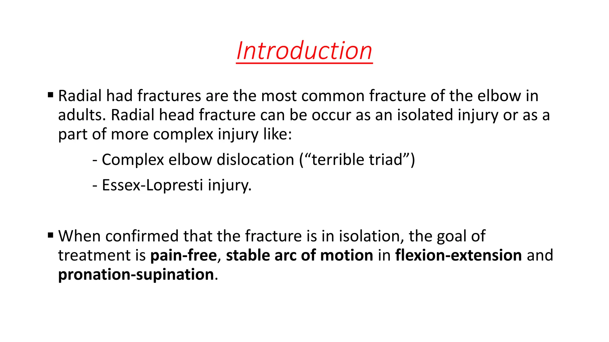
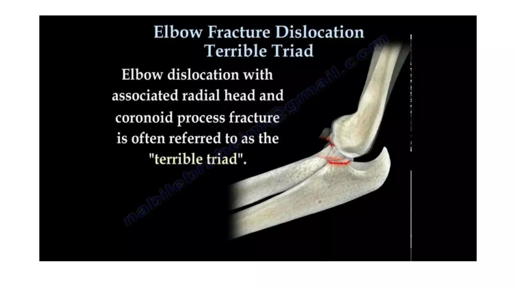
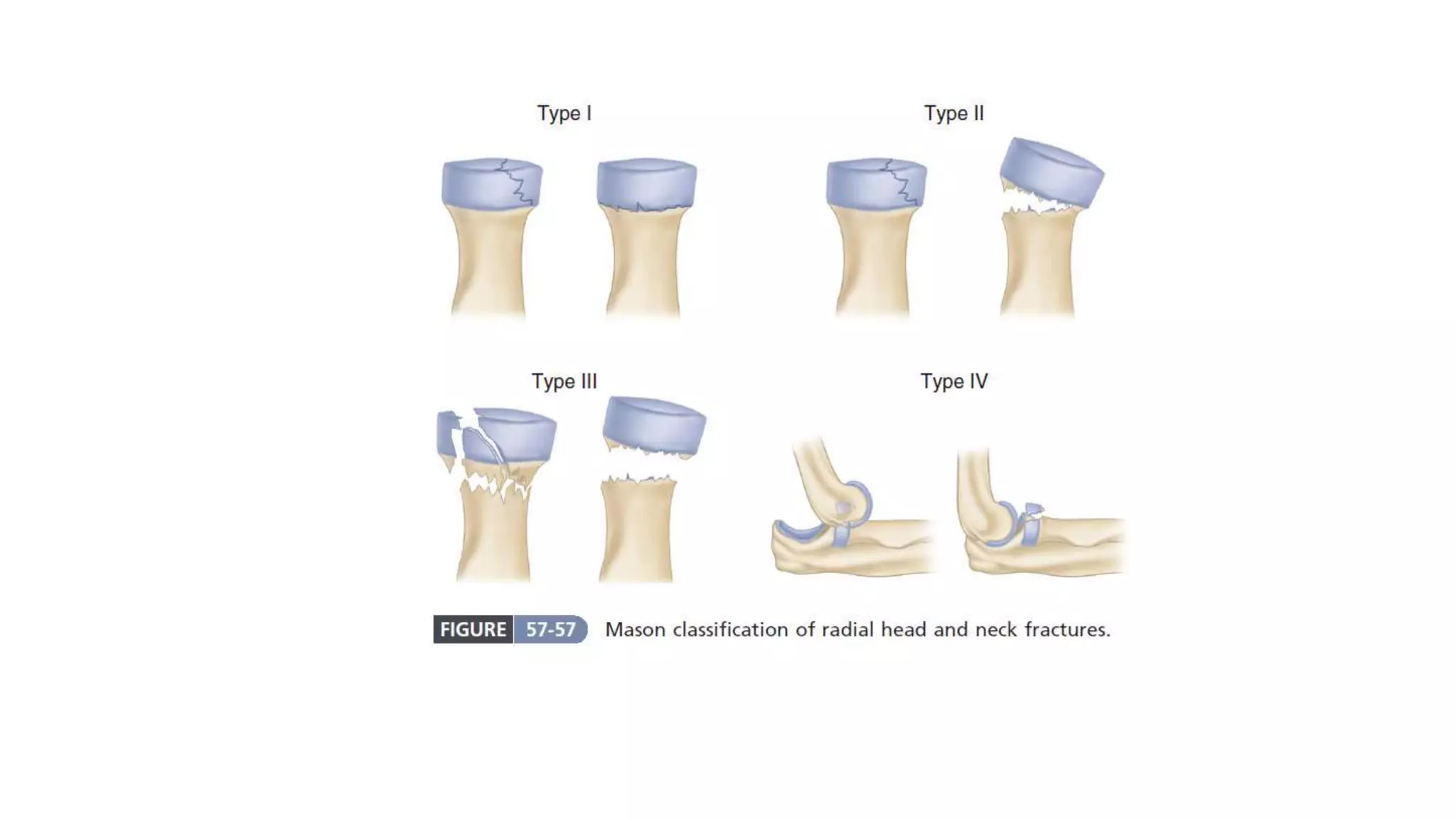
Radial Head Fracture
Dr. Ashiqur Rahman
Resident Orthopedics
Dhaka Medical college Hospital
Introduction
 Radial had fractures are the most common fracture of the elbow in
adults. Radial head fracture can be occur as an isolated injury or as a
part of more complex injury like:
- Complex elbow dislocation (“terrible triad”)
- Essex-Lopresti injury.
 When confirmed that the fracture is in isolation, the goal of
treatment is pain-free, stable arc of motion in flexion-extension and
pronation-supination.
 Mechanism of injury:
- Fall on the elbow
- Fall on an outstretched hand with the elbow extended.
 Associated Injuries with Radial Head Fractures:
- Tears of the LCLs and/or MCLs
- Dislocations of the elbow
- Fractures of the coronoid, capitellum, olecranon, and proximal
ulna
- Rupture of the interosseous membrane
Three types of fracture are identified and classified by Mason:
(i) Type I – Undisplaced partial articular fracture of the radial
head.
(ii) Type II – displaced (>2 mm) partial articular fracture of the
radial head
(iii) Type III – comminuted radial head fracture.
Additional type has been proposed:
(iv)Type IV: Radial Head Fracture with elbow dislocation
 CT or MRI may be indicated to better understand the injury; the
associated lateral ligament avulsion
Clinical features:
-The Dx is often missed, but tenderness on pressure over the
radial head.
- And pain on pronation & supination should suggest the
diagnosis.
- A firm block on forearm rotation should not be ignored.
Applied anatomy relating to radial head #
- The radial head consists of a concave dish which articulates
with the capitellum and a flattened articular margin which
articulates with the lesser sigmoid (radial) notch of the ulna.
- A ‘safe zone’ for placement of a plate on the non-articular
margin of the proximal radius has been defined.
- ‘Safe zone’ best identified during surgery by positioning the
forearm in neutral rotation and placing the plate 10-degree
anterior to the mid-axial line.
 The radial head is not circular but
is somewhat elliptical in shape.
 Furthermore, the radiocapitellar
dish is also elliptical and typically
offset from the neck of the
radius.
TREATMENT OF MASON TYPE I #
 Undisplaced partial articular
fractures (or head splits) have a
good prognosis with non-surgical
management
 pain relief and mobilization as
comfort allows.
 Most will regain a pain-free
elbow with only a slight loss of
extension the most common
sequel.
Treatment of Mason type-II #
 Reduce the partial fracture, taking
care not to disrupt the periosteum;
tamps, dental picks, or Freer
elevators can be used as needed.
 Stabilize the reduction with one or
two small screws.
 Occasionally, a buttress plate can
be useful if the apex of the fracture
is comminuted and a large defect
remains under the articular
segment.
Treatment of Mason type-III #
 If needed for improved exposure, release the origin of the lateral
collateral ligament; this will be repaired at the end of the procedure.
 Reduce and provisionally fix the articular surface with Kirschner wires.
 Occasionally, removing the fragments and assembling them on the
back table may facilitate reduction.
 Protect the posterior interosseous nerve by pronating the forearm.
 Apply a small plate along the lateral surface of the proximal radius
with the wrist in neutral (safe zone) and secure it with lag screws as
needed.
 Bone graft the defect if needed.
 Check pronation and supination of the forearm.
Post operative care
- The arm is placed in a molded.
- Posterior plaster splint at 90 degrees.
- At 3 to 7 days, the splint is removed and the arm
is supported in a sling.
- At about that time, active and active-assisted exercises
are begun.
- The patient should discontinue the sling at 3 weeks.
- Gradually increasing the exercises as tolerated.
- Forceful manipulation of the elbow is never permitted.

Radial head fracture

  • 1.
    Radial Head Fracture Dr.Ashiqur Rahman Resident Orthopedics Dhaka Medical college Hospital
  • 2.
    Introduction  Radial hadfractures are the most common fracture of the elbow in adults. Radial head fracture can be occur as an isolated injury or as a part of more complex injury like: - Complex elbow dislocation (“terrible triad”) - Essex-Lopresti injury.  When confirmed that the fracture is in isolation, the goal of treatment is pain-free, stable arc of motion in flexion-extension and pronation-supination.
  • 5.
     Mechanism ofinjury: - Fall on the elbow - Fall on an outstretched hand with the elbow extended.  Associated Injuries with Radial Head Fractures: - Tears of the LCLs and/or MCLs - Dislocations of the elbow - Fractures of the coronoid, capitellum, olecranon, and proximal ulna - Rupture of the interosseous membrane
  • 6.
    Three types offracture are identified and classified by Mason: (i) Type I – Undisplaced partial articular fracture of the radial head. (ii) Type II – displaced (>2 mm) partial articular fracture of the radial head (iii) Type III – comminuted radial head fracture. Additional type has been proposed: (iv)Type IV: Radial Head Fracture with elbow dislocation  CT or MRI may be indicated to better understand the injury; the associated lateral ligament avulsion
  • 8.
    Clinical features: -The Dxis often missed, but tenderness on pressure over the radial head. - And pain on pronation & supination should suggest the diagnosis. - A firm block on forearm rotation should not be ignored.
  • 9.
    Applied anatomy relatingto radial head # - The radial head consists of a concave dish which articulates with the capitellum and a flattened articular margin which articulates with the lesser sigmoid (radial) notch of the ulna. - A ‘safe zone’ for placement of a plate on the non-articular margin of the proximal radius has been defined. - ‘Safe zone’ best identified during surgery by positioning the forearm in neutral rotation and placing the plate 10-degree anterior to the mid-axial line.
  • 10.
     The radialhead is not circular but is somewhat elliptical in shape.  Furthermore, the radiocapitellar dish is also elliptical and typically offset from the neck of the radius.
  • 11.
    TREATMENT OF MASONTYPE I #  Undisplaced partial articular fractures (or head splits) have a good prognosis with non-surgical management  pain relief and mobilization as comfort allows.  Most will regain a pain-free elbow with only a slight loss of extension the most common sequel.
  • 12.
    Treatment of Masontype-II #  Reduce the partial fracture, taking care not to disrupt the periosteum; tamps, dental picks, or Freer elevators can be used as needed.  Stabilize the reduction with one or two small screws.  Occasionally, a buttress plate can be useful if the apex of the fracture is comminuted and a large defect remains under the articular segment.
  • 13.
    Treatment of Masontype-III #  If needed for improved exposure, release the origin of the lateral collateral ligament; this will be repaired at the end of the procedure.  Reduce and provisionally fix the articular surface with Kirschner wires.  Occasionally, removing the fragments and assembling them on the back table may facilitate reduction.  Protect the posterior interosseous nerve by pronating the forearm.
  • 14.
     Apply asmall plate along the lateral surface of the proximal radius with the wrist in neutral (safe zone) and secure it with lag screws as needed.  Bone graft the defect if needed.  Check pronation and supination of the forearm.
  • 15.
    Post operative care -The arm is placed in a molded. - Posterior plaster splint at 90 degrees. - At 3 to 7 days, the splint is removed and the arm is supported in a sling. - At about that time, active and active-assisted exercises are begun. - The patient should discontinue the sling at 3 weeks. - Gradually increasing the exercises as tolerated. - Forceful manipulation of the elbow is never permitted.