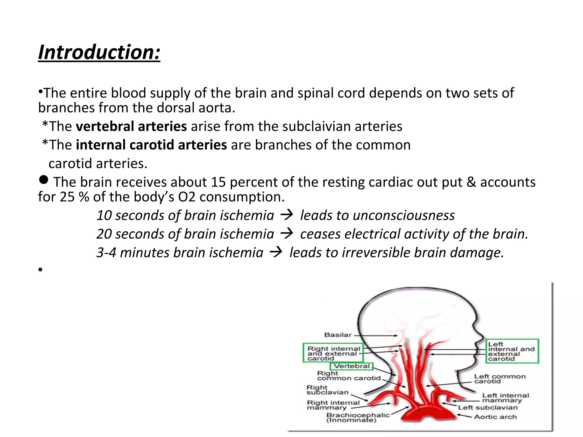
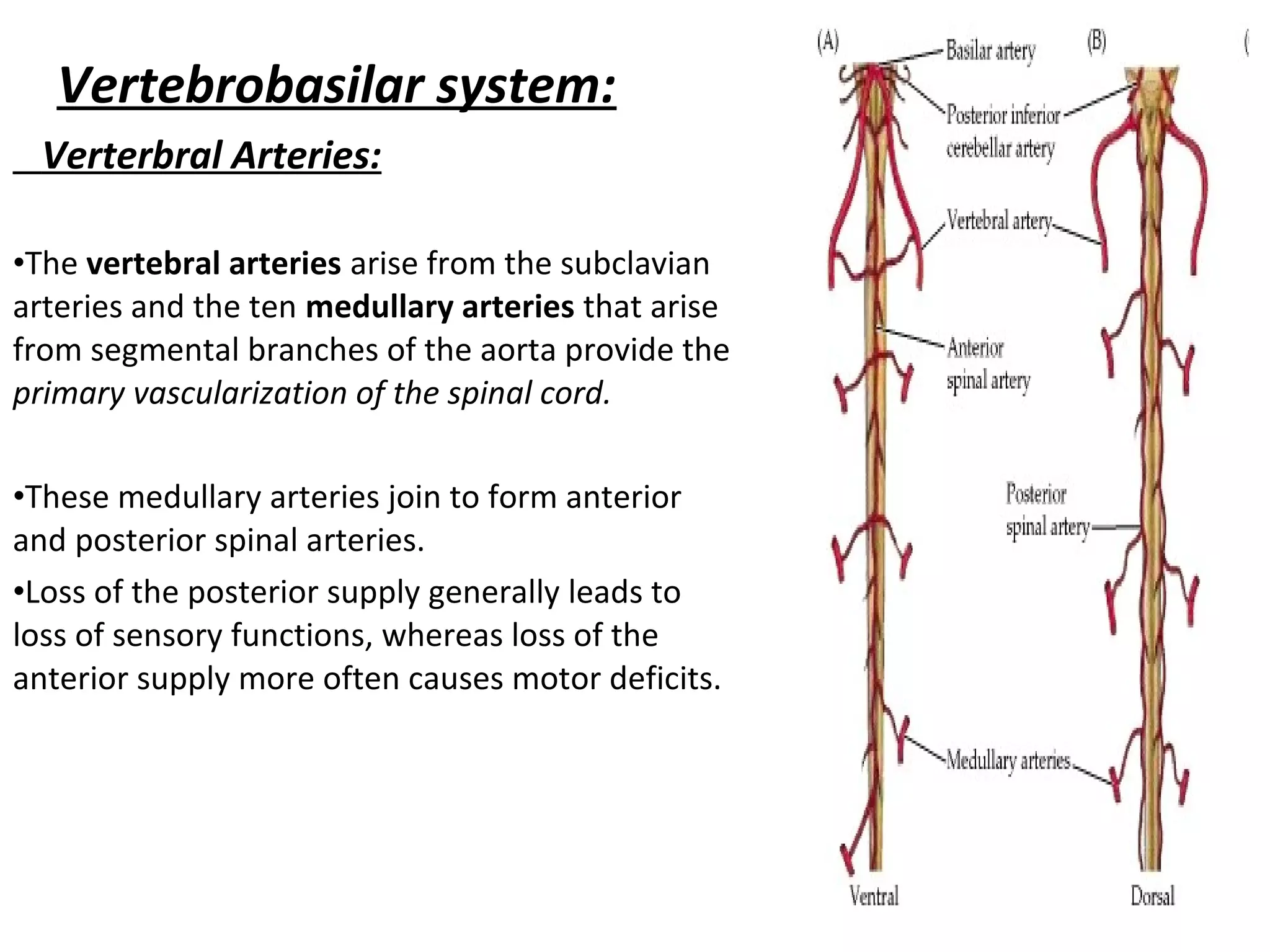
The document summarizes the arterial blood supply and venous drainage of the brain. It discusses the two main sources of arterial blood - the internal carotid and vertebral arteries. It describes the branches of these arteries and their territories. It also discusses the clinical consequences of occlusions in different arteries. The circle of Willis and venous drainage routes are also summarized.
Pyramidal tract by Sunita.M.Tiwale,Prof. Dept of physiology,D.Y.Patil Medical...Physiology Dept
Specific Learning Objectives:
At the end of session the students should be able to :
Enumerate the descending tracts.
Describe the origin, course, termination, collaterals of Pyramidal tract.
Describe the functions of the pyramidal tract.
here i am to explain the Anatomy and physiology of part of the Pyramidal tract, that is the corticospinal tract. I also added the clinical significance of corticospinal tract. The course of the corticospinal tract are well explained.
Largest part of hind brain.
Called “ silent area/Little Brain ”
Weight- 150 gms.
Cerebellar cortex is a large folded sheet, each fold is called Folium.
Connected to brain stem by 3 pairs of peduncles- Superior (Brachium conjunctiva), Middle (Brachium Pontis) & Inferior (Restiform body) peduncle.
cerebrum, sulci and gyri of cerebrum, lobes of cerebrum, frontal lobe , parietal lobe, temporal lobe and occipital lobe, sulci and gyri presnet in each lobes, and the functional areas , of cerebrum, brodmann areas of cerebrum, borders and surfaces of cerebrum, insula,
This lecture is all about the recognition of an abnormal EEG, its characteristics, its appearance and all about how to differentiate the abnormal activity with normal EEG background.
The detail description about peripheral nervous system, neuron, its covering, types of neuron, synapses, spinal nerves, plexus, and more about cranial nerves at last not the least about somatic and autonomic nervous system. you may also find the information about types of peripheral nervous system in detail.
Flu Vaccine Alert in Bangalore Karnatakaaddon Scans
As flu season approaches, health officials in Bangalore, Karnataka, are urging residents to get their flu vaccinations. The seasonal flu, while common, can lead to severe health complications, particularly for vulnerable populations such as young children, the elderly, and those with underlying health conditions.
Dr. Vidisha Kumari, a leading epidemiologist in Bangalore, emphasizes the importance of getting vaccinated. "The flu vaccine is our best defense against the influenza virus. It not only protects individuals but also helps prevent the spread of the virus in our communities," he says.
This year, the flu season is expected to coincide with a potential increase in other respiratory illnesses. The Karnataka Health Department has launched an awareness campaign highlighting the significance of flu vaccinations. They have set up multiple vaccination centers across Bangalore, making it convenient for residents to receive their shots.
To encourage widespread vaccination, the government is also collaborating with local schools, workplaces, and community centers to facilitate vaccination drives. Special attention is being given to ensuring that the vaccine is accessible to all, including marginalized communities who may have limited access to healthcare.
Residents are reminded that the flu vaccine is safe and effective. Common side effects are mild and may include soreness at the injection site, mild fever, or muscle aches. These side effects are generally short-lived and far less severe than the flu itself.
Healthcare providers are also stressing the importance of continuing COVID-19 precautions. Wearing masks, practicing good hand hygiene, and maintaining social distancing are still crucial, especially in crowded places.
Protect yourself and your loved ones by getting vaccinated. Together, we can help keep Bangalore healthy and safe this flu season. For more information on vaccination centers and schedules, residents can visit the Karnataka Health Department’s official website or follow their social media pages.
Stay informed, stay safe, and get your flu shot today!
TEST BANK for Operations Management, 14th Edition by William J. Stevenson, Ve...kevinkariuki227
TEST BANK for Operations Management, 14th Edition by William J. Stevenson, Verified Chapters 1 - 19, Complete Newest Version.pdf
Recomendações da OMS sobre cuidados maternos e neonatais para uma experiência pós-natal positiva.
Em consonância com os ODS – Objetivos do Desenvolvimento Sustentável e a Estratégia Global para a Saúde das Mulheres, Crianças e Adolescentes, e aplicando uma abordagem baseada nos direitos humanos, os esforços de cuidados pós-natais devem expandir-se para além da cobertura e da simples sobrevivência, de modo a incluir cuidados de qualidade.
Estas diretrizes visam melhorar a qualidade dos cuidados pós-natais essenciais e de rotina prestados às mulheres e aos recém-nascidos, com o objetivo final de melhorar a saúde e o bem-estar materno e neonatal.
Uma “experiência pós-natal positiva” é um resultado importante para todas as mulheres que dão à luz e para os seus recém-nascidos, estabelecendo as bases para a melhoria da saúde e do bem-estar a curto e longo prazo. Uma experiência pós-natal positiva é definida como aquela em que as mulheres, pessoas que gestam, os recém-nascidos, os casais, os pais, os cuidadores e as famílias recebem informação consistente, garantia e apoio de profissionais de saúde motivados; e onde um sistema de saúde flexível e com recursos reconheça as necessidades das mulheres e dos bebês e respeite o seu contexto cultural.
Estas diretrizes consolidadas apresentam algumas recomendações novas e já bem fundamentadas sobre cuidados pós-natais de rotina para mulheres e neonatos que recebem cuidados no pós-parto em unidades de saúde ou na comunidade, independentemente dos recursos disponíveis.
É fornecido um conjunto abrangente de recomendações para cuidados durante o período puerperal, com ênfase nos cuidados essenciais que todas as mulheres e recém-nascidos devem receber, e com a devida atenção à qualidade dos cuidados; isto é, a entrega e a experiência do cuidado recebido. Estas diretrizes atualizam e ampliam as recomendações da OMS de 2014 sobre cuidados pós-natais da mãe e do recém-nascido e complementam as atuais diretrizes da OMS sobre a gestão de complicações pós-natais.
O estabelecimento da amamentação e o manejo das principais intercorrências é contemplada.
Recomendamos muito.
Vamos discutir essas recomendações no nosso curso de pós-graduação em Aleitamento no Instituto Ciclos.
Esta publicação só está disponível em inglês até o momento.
Prof. Marcus Renato de Carvalho
www.agostodourado.com
Ethanol (CH3CH2OH), or beverage alcohol, is a two-carbon alcohol
that is rapidly distributed in the body and brain. Ethanol alters many
neurochemical systems and has rewarding and addictive properties. It
is the oldest recreational drug and likely contributes to more morbidity,
mortality, and public health costs than all illicit drugs combined. The
5th edition of the Diagnostic and Statistical Manual of Mental Disorders
(DSM-5) integrates alcohol abuse and alcohol dependence into a single
disorder called alcohol use disorder (AUD), with mild, moderate,
and severe subclassifications (American Psychiatric Association, 2013).
In the DSM-5, all types of substance abuse and dependence have been
combined into a single substance use disorder (SUD) on a continuum
from mild to severe. A diagnosis of AUD requires that at least two of
the 11 DSM-5 behaviors be present within a 12-month period (mild
AUD: 2–3 criteria; moderate AUD: 4–5 criteria; severe AUD: 6–11 criteria).
The four main behavioral effects of AUD are impaired control over
drinking, negative social consequences, risky use, and altered physiological
effects (tolerance, withdrawal). This chapter presents an overview
of the prevalence and harmful consequences of AUD in the U.S.,
the systemic nature of the disease, neurocircuitry and stages of AUD,
comorbidities, fetal alcohol spectrum disorders, genetic risk factors, and
pharmacotherapies for AUD.
Basavarajeeyam is an important text for ayurvedic physician belonging to andhra pradehs. It is a popular compendium in various parts of our country as well as in andhra pradesh. The content of the text was presented in sanskrit and telugu language (Bilingual). One of the most famous book in ayurvedic pharmaceutics and therapeutics. This book contains 25 chapters called as prakaranas. Many rasaoushadis were explained, pioneer of dhatu druti, nadi pareeksha, mutra pareeksha etc. Belongs to the period of 15-16 century. New diseases like upadamsha, phiranga rogas are explained.
micro teaching on communication m.sc nursing.pdfAnurag Sharma
Microteaching is a unique model of practice teaching. It is a viable instrument for the. desired change in the teaching behavior or the behavior potential which, in specified types of real. classroom situations, tends to facilitate the achievement of specified types of objectives.
1. BLOOD suppLy TO THE
BRAIN
Shehzad Hussain
Technologist
07/08-11-2017
2. Introduction:
•The entire blood supply of the brain and spinal cord depends on two sets of
branches from the dorsal aorta.
*The vertebral arteries arise from the subclaivian arteries
*The internal carotid arteries are branches of the common
carotid arteries.
The brain receives about 15 percent of the resting cardiac out put & accounts
for 25 % of the body’s O2 consumption.
10 seconds of brain ischemia leads to unconsciousness
20 seconds of brain ischemia ceases electrical activity of the brain.
3-4 minutes brain ischemia leads to irreversible brain damage.
3.
4. ARTERIAL SUPPLY TO THE BRAIN:
• The arterial blood reaches the brain through
the pair of;
1-internal carotid arteries.{80% supply of
telencephalon & diencephalon}
2-vertebral arteries.{20% supply to the
brainstem & cerebellum alongwith some
cortical regions}
5. The internal carotid arteries:
It arise at the point in the neck
(foramen lacerum) and enters into cavernous
sinus where the common carotid arteries
bifurcate into external & internal carotid
arteries.
•External carotid artery supplies blood to the
facial muscles.
•Internal carotid artery enters the cavernous
sinus through the foramen lacerum enters the
subarachnoid space by piercing the arachnoid
mater and lies lateral to the optic chiasma.
6. Various Branches of Internal Carotid
Artery:
• The internal carotid arteries branch to form two
major cerebral arteries, the anterior and middle
cerebral arteries
• As it leaves the cavernous sinus it gives rise to
the ophthalmic artery & bifurcates into anterior
& middle cerebral arteries
• It also gives rise to
• a) anterior coroidal artery
• b) posterior communicating artery.
9. 1) Ophthalmic artery:
It passes into the orbit through the optic foramen. It supplies
the structures of the orbit, frontal part of the scalp and dorsum
of the nose.
2) Anterior choroidal artery:
It supplies the optic tract, choroid plexus of the lateral ventricle,
hippocampus and some of the deep structures of the
hemisphere, including the internal capsule.
3) Posterior communicating artery:
It passes posteriorly inferior to the optic tract & joins the
posteior cerebral artery with the middle cerebral artery.
10. • 4) Anterior cerebral artery:
It passes medially above the optic nerve and then passes into
the great longitudinal fissure between the frontal lobes
where it joins the corresponding vessels of the opposite side
by anterior communicating artery.
• It ramify the medial surface of the frontal and parietal lobes
and supplies them. Also, its branches extend out of the great
longitudinal fissure to supply a narrow lateral band of frontal
and parietal cortices i.e. 2-2.5 cm laterally.
• The territory supplied by it includes the motor and sensory
cortices for the lower limb.
11.
12.
13. • Anterior coroidal artery occlusion:
• 1) amnesia
• 2) Alzheimer’s disease
• 3) mid temporal lobe epilepsy
Middle cerebral artery occlusion:
1) Contra lateral hemi paresis
2) hemi sensory loss involving mainly the face and the arm ( precentral and post
central gyri)
3) Aphasia.
4) Contra lateral homonymous heminopia (damage to the optic radiation)
14.
15. Vertebrobasilar system:
Verterbral Arteries:
•The vertebral arteries arise from the subclavian
arteries and the ten medullary arteries that arise
from segmental branches of the aorta provide the
primary vascularization of the spinal cord.
•These medullary arteries join to form anterior
and posterior spinal arteries.
•Loss of the posterior supply generally leads to
loss of sensory functions, whereas loss of the
anterior supply more often causes motor deficits.
16.
17. • These arteries enter the cranial cavity through
the foramen magnum.
• Each of it gives rise to the 3 main branches ;
• 1) the posterior spinal artery
• 2) anterior spinal artery
• 3) posterior inferior cerebral artery (supplied
post. & infero aspect of cerebellum).
18. • Basilar artery:
• Anterior & posterior vertebral arteries unite at the junction
between medulla and pons to form the basilar artery.
It gives rise to the following branches;
• 1- Anterior inferior cerebral artery.
• 2- superior cerebral artery
• 3- pontine arteries
• 4- labyrinthine arteries
• 5- posterior cerebral artery
*Imp. The brain stem, cerebellum and occipital lobe are
supplied by the vertebrobasilar system
19.
20. Branches of Basilar Artery:
1.Pontine arteries: It supplies pons.
2.Anterior inferior cerebellar artery:
It supplies the anterior and inferior portion of the cerebellum.
3. Labyrinthine artery
It supplies the inner ear. Its occlusion leads to vertigo & ipsilateral deafness.
4. Superior cerebellar artery
It supplies the superior aspect of the cerebellum.
5. Posterior cerebral artery:
It curves around the midbrain to supply the visual cortex of the occipital lobe and the
infer-o-medial aspect of the temporal lobe.
21. Anterior cerebral artery occlusion
•If occlusion is distal to ACA, the collateral circulation is usually
adequate to preserve the circulation.
•If proximal to the ACA then may produce the following signs and
symptoms.
1) Contra lateral hemi paresis
2) Hemi sensory loss mainly involving the legs and foots.
3) Inability to identify objects correctly
4) personality changes.(frontal and parietal lobes)
22. • Middle cerebral artery:
• It is the largest branch of the 3 cerebral arteries. It passes
laterally to enter the lateral fissure within which it
subdivides.
• Its branches supply the whole of the lateral surface of the
frontal, parietal and temporal lobes except those areas which
are supplied by the ACA.
• It supplies the primary motor and sensory cortices for the
whole body excluding the lower limb. The auditory cortex
and the insula in the depth of the lateral fissure.
• It gives rise to a branch called lenticulostriate arteries which
supply diencephalon & telencephalon.
24. Circle of Willis
• It is an arterial circle at the base of the brain in the
interpeduncular fossa.
• It is formed by anterior and middle cerebral branches of the
internal carotid artery & posterior cerebral branches of
basilar artery.
• The two anterior cerebral arteries are connected by anterior
communicating artery.
• The middle and posterior communicating artery of same side
are united by posterior communicating artery.
25.
26. Venous Drainage of the Brain
It includes a External and Internal cerebral veins
•External cerebral veins:
1- Superior cerebral veins that drain superolateral surface of cerebral hemisphere
terminate into superior sagittal veins.
2- Superficial middle cerebral vein drains area around lateral sulcus they terminate into
cavernous sinus.
•Internal cerebral vein
1- Great cerebral veins : It is formed by union of two internal cerebral veins. It terminates
in the straight sinus drains the deep
structures of the forebrain.
2- Basal veins : It is formed by union of deep middle cerebral vein and anterior cerebral
vein and terminates by joining great cerebral vein.
27. 3- Dural venous sinuses:
•The Dural venous sinuses are connected to extra cranial veins
via emissary (representative) veins.
•Cerebral damage caused by venous infarction manifests as
epileptic attacks and focal paralysis of the limbs.