Cerebral Cortex
Dr. E.Muralinath, Dr.C. Kalyan Chakravathi Dr. M.
Guruprasad, Dr. K. Sravani Pragna, Dr. P. Manjari,
Dr. D. Kusumalatha, Dr. K. Sridevi , Dr. Ch. Ramya
Sudha and R. Gnana Lahari
2.
• INTRODUCTION
• Cerebralcortex is also termed as pallidum and it contains two hemispheres.
• Surface area of cerebral cortex in human beings is 2.2 sq m.
• Both the cerebral hemispheres are separated by a deep vertical fissure (deep furrow or
groove).
• The separation is completed on an anterior side and posterior side also.
•
3.
• HISTOLOGY OFCEREBRAL CORTEX
• LAYERS OF CEREBRAL CORTEX
• Cerebral cortex contains gray matter that encircles the deeper white matter.
• It is formed by different types of nerve cells along with their processes and neuroglia.
• It is not uniform throughout. It is thickest, i.e. 4.5 cm at the precentral gyrus and thinnest at frontal and occipital poles. :
• 1. Molecular or Plexiform Layer
• Molecular layer consists of few small fusiform cells. It also consists of dendrites or axons from cells of deeper layers.
• 2. External Granular Layer
• External granular layer has large number of closely packed small cells, which are round, polygonal or triangular in shape. Dendrites of these cells pass
into molecular layer. Axons terminate in the deeper layers. Some axons gain an entry into white substance of the hemisphere.
• 3. Outer Pyramidal Layer
• The formation of outer pyramidal layer happens by pyramidal cells, which are of two sizes. Medium sized pyramidal cells are in the outer portion and
larger pyramidal cells are in
• deeper portion. This part forms the major portion of cerebral cortex. Part of the cerebral cortex that has all six layers of structures is also termed as
neocortex
• 4. Internal Granular Layer
• Like external granular layer, this layer also consists of closely packed smaller cells, which are stellate type. But, the nerve fibers are more in this
layer compare to external granular layer. This layer consists of many horizontal fibers, which look like a white strip termed as outer
• strip.
4.
• 5. GanglionicLayer or Internal Pyramidal Layer
• Ganglionic layer or internal pyramidal layer contains pyramidal cells of graded sizes. It is well developed particularly in the
precentral (motor) cortex. Pyramidal cells in this region are otherwise called Betz cells or giant cells.
• 6. Fusiform Cell Layer
• Fusiform cell layer is in contact particularly with white matter of cerebral hemisphere. It consists of closely packed small
spindle-shaped cells.
• PARTS OF CEREBRAL CORTEX
• Cerebral cortex is categorized into two parts dependent upon phylogeny (evolutionary development of a species):
• 1. Neocortex
• 2. Allocortex.
• 1. Neocortex
• Neocortex is the phylogenetically new structure of cerebral cortex. It is also otherwise known as isocortex or neopallium.
gyri (singular = gyrus). Sulcus is a slight depression or groove and gyrus is a raised ridge.
• . 2. Allocortex
• Allocortex is the phylogenetically oldest structure of cerebral cortex. It contains less than six layers of structures. It is
categorized into two divisions namely, archicortex and paleocortex, which form the parts of limbic system
5.
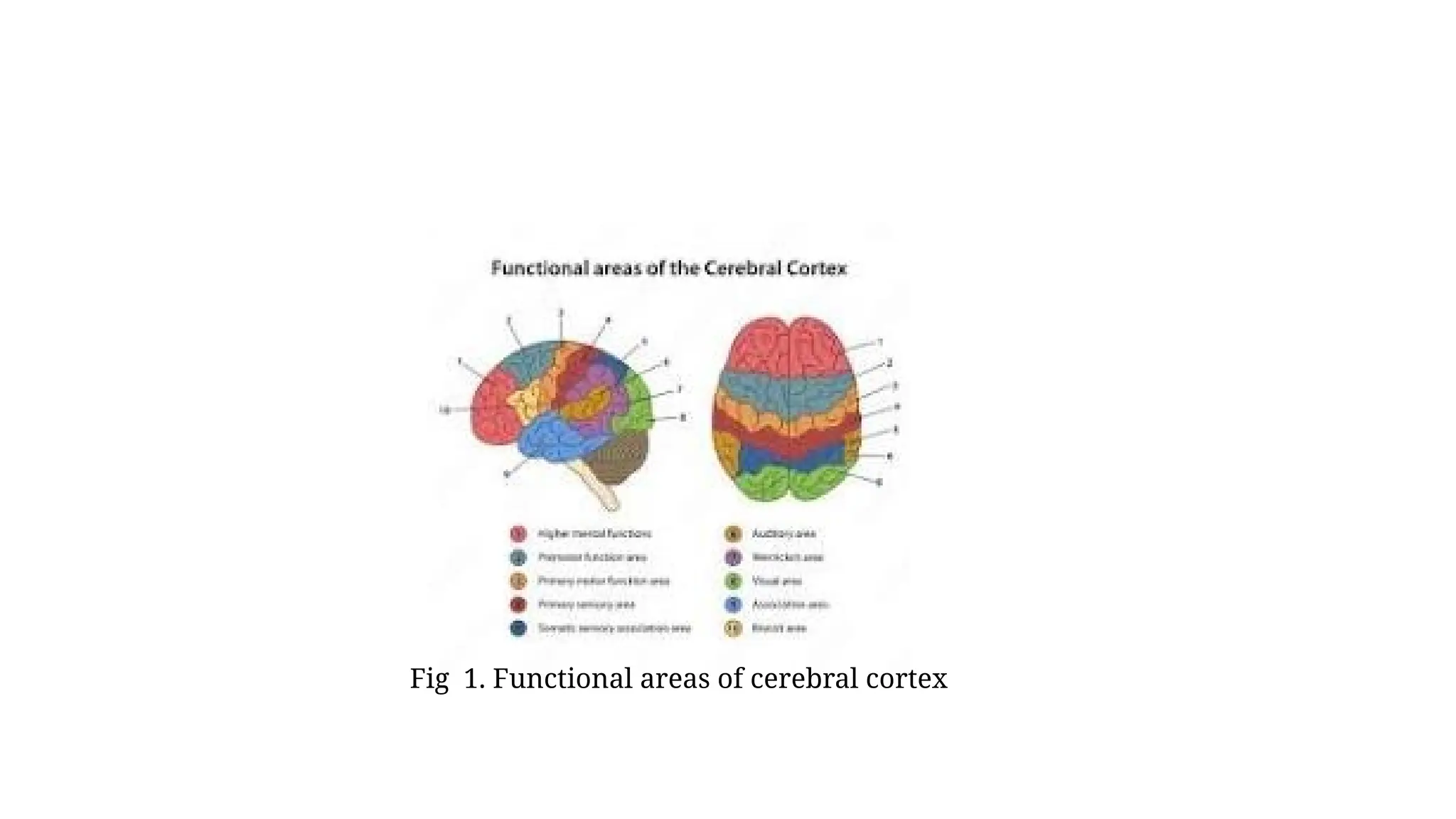
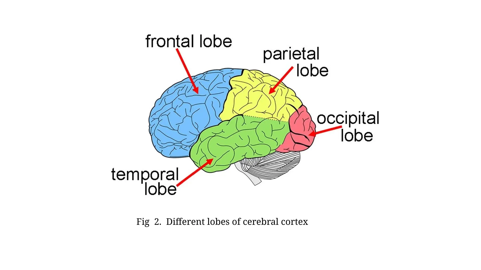
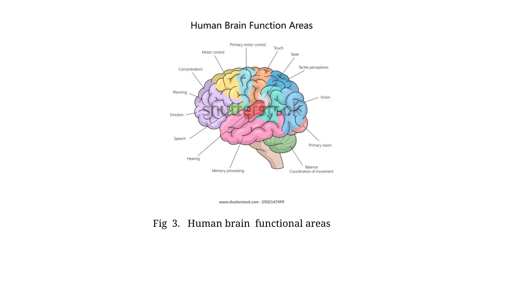
• LOBES OFCEREBRAL CORTEX
• In each hemisphere, there are three surfaces lateral, medial and inferior surfaces. Neocortex of
each cerebral hemisphere consists of four lobes .:
• 1. Frontal lobe
• 2. Parietal lobe
• 3. Occipital lobe
• 4. Temporal lobe.
• The demarcation of lobes of each hemisphere happens by four main fissures and sulci:
• 1. Central sulcus or Rolandic fissure between frontal and parietal lobes
• 2. Parieto-occipital sulcus between parietal and occipital lobe
• 3. Sylvian fissure or lateral sulcus between parietal and temporal lobes
• 4. Callosomarginal fissure between temporal lobe and limbic area.
6.
• CEREBRAL DOMINANCE
•Cerebral dominance is elucidated as the dominance of one cerebral hemisphere over the other in the control of
cerebral functions. Both the cerebral hemispheres are not functionally equivalent. Some functional asymmetries are
well known.
• CEREBRAL DOMINANCE AND HANDEDNESS
• Cerebral dominance is related to handedness, i.e. preference of the individual to use right or left hand. More than
90% of people are right handed.
• In these individuals, the left hemisphere is dominant and it regulates the analytical process and language related
functions namely speech, reading and writing.
• 3)Right hemisphere is otherwise known as representational hemisphere since it is related to artistic and
visuospatial functions namely judging the distance, detecting the direction, recognizing the tones, etc.
• 4) Lesion in dominant hemisphere results in language disorders.
• 5)Lesion in representational hemisphere leads to the occurrence of only mild effects namely astereognosis.
• 6)Left hemisphere is the dominant hemisphere in about 75% of the right-handed persons. In the remaining left-
handed persons, right hemisphere regulates the language function. Some of these persons do not have dominant
hemisphere.
7.
• BRODMANN AREAS
•Brodmann area is a region of cerebral cortex explained on the basis of its cytoarchitecture. b)Cytoarchitecture means organization of cells. Brodmann areas were
originally defined and numbered in 1909 by Korbinian Brodmann based on the laminar organization
• of neurons in the cortex.
• c)Some of these areas were given specific names based on their functions.
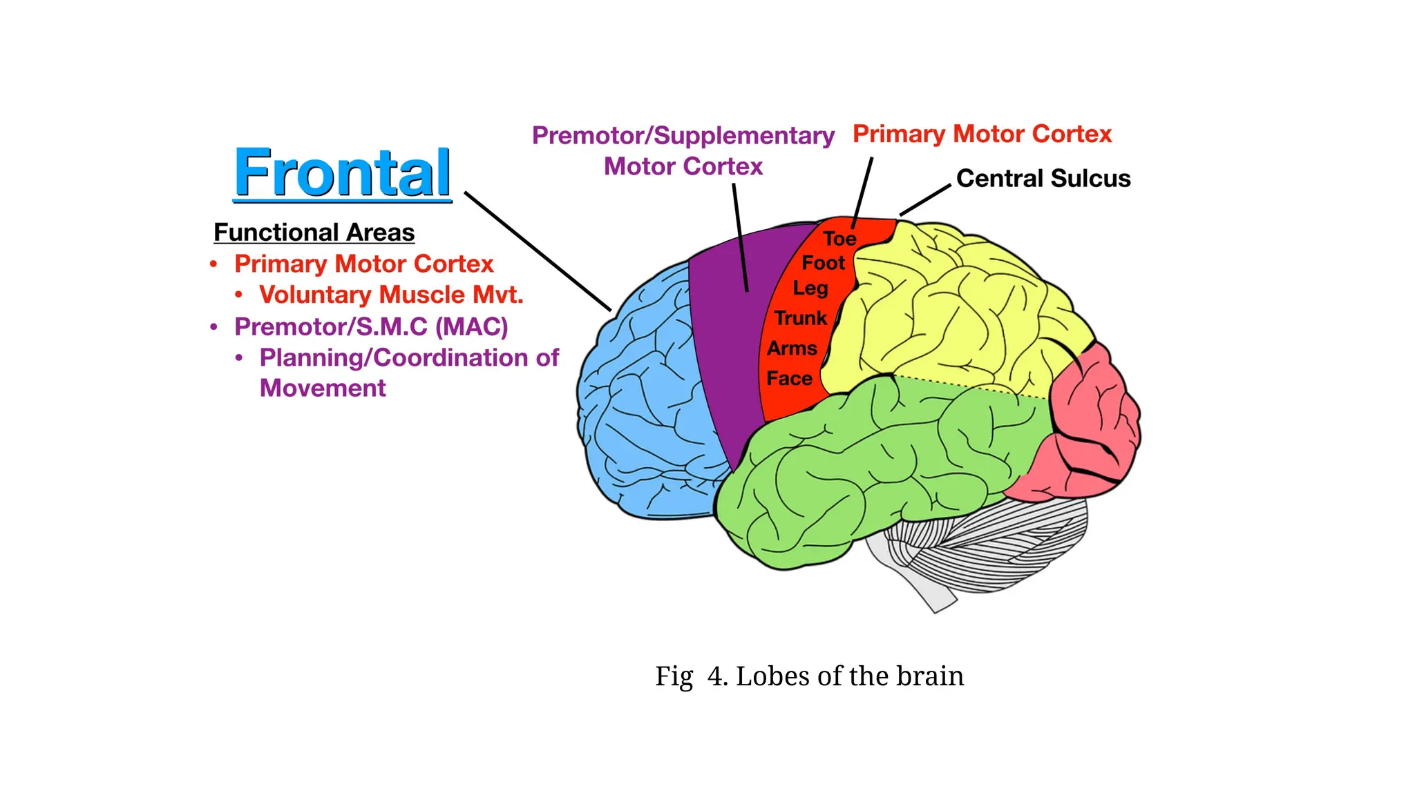
• FRONTAL LOBE OF CEREBRAL CORTEX
• Frontal lobe forms one third of the cortical surface. It expands from frontal pole to the central sulcus and restricted below by the lateral sulcus. Frontal lobe of cerebral
cortex is categorized into two parts:
• A. Precentral cortex, which is located posteriorly
• B. Prefrontal cortex, which is located anteriorly.
• PRECENTRAL CORTEX
• Precentral cortex forms the posterior part of frontal lobe.
• It includes the lip of central sulcus, whole of precentral gyrus and posterior portions of superior, middle and inferior frontal gyri.
• It also expands to the medial surface.
• This part of cerebral cortex is also known as excitomotor cortex or area, since the activation of different points in this area causes activity of discrete skeletal muscle.
• Precentral cortex is further categorized into three functional areas :
• 1. Primary motor area
• 2. Premotor area
• 3. Supplementary motor area.
•
8.
• 1. PrimaryMotor Area
• Primary motor area expands throughout the precentral gyrus and the adjoining lip of central sulcus. Areas 4 and 4S are observed here.
• Structure of primary motor area
• Even though this area has all the six layers, the granular layer is thin. Special structural feature of this layer is the presence of giant pyramidal cells termed as Betz cells in
• ganglionic layer.
•
• Connections of primary motor area
• Efferent connections
• i. Fibers of pyramidal tracts arise from the Betz cells. These fibers synapse along with motor neurons in anterior gray horn of opposite side (few fibers attain the same side motor neurons)
in spinal cord
• ii. Frontopontine fibers from this area attain pontine nuclei of same side
• iii. Fibers are also projected to corpus striatum, red nucleus, thalamus, subthalamus and reticular formation
• iv. Association fibers connect the primary motor area to other areas of cortex.
• Afferent connections
• Primary motor area receives fibers from dentate nucleus (cerebellum) via red nucleus and thalamus.
• Functions of primary motor area
• Primary motor area is associated with initiation of voluntary movements and speech.
•
• Area 4
• It is a tapering strip of area located in precentral gyrus of frontal lobe. Broad end lies superiorly at the upper border of hemisphere and most of the efferent fibers of
• primary motor area arise from this area .
•
9.
• Function ofarea 4
• Area 4 is the center for movement, as it sends all efferent (corticospinal) fibers of primary motor
area. Through the fibers of corticospinal tracts, area 4 stimulates the lower motor neurons
particularly in the spinal cord. It stimulates both α-motor neurons and γ-motor neurons
simultaneously by the process called coactivation
• .
• Stimulation of α-motor neurons results in contraction of extrafusal fibers of the muscles.
Stimulation of γ-motor neurons causes contraction of intrafusal fibers lresulting in an
enhancemernt of muscle tone.
•
• Effect of stimulation of area 4
• Electrical activation of area 4 causes discrete isolated movements in the opposite side of the
body. The groups of muscles or single isolated muscle may be activated based on the area
stimulated.
10.
• Localization –homunculus
• Muscles of various parts of the body are represented in area 4 in an inverted way from medial to
lateral surface.
• Lower parts of body are represented in medial surface and upper parts of the body are represented
in the lateral surface.
• Order of representation from medial to lateral surface: Toes, ankle, knee, hip, trunk, shoulder, arm,
elbow, wrist, hand fingers and face.
• Whatever it may be, parts of the face are not represented in inverted manner.
• Area 4 is associated with contraction of discrete muscles. It sends motor signals to the facial
muscles of both sides (bilateral) and the other muscles of the opposite side (contralateral).
•
11.
• Effect oflesion of area 4
• Effect of lesion or ablation of area 4 differs in different species.
• In cats, the ability to walk is not impacted.
• In monkeys, there is contralateral flaccid paralysis, hypotonia and loss of reflexes.
• Myotatic reflexes reappear in a short time
• In man, the symptoms are severe compare to monkeys. In unilateral lesion, paralysis happens in contralateral side.
• Complete paralysis is rare. If both sides are affected, the effect is more severe.
• Recovery happens very slowly. During recovery, upper parts of body recover first.
• If area 4 is affected along with area 6, the effect is very severe, causing hemiplegia with spastic paralysis.
• j)Hemiplegia means the paralysis in one half of the body. In spastic paralysis, the muscles undergo spastic
contraction because of an enhanced muscle tone.
• Area 4S
• Area 4S is known as suppressor area. It forms a narrow strip anterior to area 4. It scrutinizes and suppresses the
extra impulses produced by area 4 and stops exaggeration of movements.
12.
• 2. PremotorArea
• Premotor area includes areas 6, 8, 44 and 45. The premotor area is anterior to primary motor area in the precentral cortex.
The premotor area is related to control of postural movements by sending motor signals to axial muscles (muscles near
the midline of the body).
• Structure of premotor area
• Premotor area is similar to primary motor area in structure except for the absence of giant pyramidal cells particularly in
ganglionic layer.
• Area 6
• Area 6 is in the posterior portions of superior, middle and inferior frontal gyri. It is subcategorized into 6a and 6b. It gives
origin to some of the pyramidal tract fibers. The
• other connections are similar to those of area 4.
• Functions of area 6
• Area 6 has two functions:
• i. It is associated with coordination of movements initiated by area 4. It assists in making the skilled movements more
accurate and smooth.
• ii. It is believed to be the cortical center particularly for extrapyramidal system.
13.
• Effect ofstimulation of area 6
• Electrical activation of area 6a in human being results in the same effects as the activation of area 4. Whatever it may be,
the stimulus must be stronger to cause response from
• area 6. The effects of stimulation of this area are:
• i. Activation of area 6a leads to generalized pattern of movements such as rotation of head, eyes and trunk
towards the opposite side
• ii. Activation of 6b leads to the occurrence of rhythmic, complex coordinated movements involving the muscles of
face, buccal cavity, larynx as well as pharyn
• Effect of lesion of area 6
• Lesion or removal of area 6 in monkeys results in loss of skilled movements. After the lesion, the recovery may happen;
but the movements become awkward. It also produces grasping reflexes. Lesion involving areas 6 and area 4 causes
severe symptoms of hemiplegia along
• with spastic paralysis.
• Area 8
• Area 8 is otherwise known as frontal eye field. It lies anterior to area 6 in the precentral cortex. It is associated with
movements of eyeball. This area receives afferent fibers from dorsomedial nucleus of thalamus and occipital lobe.
14.
• Function ofarea 8
• Frontal eye field is associated with conjugate movement of eyeballs . This area causes voluntary
scanning movements of eyeballs and it is independent of visual stimuli. It is also responsible for opening and
closing of eyelids, pupillary dilatation and
• lacrimation.
•
• Effect of activation of area 8
• An activation of this area leads to the occurrence of conjugate movements of eyeballs to the opposite side.
•
• Effect of lesion of area 8
• Lesion of this area turns the eyes to the affected side. Conjugate movements of eyes are lost. Whatever it
may be, pupils and eyelids are not affected. In animals, while walking, circular movements happen towards
the affected side.
•
15.
• Broca area
•Broca area is the motor area for speech.
• It includes areas 44 and 45. Broca area is observed in left hemisphere (dominant hemisphere) of right-
handed persons and in the right hemisphere of left-handed persons.
• t is a special region of premotor cortex located in inferior frontal gyrus.
• Area 44 is located in pars triangularis and 45 in pars opercularis of this gyrus.
•
• Function of Broca area
• Broca area plays an important role regarding the movements of tongue, lips and larynx which are
involved in speech.
•
• Effect of lesion of Broca area Lesion in Broca area leads to aphasia .
•
16.
• 3. SupplementaryMotor Area
• a) Supplementary motor area is located in medial surface of frontal lobe rostral to primary motor area.
• b)Various motor movements are elicited because of electrical stimulation of this area namely raising the
contra lateral arm, turning the head and eye and movements of synergistic muscles of trunk and legs.
•
• Function of supplementary motor area
• Exact function of this area is not understood in a clear manner. It is suggested that it is associated with
coordinated skilled movements.
•
• Effect of lesion of supplementary motor area
• During lesion in this area of human being, the head and eyeballs turn towards the affected side. Destruction
of this area in monkeys causes weak grasping reflexes particularly in contralateral side and bilateral
hypertonia of shoulder muscles. But, paralysis is not
• observed.
17.
PREFRONTAL CORTEX ORORBITOFRONTAL CORTEX
• Prefrontal cortex is the anterior part of frontal lobe of cerebral cortex, in front of areas 8 and 44. It occupies the medial, lateral and inferior surfaces and
includes orbital gyri, medial frontal gyrus and the anterior portions of superior, middle and inferior frontal gyri.
• Areas present in prefrontal cortex are 9, 10, 11, 12, 13, 14, 23, 24, 29 and 32. Areas 12, 13, 14, 23, 24, 29 and 32 are in medial surface . Areas 9, 10
• and 11 are in lateral surface.
•
• Connections of Prefrontal Cortex
• Afferent fibers
• Afferent fibers of prefrontal cortex come from:
• 1. Dorsomedial nucleus of thalamus
• 2. Hypothalamus
• 3. Corpus striatum
• 4. Amygdala
• 5. Midbrain.
• Areas 23, 24, 29 and 32 attain fibers particularly from anterior nucleus of thalamus. Area 32 receives fibers from suppressor area of precentral cortex
also.
•
•
18.
• Efferent fibers
•Efferent fibers are projected to:
• 1. Thalamus
• 2. Hypothalamus
• 3. Tegmentum
• 4. Caudate nucleus
• 5. Pons
• 6. Temporal lobe of cerebral cortex.
• Area 13, along with hippocampus, uncus and amygdala sends fibers to mamillary body of
hypothalamus via fornix. This area is associated with emotional reactions.
19.
• Functions ofPrefrontal Cortex
• Earlier, this area was considered as inexcitable to electrical stimulation. Hence, it was known as the
silent area or association area. But, now it is known that the activation of this area along with low
voltage electrical stimulus leads to the occurrence of changes in the activity of digestive, cardiovascular,
respiratory and excretory systems and other autonomic functions. It also causes fear. Various functions of
prefrontal cortex are:
• 1. It forms the center for the higher functions such as emotion, learning, memory and social behavior.
Short-term memories are registered here.
• 2. It is the center for planned actions
• 3. This area is the seat of intelligence; so, it is also termed as the organ of mind
• 4. It is responsible for the personality of the individuals
• 5. autonomic changes
• Prefrontal cortex is responsible for the various during emotional conditions,
• Due to its connections along with hypothalamus and brainstem.
20.
• Effect ofLesion of Prefrontal Cortex
• Bilateral lesion or removal of prefrontal cortex in human beings does not cause paralysis.
• It leads to the occurrence of initiation and loss of mental alertness.
• Very little or no change happend in memory, judgment and intelligence.
•
• APPLIED PHYSIOLOGY – FRONTAL LOBE SYNDROME
•
• Injury or ablation of prefrontal cortex results in a condition termed as frontal lobe syndrome.
• Features of this syndrome are:
• 1. Emotional instability: There is lack of restraint, resulting in hostility, aggressiveness as well as restlessness
• 2.. Lack of concentration There is lack of initiation and lack of fixing attention and difficulty in planning any
course of action
• 3.. Impairment of recent memory happens. Whatever it may be, the memory of remote events is not lost.
21.
• 4.. Lossof moral and social sense is common and there is loss of love for family and friends
• 5.. There is failure to realize the seriousness of the condition. The subject has the sense of well-
being and also has flight of ideas.
• 6.. Apart from mental defects, there are some functional abnormalities also:
• i. Hyperphagia (increased food intake)
• ii. Loss of control particularly over sphincter of the urinary bladder or rectum
• iii. Disturbances in orientation
• iv. Slight tremor.
22.
• PARIETAL LOBE
•Parietal lobe expands from central sulcus and merges along with occipital lobe behind and temporal lobe below.
• This lobe is separated from occipital lobe by parieto-occipital sulcus and from temporal lobe by Sylvian sulcus. Parietal lobe is categorized into three functional areas:
• A. Somesthetic area I
• B. Somesthetic area II
• C. Somesthetic association area.
• In addition to these three areas, a part of sensory motor area is also located in parietal lobe .
• SOMESTHETIC AREA I
• Somesthetic area I is also known as somatosensory area I or primary somesthetic or primary sensory area.
• It is observed particularly in the posterior lip of central sulcus, in the postcentral gyrus and in the paracentral lobu
• Areas of Somesthetic Area I
• Somesthetic area I consists of three areas, which are known as areas 3, 1 and 2. Anterior part of this forms area 3 and posterior part forms areas 1 and 2.
•
• Connections of Somesthetic Area I
• Somesthetic area I attains sensory fibers from thalamus via parietal part of thalamic radiation.
• Localization – Homunculus
• Different sensory areas of the body are represented in postcentral gyrus (primary sensory area) in an inverted manner as in the motor area.
• Toes are represented in lowest part of medial surface, legs at the upper border of hemispheres, then from above downwards knee, thigh, hip, trunk, upper limb, neck
and face. C)Representation of face is not inverted. Representation of parts of face from above downwards is eyelids, nose, cheek, upper lip and lower lip .
23.
• Functions ofSomesthetic Area I
• 1. Somesthetic area I is responsible for perception and integration of cutaneous and kinesthetic sensations. It receives sensory impulses from
cutaneous receptors (touch, pressure, pain, temperature) and proprioceptors of opposite side through thalamic radiation. Area 1 is concerned with sensory
perception. Areas 3 and 2 are involved in the integration of
• these sensations.
• 2. This area sends sensory feedback to the premotor area
• 3. This area is also concerned with the movements of head and eyeballs
• 4. Discriminative functions: In addition to perception of cutaneous and kinesthetic sensation, this area is also responsible for recognizing the discriminative
features of sensations.
• Discriminative functions are:
• i. Spatial recognition: Tactile localization, two point discrimination and recognition of position
• and passive movements of limbs
• ii. Recognition of intensity of different stimuli
• iii. Recognition of similarities and differences between the stimuli.
• Effect of Stimulation of Somesthetic Area I
• Electrical stimulation of somesthetic area I produces vague sensations such as numbness and tingling.
• Effects of Lesion of Somesthetic Area I
• If thalamus also is influenced by lesion, there is loss of sensations in the opposite side of the body.
•
24.
• SOMESTHETIC AREAII
• Somesthetic area II is located in postcentral gyrus below the area of face of somesthetic area I. A
part of this is buried in Sylvian sulcus. It is also termed as secondary somesthetic area or
somatosensory area II.
• Functions of Somesthetic Area II
• Somesthetic area II gains sensory impulses from somesthetic area I and from thalamus in a direct
manner.
• Even though the exact role of this area is not clear, it is associated with perception of sensation.
• Thus, the sensory parts of body consist of two representations, in somesthetic area
• I and area II.
25.
• SOMESTHETIC ASSOCIATIONAREA
• Somesthetic association area is located posterior to postcentral gyrus, above the auditory cortex and in front of visual cortex. It
consists of two areas, 5 and 7.
•
• Functions of Somesthetic Association Area
• Somesthetic association area is associated with synthesis of various sensations perceived by somesthetic area I.
• Thus, the somesthetic association area forms the center for combined sensations namely stereognosis. Lesion of this area leads to
astereognosis.
•
• Sensory Motor Area
• Sensory area of cortex is not restricted to postcentral gyrus in parietal lobe.
• It expands anteriorly into motor area in precentral gyrus of frontal lobe.
• Similarly, the motor area is expanded from precentral gyrus posteriorly into postcentral gyrus.
• Thus, the precentral and postcentral gyri are knit together by association neurons and are
• inter-related on functional basis.
• e)So, this area is otherwise known as sensory motor area.
26.
• APPLIED PHYSIOLOGY
•Lesion or ablation of parietal lobe (sensory cortex) leads to the following disturbances:
• 1. Contralateral disturbance of cutaneous sensations
• 2. Disturbances in kinesthetic sensations
• 3. Loss of tactile localization as well as discrimination.
• TEMPORAL LOBE
• Temporal lobe of cerebral cortex consists of three functional areas :
• A. Primary auditory area
• B. Secondary auditory area or auditopsychic area
• C. Area for equilibrium.
27.
• PRIMARY AUDITORYAREA
• Primary auditory area includes:
• 1. Area 41
• 2. Area 42
• 3. Wernicke area.
• Areas 41 and 42 are located in anterior transverse gyrus and lateral surface of superior temporal gyrus. Wernicke area is in upper part
of superior temporal gyrus posterior to areas 41 and 42.
• Connections of Primary Auditory Area
• Afferent connections
• Primary auditory area receives afferent fibers from:
• 1. Medial geniculate body via auditory radiation
• 2. Pulvinar of thalamus.
• Efferent connections
• This area sends efferent fibers to:
• 1. Medial geniculate body
• 2. Pulvinar.
28.
• Functions ofPrimary Auditory Area
• Primary auditory area is associated with perception of auditory impulses, analysis of pitch and
detection of intensity and source of sound.
• Areas 41 and 42 are associated only with the perception of auditory sensation (sound). Wernicke
area is responsible for the interpretation of auditory sensation.
• It executes this function along with the help of secondary auditory area (area 22). d)Wernicke area
is also essential for understanding the auditory information regarding any word and sending the
information to Broca area .
•
29.
• SECONDARY AUDITORYAREA
• Secondary auditory area occupies the superior temporal gyrus.
• It is also termed as auditopsychic area or auditory association area.
• It includes area 22. This area is associated with interpretation of auditory sensation along with
Wernicke area.
• It is also related to the storage of memories of spoken words
•
• AREA FOR EQUILIBRIUM
• Area for equilibrium is in the posterior part of superior temporal gyrus.
• It is associated with the maintenance of equilibrium of the body.
• Inactivation of this area leads to dizziness, swaying, falling as well as feeling of rotation.
30.
• APPLIED PHYSIOLOGY– TEMPORAL LOBE SYNDROME
• Temporal lobe syndrome is otherwise termed as KluverBucy syndrome.
• It is observed in animals, especially monkeys after the bilateral ablation of temporal lobe along with
amygdala and uncus
• . It happens in human Beings particularly bilateral lesions of these structures.
• Manifestations of this syndrome are:
• 1. Aphasia (disturbance in speech).
• 2. Auditory disturbances like frequent attacks of tinnitus, auditory hallucinations with sounds
• like buzzing, ringing or humming. Tinnitus means noise in the ear. Hallucination means feeling
of a particular type of sensation without any stimulus.
• 3. Disturbances in smell and taste sensations
• 4. Dreamy states: The patients are not aware of their own activities and have the feeling of unreality
31.
• OCCIPITAL LOBE
•Occipital lobe is alo otherwise known as the visual cortex.
• Areas and connections of temporal lobe
• Primary auditory areas 41, 42, Wernicke area
• 1. Medial geniculate body via auditory
• radiation
• 2. Pulvinar
• 1. Medial geniculate body
• 2. Pulvinar
• Auditopsychic area 22
• Area for equilibrium
32.
• Areas andconnections of parietal lobe
• Somesthetic area I – 3, 1, 2 (Primary somesthetic area) Thalamus
• Premotor area
• Somesthetic area II
• Somesthetic area I
• Thalamus
• Motor area
• Somesthetic association areas 5, 7
• Somesthetic area I
• Somesthetic area
•
33.
• AREAS OFVISUAL CORTEX
• Occipital lobe contains three functional areas:
• 1. Primary visual area (area 17)
• 2. Secondary visual area or visuopsychic area (area
• 18)
• 3. Occipital eye field (area 19).
• Connections of Occipital Lobe
• Occipital lobe receives afferent fibers from lateral geniculate body. It sends efferent fibers to superior colliculus and lateral geniculate
body.
• Functions of Occipital Lobe
• 1. Primary visual area (area 17) is associated with perception of visual sensation
• 2. Secondary visual area (area 18) is associated with interpretation of visual sensation and storage of memories of visual symbols
• 3. Occipital eye field (area 19) is associated with reflex movement of eyeballs. It is also associated with associated movements of
eyeballs while following a moving object. .
• APPLIED PHYSIOLOGY
• Lesion in the upper or lower part of visual cortex leads to hemianopia. Bilateral lesion lresults in total blindness.
34.
• METHODS TOSTUDY CORTICAL CONNECTIONS AND FUNCTIONS
• BY CUTTING OR DESTRUCTION OF NERVE CELL
• 1. If the destruction of the nerve cell body occurs, degenerative changes happen throughout the
axon arising from
• it. By using Marchi staining technique, course of the nerve fiber could be traced. If any part of
motor area is destroyed, the degeneration of the fibers in the pyramidal tracts can be tracedin an
easy manner. When arm fibers are involved, the degeneration happens up to lower cervical and
upper thoracic level.
• 2. If an axon is cut, the nerve cell body (from which the axon arises) undergoes chromatolysis. If
any fiber in pyramidal tract is cut, the chromatolysis takes place in
• nerve cell body located in motor cortex. Thus, this method is used to study connections and
• localization in motor cortex. It is also used for the study of connections of different parts of cerebral
cortex.
•
35.
• BY RECORDINGELECTRICAL ACTIVITY – EVOKED POTENTIAL
• If an impulse passes through a nerve, its route and the termination can be detected by recording
the electrical potentials with the help of microelectrodes at different
• points along the course of the nerve fiber. This method is used to trace certain pathways from or to
the cortex, especially auditory pathway and pyramidal tract.
• Evoked Potential
• Evoked potential is the electrical potential or electrical response in a neuron or group of neurons
particularly in the brain produced by an external stimulus.
• It is also otherwise known as evoked cortical potential.
• If any receptor of skin or a sense organ (eye or ear) is activated, the impulses pass through the
afferents and attain cerebral cortex.
• By using scalp electrodes, the potentials developed particularly in cortical areas can be recorded.
•
36.
• This methodalso plays an important role regarding the determination of determine the functions of
various cortical areas.
• It is also used to map out the cortical representation of body (localization) for sensory function.
• Evoked potential is recorded by placing the exploring electrode on the surface of the head over the
primary cortical area of the particular sensation.
• Indifferent electrode is placed on a distant area of head. In human beings small disk like electrodes
are placed on different areas of head with the help of a tape or washable paste.
• Electrode cap, which is placed over the head can also be used. Analysis and interpretation of the
potential is performed with the help of computer. Evoked potential is manifested by two types of
response.
•
37.
• Areas andconnections of occipital lobe
• Primary visual area – 17
• Lateral geniculate body
• Superior colliculus
• Lateral geniculate body
• Visual association area – 18
• Occipital eye field – 19
• Functions of cortical lobes
• Precentral cortex
• Primary motor area
• Area 4
• Initiates of movements
•
• Area 4S
• Stops exaggeration of movements initiated by area 4
38.
•
• Premotor area
•Area 6
• Coordinates movements initiated by area 4
• Acts as higher center for extrapyramidal system
•
• Area 8
• Frontal eye field
• Associated with conjugate movements of eyeballs
• Associated with voluntary movements of eyeballs
• Broca area:
•
• Areas 44 and 45
• Initiates movements involved in speech; motor speech area
39.
•
• Supplementary motorarea
• Associated with coordinated skilled movements
• Prefrontal cortex Areas 9, 10, 11, 12, 13, 14, 23, 24, 29 and 32
• Associated with emotion, learning, memory and social behavior
• Act as the center for planned actions
• Form seat of intelligence
• Initiate autonomic changes partricularly during emotional conditions
• Somesthetic area I
• Perceives cutaneous and kinesthetic sensations
• Areas 3 and 2
• Integrate cutaneous and kinesthetic sensations
• Areas 3, 2 and 1
• Send feedback to premotor area
• Associated with movements of head and eyeballs
• Associated with recognition of discriminative features of sensations
40.
• Perceives cutaneousand kinesthetic sensations
•
• Somesthetic association area
• Areas 5 and 7
• Synthesize sensations perceived by somesthetic area I
• (forms the center for combined sensations)
•
• Primary auditory area
• Areas 41 and 42
• Perceive auditory sensation
• Wernicke area
• Interprets auditory sensation (along with area 22)
•
• Secondary auditory area
• Area 22
• Interprets auditory sensation (along with Wernicke area)
•
•
41.
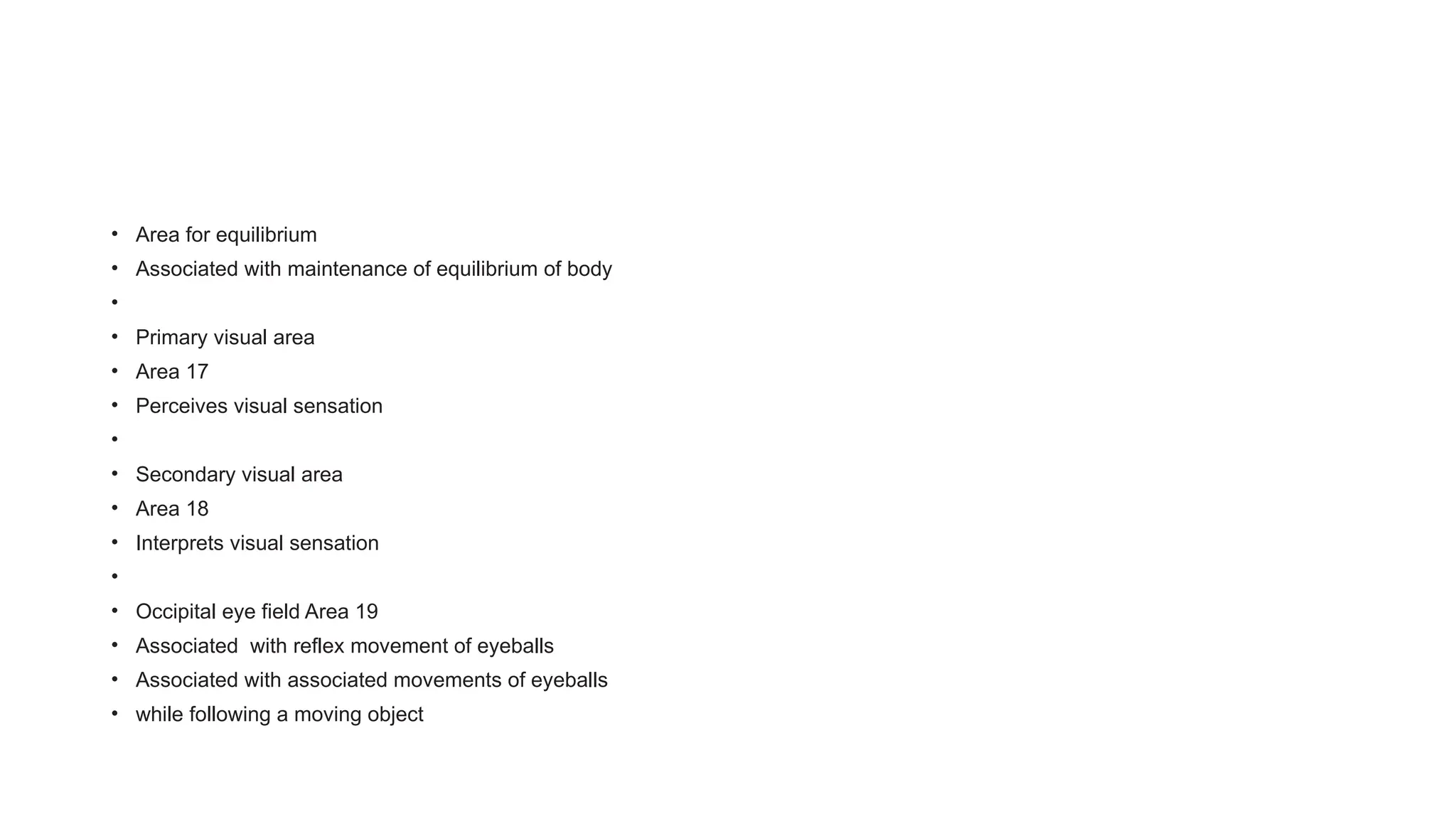
• Area forequilibrium
• Associated with maintenance of equilibrium of body
•
• Primary visual area
• Area 17
• Perceives visual sensation
•
• Secondary visual area
• Area 18
• Interprets visual sensation
•
• Occipital eye field Area 19
• Associated with reflex movement of eyeballs
• Associated with associated movements of eyeballs
• while following a moving object
42.
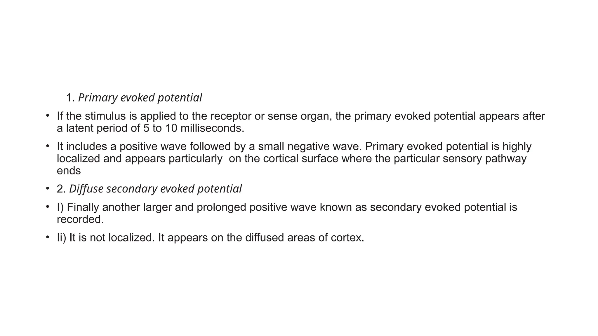
• O1. Primaryevoked potential
• If the stimulus is applied to the receptor or sense organ, the primary evoked potential appears after
a latent period of 5 to 10 milliseconds.
• It includes a positive wave followed by a small negative wave. Primary evoked potential is highly
localized and appears particularly on the cortical surface where the particular sensory pathway
ends
• 2. Diffuse secondary evoked potential
• I) Finally another larger and prolonged positive wave known as secondary evoked potential is
recorded.
• Ii) It is not localized. It appears on the diffused areas of cortex.
43.
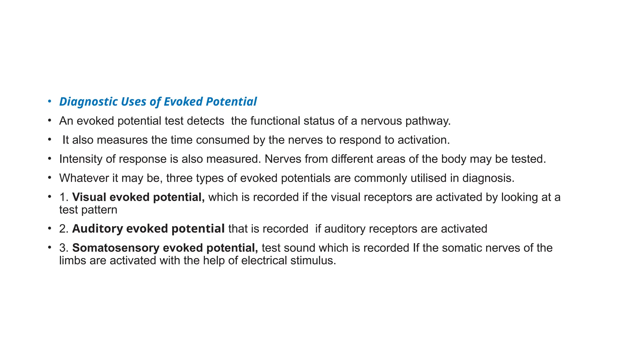
• Diagnostic Usesof Evoked Potential
• An evoked potential test detects the functional status of a nervous pathway.
• It also measures the time consumed by the nerves to respond to activation.
• Intensity of response is also measured. Nerves from different areas of the body may be tested.
• Whatever it may be, three types of evoked potentials are commonly utilised in diagnosis.
• 1. Visual evoked potential, which is recorded if the visual receptors are activated by looking at a
test pattern
• 2. Auditory evoked potential that is recorded if auditory receptors are activated
• 3. Somatosensory evoked potential, test sound which is recorded If the somatic nerves of the
limbs are activated with the help of electrical stimulus.
44.
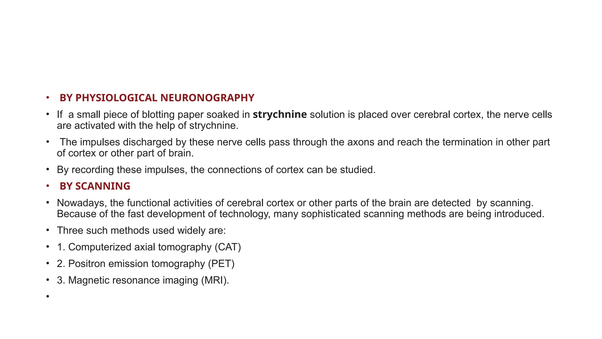
• BY PHYSIOLOGICALNEURONOGRAPHY
• If a small piece of blotting paper soaked in strychnine solution is placed over cerebral cortex, the nerve cells
are activated with the help of strychnine.
• The impulses discharged by these nerve cells pass through the axons and reach the termination in other part
of cortex or other part of brain.
• By recording these impulses, the connections of cortex can be studied.
• BY SCANNING
• Nowadays, the functional activities of cerebral cortex or other parts of the brain are detected by scanning.
Because of the fast development of technology, many sophisticated scanning methods are being introduced.
• Three such methods used widely are:
• 1. Computerized axial tomography (CAT)
• 2. Positron emission tomography (PET)
• 3. Magnetic resonance imaging (MRI).
•
• References
• 1.
•Hartmann P, Ramseier A, Gudat F, Mihatsch MJ, Polasek W. [Normal weight of the brain in adults in relation to age, sex,
body height and weight]. Pathologe. 1994 Jun;15(3):165-70. [PubMed]
• 2.
• Neulinger K, Oram J, Tinson H, O'Gorman J, Shum DH. Prospective memory and frontal lobe function. Neuropsychol
Dev Cogn B Aging Neuropsychol Cogn. 2016;23(2):171-83. [PubMed]
• 3.
• Flinker A, Korzeniewska A, Shestyuk AY, Franaszczuk PJ, Dronkers NF, Knight RT, Crone NE. Redefining the role of
Broca's area in speech. Proc Natl Acad Sci U S A. 2015 Mar 03;112(9):2871-5. [PMC free article] [PubMed]
• 4.
• Barrash J, Stuss DT, Aksan N, Anderson SW, Jones RD, Manzel K, Tranel D. "Frontal lobe syndrome"? Subtypes of
acquired personality disturbances in patients with focal brain damage. Cortex. 2018 Sep;106:65-80. [PMC free article] [
PubMed]
• 5.
• Collins A, Koechlin E. Reasoning, learning, and creativity: frontal lobe function and human decision-making. PLoS Biol.
2012;10(3):e1001293. [PMC free article] [PubMed]
50.
•
• 6.
• ChouinardPA, Paus T. The primary motor and premotor areas of the human cerebral cortex. Neuroscientist. 2006
Apr;12(2):143-52. [PubMed]
• 7.
• Berlucchi G, Vallar G. The history of the neurophysiology and neurology of the parietal lobe. Handb Clin Neurol.
2018;151:3-30. [PubMed]
• 8.
• Klingner CM, Witte OW. Somatosensory deficits. Handb Clin Neurol. 2018;151:185-206. [PubMed]
• 9.
• Kiernan JA. Anatomy of the temporal lobe. Epilepsy Res Treat. 2012;2012:176157. [PMC free article] [PubMed]
• 10.
• Nourski KV. Auditory processing in the human cortex: An intracranial electrophysiology perspective. Laryngoscope
Investig Otolaryngol. 2017 Aug;2(4):147-156. [PMC free article] [PubMed]
•
51.
• 11.
• BinderJR. Current Controversies on Wernicke's Area and its Role in Language. Curr Neurol Neurosci Rep.
2017 Aug;17(8):58. [PubMed]
• 12.
• Xu J, Wang J, Fan L, Li H, Zhang W, Hu Q, Jiang T. Tractography-based Parcellation of the Human Middle
Temporal Gyrus. Sci Rep. 2015 Dec 22;5:18883. [PMC free article] [PubMed]
• 13.
• Conway BR. The Organization and Operation of Inferior Temporal Cortex. Annu Rev Vis Sci. 2018 Sep
15;4:381-402. [PMC free article] [PubMed]
• 14.
• Clark RE. Current Topics Regarding the Function of the Medial Temporal Lobe Memory System. Curr Top
Behav Neurosci. 2018;37:13-42. [PubMed]
• 15.
• Huff T, Mahabadi N, Tadi P. StatPearls [Internet]. StatPearls Publishing; Treasure Island (FL): Aug 14, 2023.
Neuroanatomy, Visual Cortex. [PubMed]
![• References
• 1.
• Hartmann P, Ramseier A, Gudat F, Mihatsch MJ, Polasek W. [Normal weight of the brain in adults in relation to age, sex,
body height and weight]. Pathologe. 1994 Jun;15(3):165-70. [PubMed]
• 2.
• Neulinger K, Oram J, Tinson H, O'Gorman J, Shum DH. Prospective memory and frontal lobe function. Neuropsychol
Dev Cogn B Aging Neuropsychol Cogn. 2016;23(2):171-83. [PubMed]
• 3.
• Flinker A, Korzeniewska A, Shestyuk AY, Franaszczuk PJ, Dronkers NF, Knight RT, Crone NE. Redefining the role of
Broca's area in speech. Proc Natl Acad Sci U S A. 2015 Mar 03;112(9):2871-5. [PMC free article] [PubMed]
• 4.
• Barrash J, Stuss DT, Aksan N, Anderson SW, Jones RD, Manzel K, Tranel D. "Frontal lobe syndrome"? Subtypes of
acquired personality disturbances in patients with focal brain damage. Cortex. 2018 Sep;106:65-80. [PMC free article] [
PubMed]
• 5.
• Collins A, Koechlin E. Reasoning, learning, and creativity: frontal lobe function and human decision-making. PLoS Biol.
2012;10(3):e1001293. [PMC free article] [PubMed]](https://image.slidesharecdn.com/cerebralcortex-250805144725-bf20f76f/75/Cerebral-Cortex-ppt-by-Dr-Muralinath-sir-49-2048.jpg)
![•
• 6.
• Chouinard PA, Paus T. The primary motor and premotor areas of the human cerebral cortex. Neuroscientist. 2006
Apr;12(2):143-52. [PubMed]
• 7.
• Berlucchi G, Vallar G. The history of the neurophysiology and neurology of the parietal lobe. Handb Clin Neurol.
2018;151:3-30. [PubMed]
• 8.
• Klingner CM, Witte OW. Somatosensory deficits. Handb Clin Neurol. 2018;151:185-206. [PubMed]
• 9.
• Kiernan JA. Anatomy of the temporal lobe. Epilepsy Res Treat. 2012;2012:176157. [PMC free article] [PubMed]
• 10.
• Nourski KV. Auditory processing in the human cortex: An intracranial electrophysiology perspective. Laryngoscope
Investig Otolaryngol. 2017 Aug;2(4):147-156. [PMC free article] [PubMed]
•](https://image.slidesharecdn.com/cerebralcortex-250805144725-bf20f76f/75/Cerebral-Cortex-ppt-by-Dr-Muralinath-sir-50-2048.jpg)
![• 11.
• Binder JR. Current Controversies on Wernicke's Area and its Role in Language. Curr Neurol Neurosci Rep.
2017 Aug;17(8):58. [PubMed]
• 12.
• Xu J, Wang J, Fan L, Li H, Zhang W, Hu Q, Jiang T. Tractography-based Parcellation of the Human Middle
Temporal Gyrus. Sci Rep. 2015 Dec 22;5:18883. [PMC free article] [PubMed]
• 13.
• Conway BR. The Organization and Operation of Inferior Temporal Cortex. Annu Rev Vis Sci. 2018 Sep
15;4:381-402. [PMC free article] [PubMed]
• 14.
• Clark RE. Current Topics Regarding the Function of the Medial Temporal Lobe Memory System. Curr Top
Behav Neurosci. 2018;37:13-42. [PubMed]
• 15.
• Huff T, Mahabadi N, Tadi P. StatPearls [Internet]. StatPearls Publishing; Treasure Island (FL): Aug 14, 2023.
Neuroanatomy, Visual Cortex. [PubMed]](https://image.slidesharecdn.com/cerebralcortex-250805144725-bf20f76f/75/Cerebral-Cortex-ppt-by-Dr-Muralinath-sir-51-2048.jpg)


![Apporach to lung biopsy [Auto-saved].pptx latest](https://cdn.slidesharecdn.com/ss_thumbnails/apporachtolungbiopsyauto-saved-251211225655-93258539-thumbnail.jpg?width=640&height=640&fit=bounds)


