Downloaded 257 times


This document discusses cell junctions and their roles in various biological processes. It defines the main types of cell junctions, including tight junctions, gap junctions, focal adhesions, adherens junctions, and desmosomes. It provides examples of how cell junction gene expression changes during epithelial-to-mesenchymal transition, stem cell differentiation, cancer, atopic dermatitis, and the effects of probiotic bacteria on intestinal barriers. PCR array tools are presented as ways to study cell junctions and related pathways at the gene expression level to better understand their functions in normal and disease states.
Introduction to the cell junctions topic by Dr. George J. Quellhorst, emphasizing their significance in cell biology.
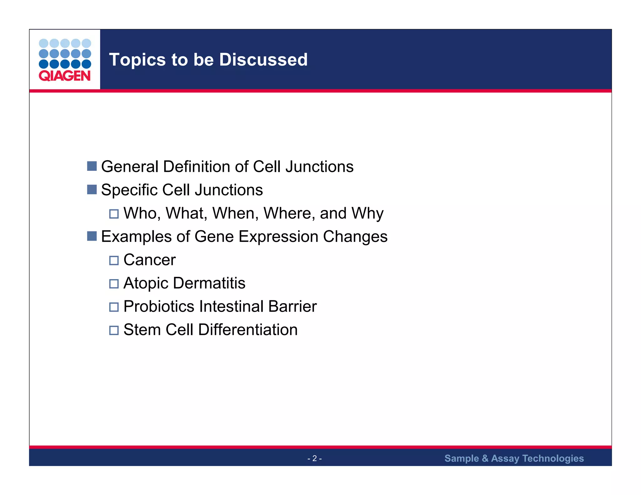
Outline of presentation topics such as definitions, types of cell junctions, and their roles in diseases.
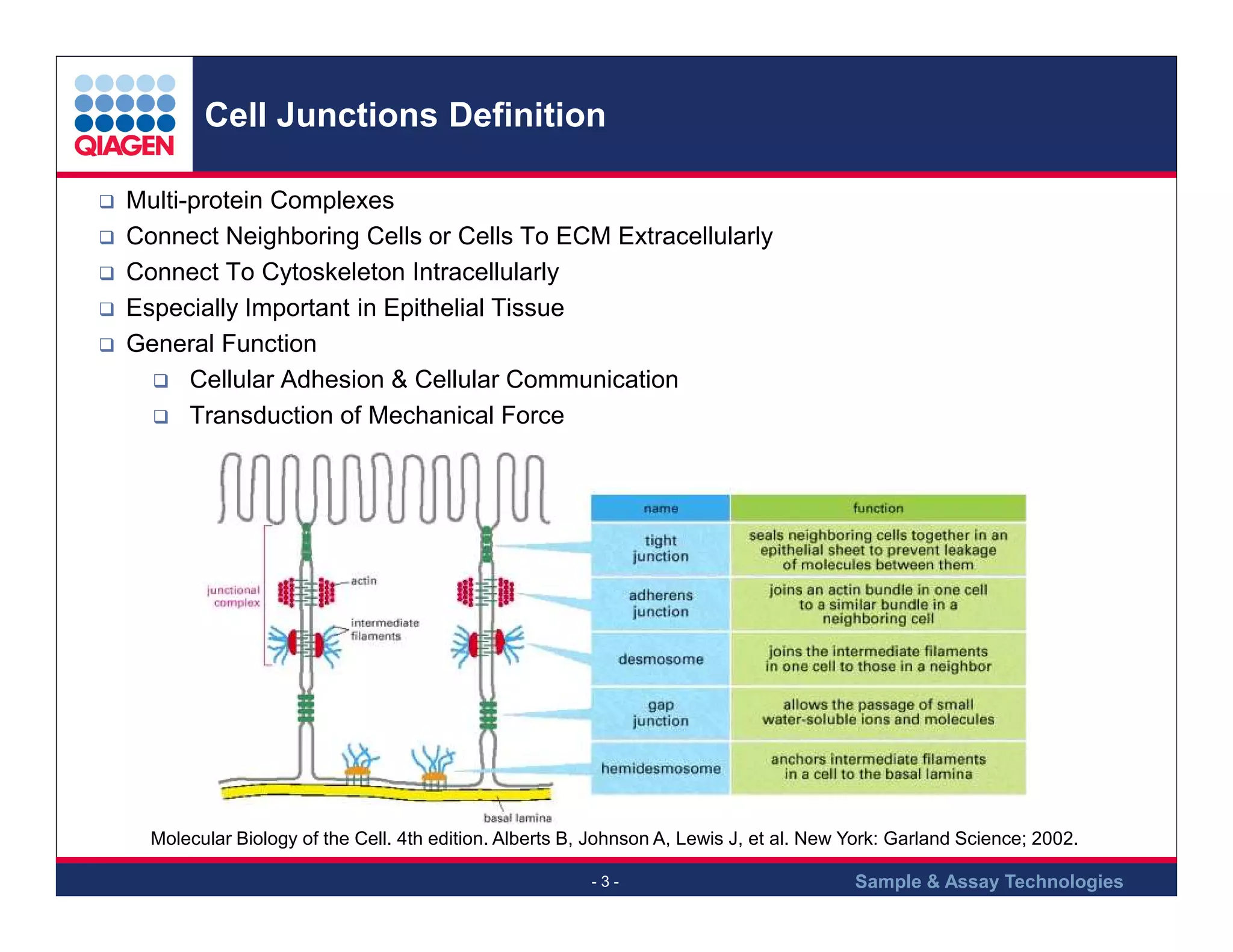
Cell junctions are multi-protein complexes important for cell adhesion, communication, and force transduction.
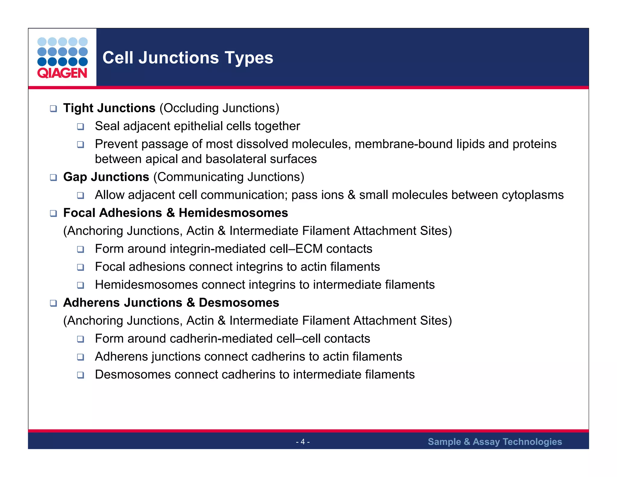
Overview of main cell junction types: Tight junctions, Gap junctions, Focal Adhesions, and their specific functions.
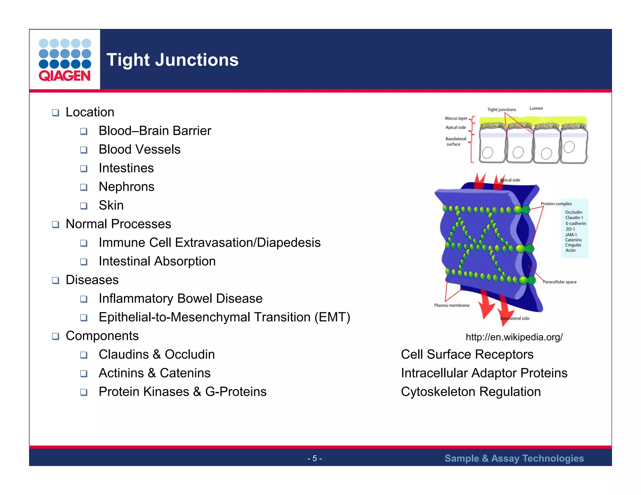
Tight junctions prevent molecule passage, located in areas like the blood-brain barrier and involved in disease processes.
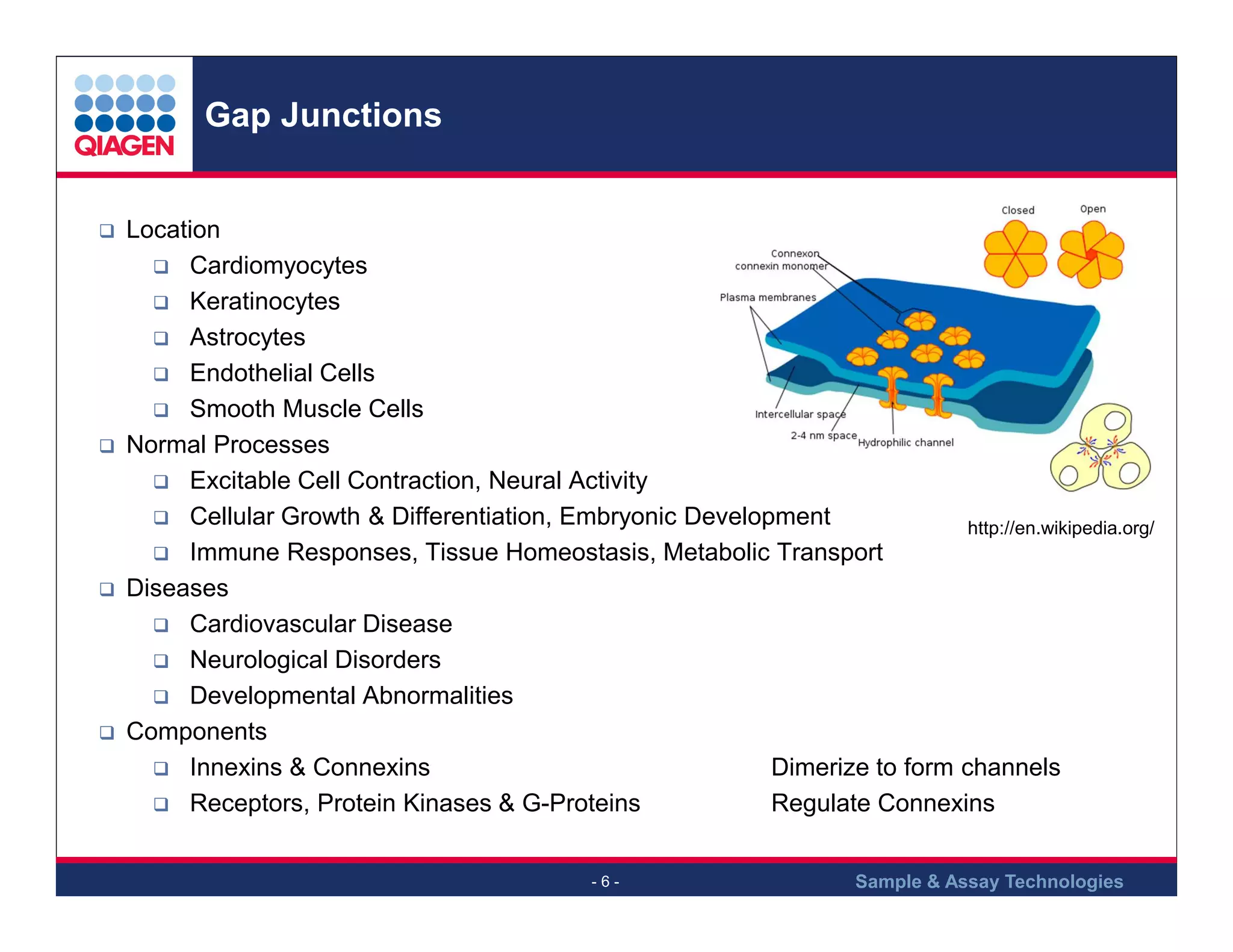
Gap junctions facilitate cell communication in diverse tissues and their role in processes like immune responses.
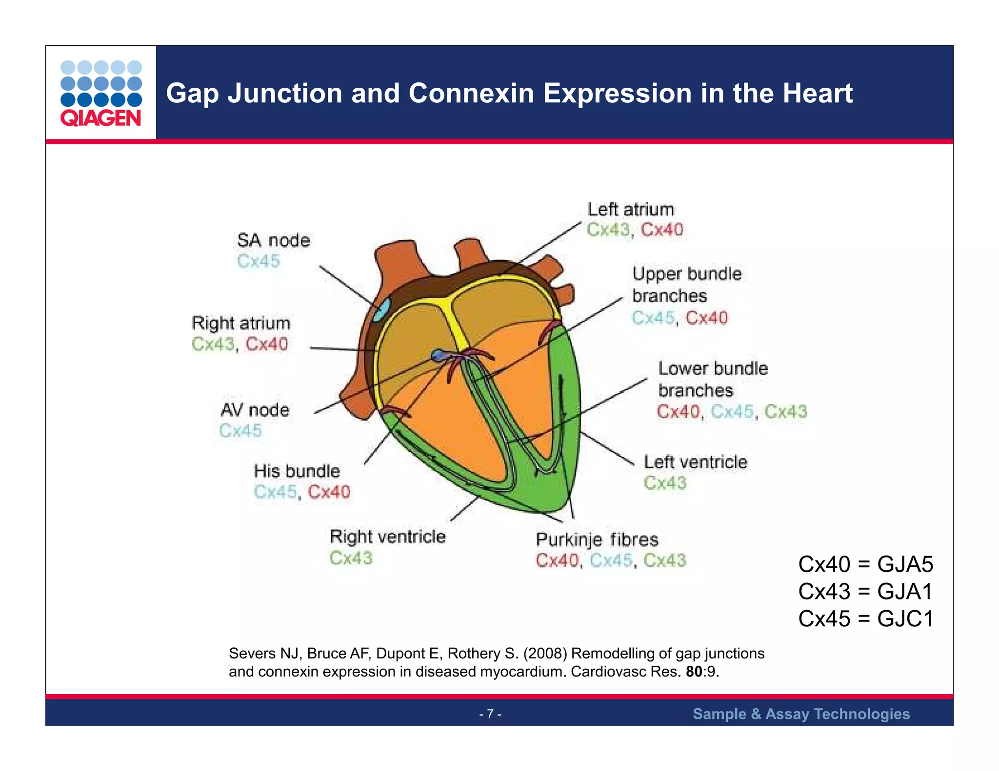
Details on connexin types in cardiac cells and their relevance in heart disease.
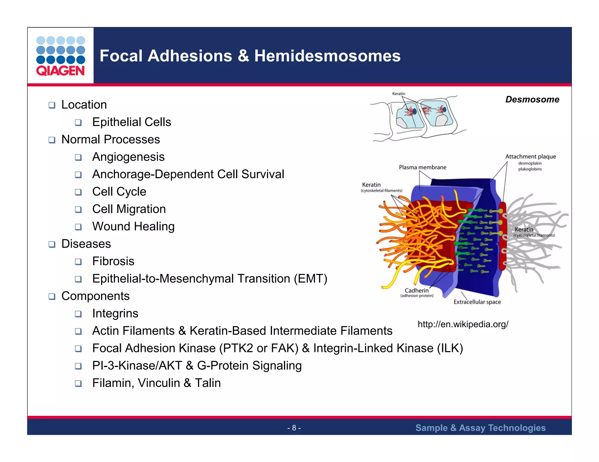
These junctions link cells to the extracellular matrix, aiding in cell survival and migration.
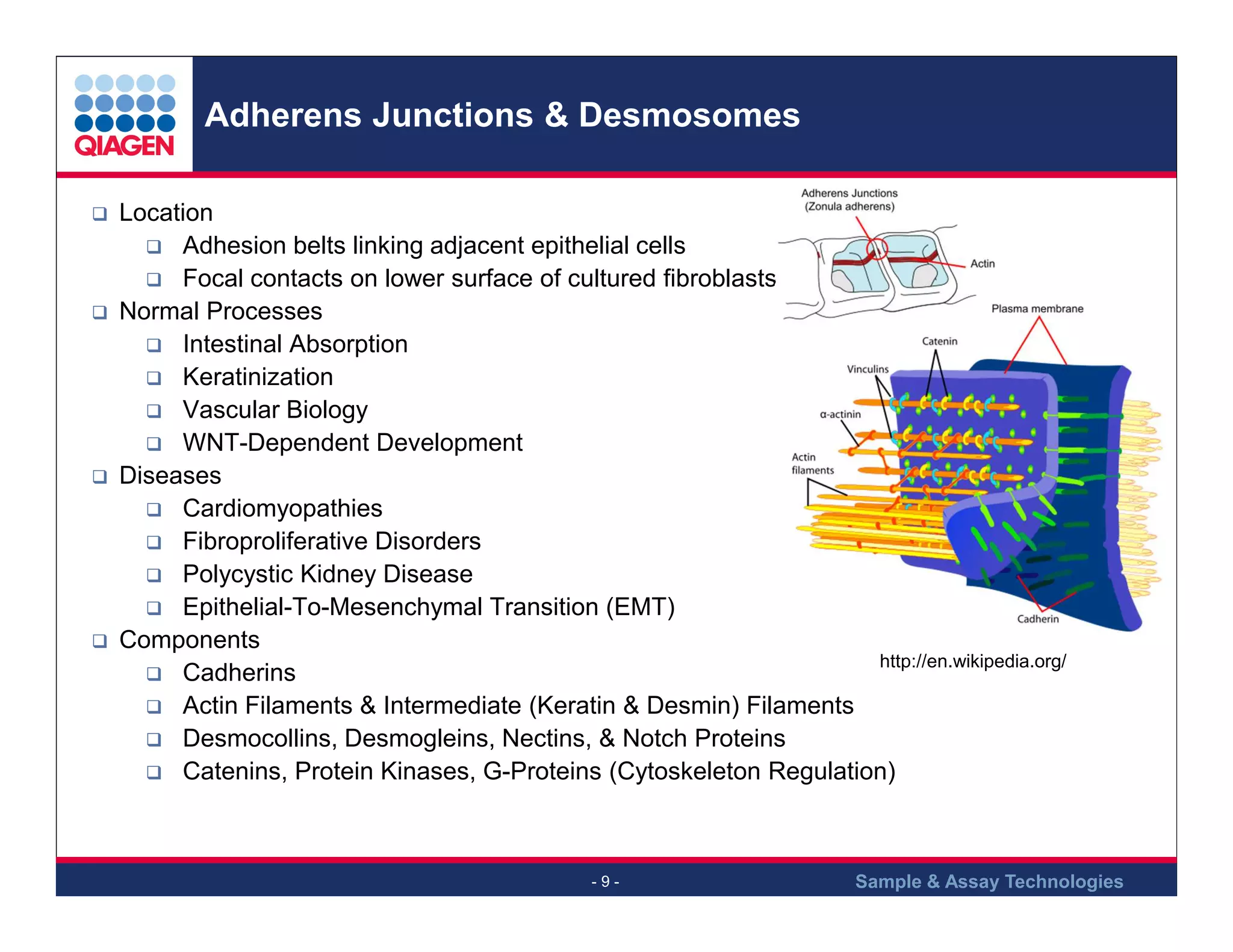
Roles of adherens junctions and desmosomes in maintaining cell adhesion and tissue integrity.
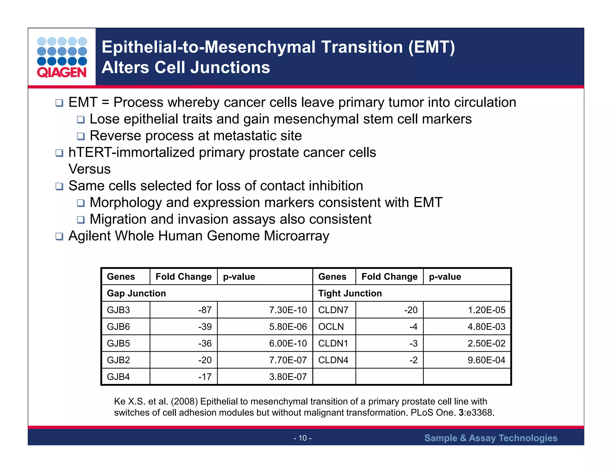
EMT alters cell junctions, contributing to cancer metastasis, with changes in gene expression.
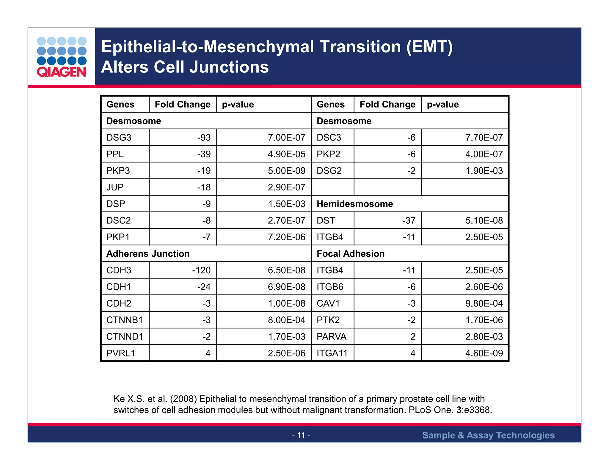
Changes in gene expression related to different types of junctions influenced by EMT.
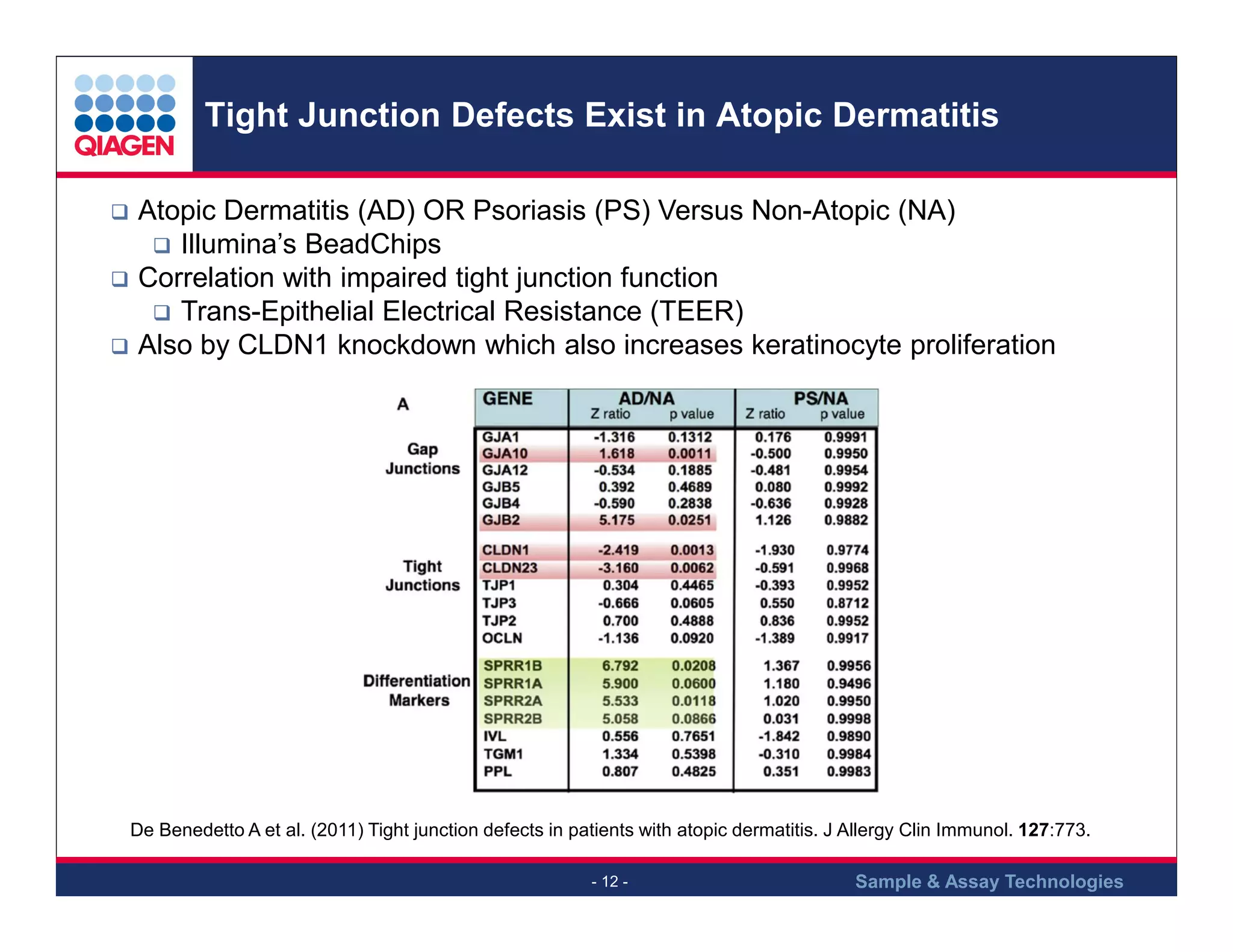
Atopic dermatitis is associated with tight junction issues, affecting barrier function.
Probiotic bacteria like Lactobacillus plantarum can improve tight junctions and intestinal barrier function.
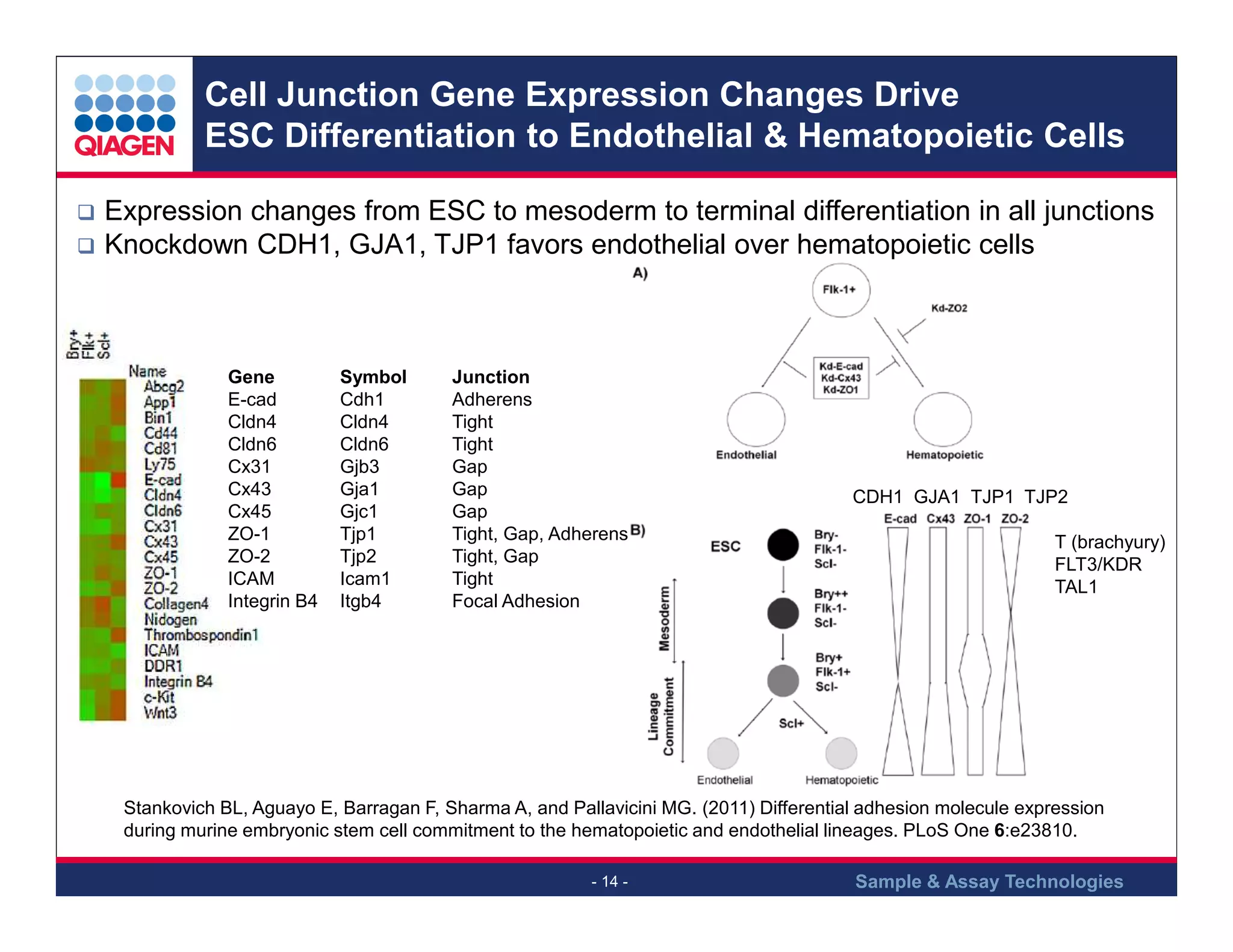
Gene expression changes in junctions influence differentiation of stem cells into specific lineages.
Summary of cell junctions' functions across cell types and their importance in various biological processes.
Introduction to RT2 Profiler PCR Arrays available for studying various cell junctions.
Promotional offer for two free PCR arrays with purchase, inviting questions about research support.
Closing remarks and invitation for questions regarding cell junctions and their biological importance.


