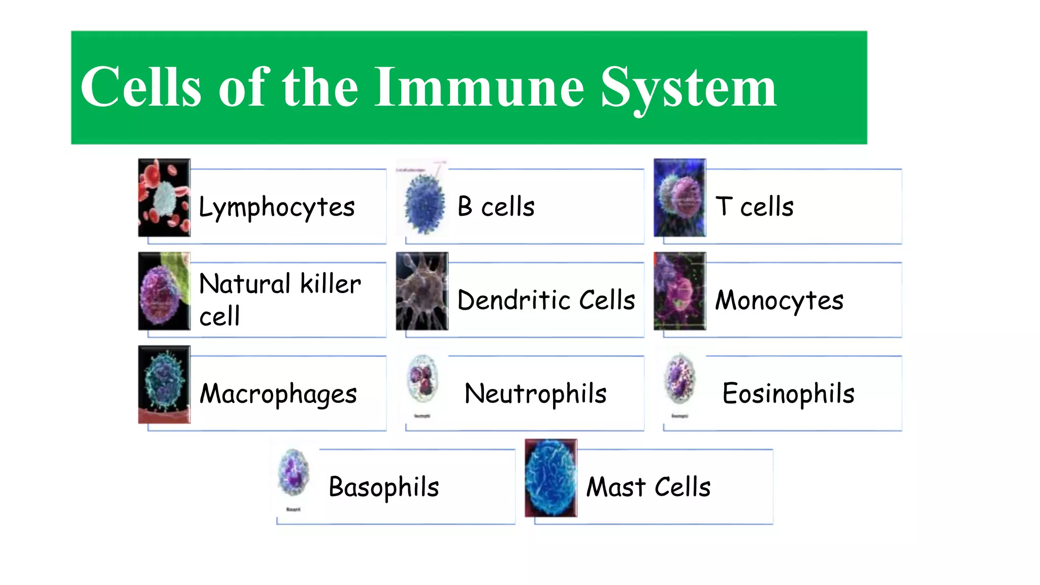
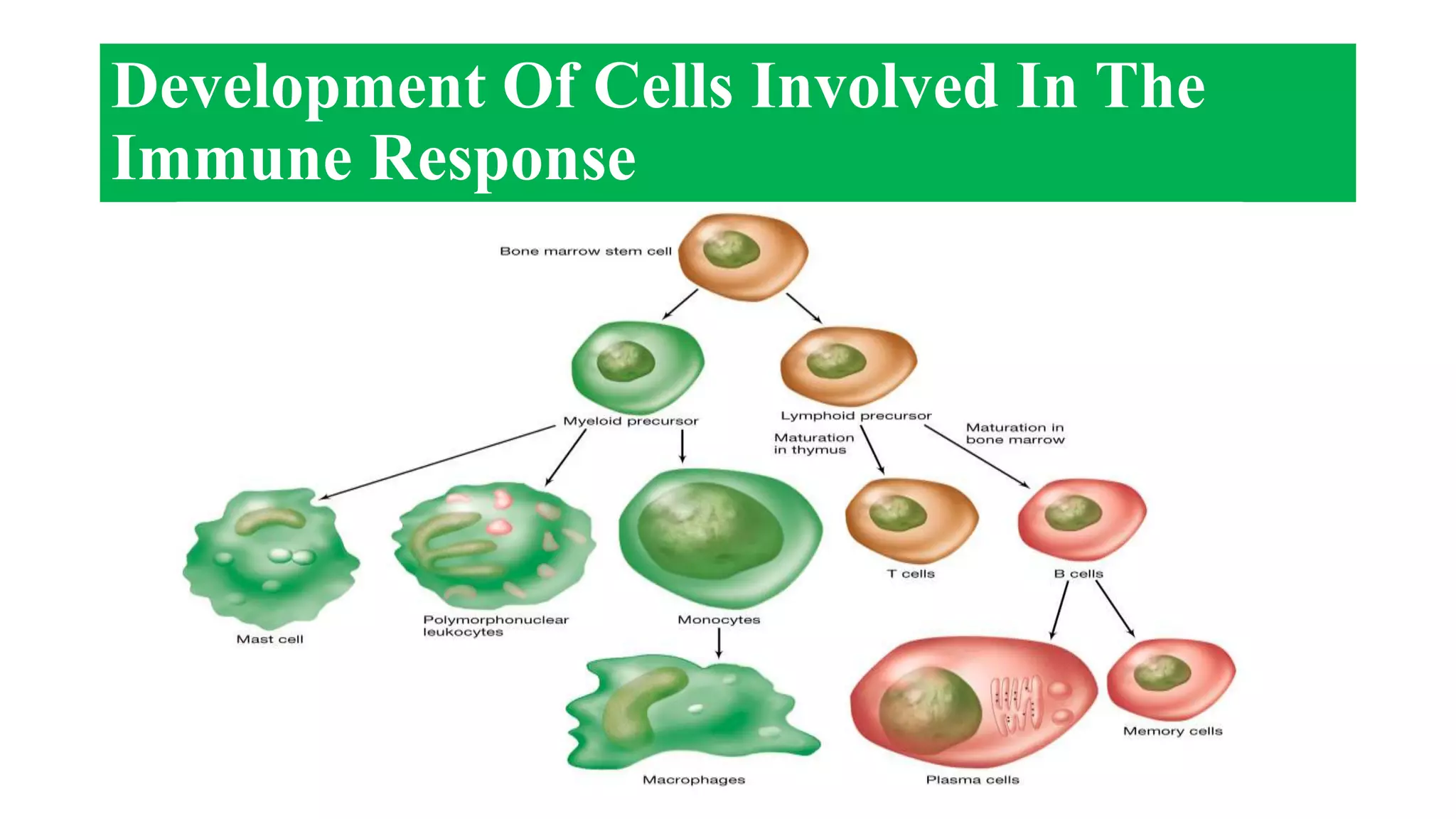
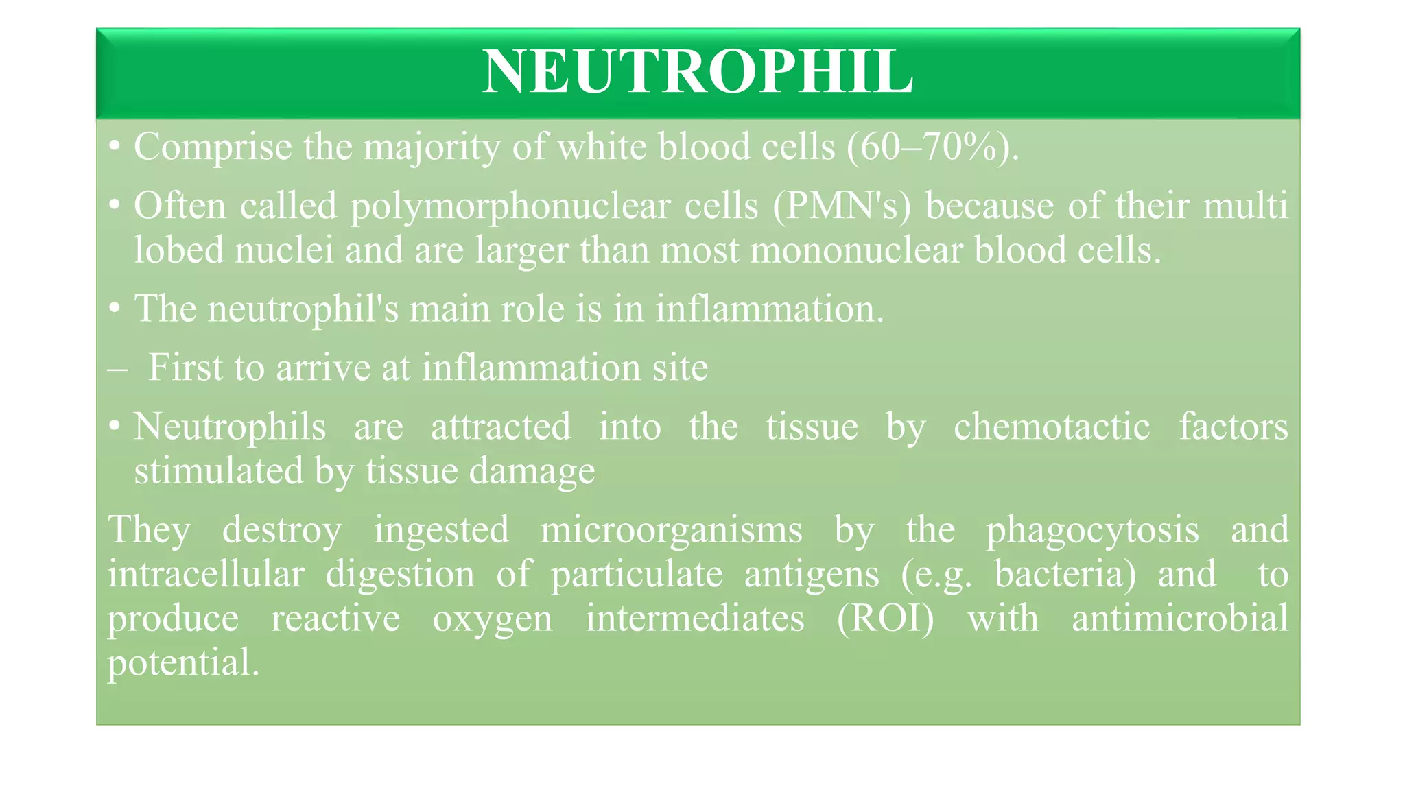
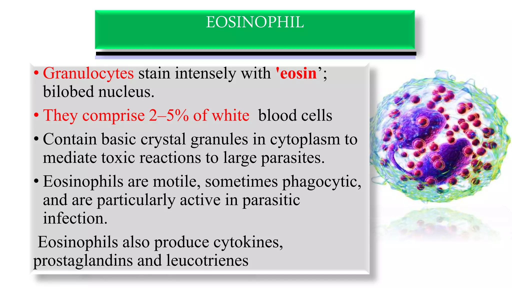
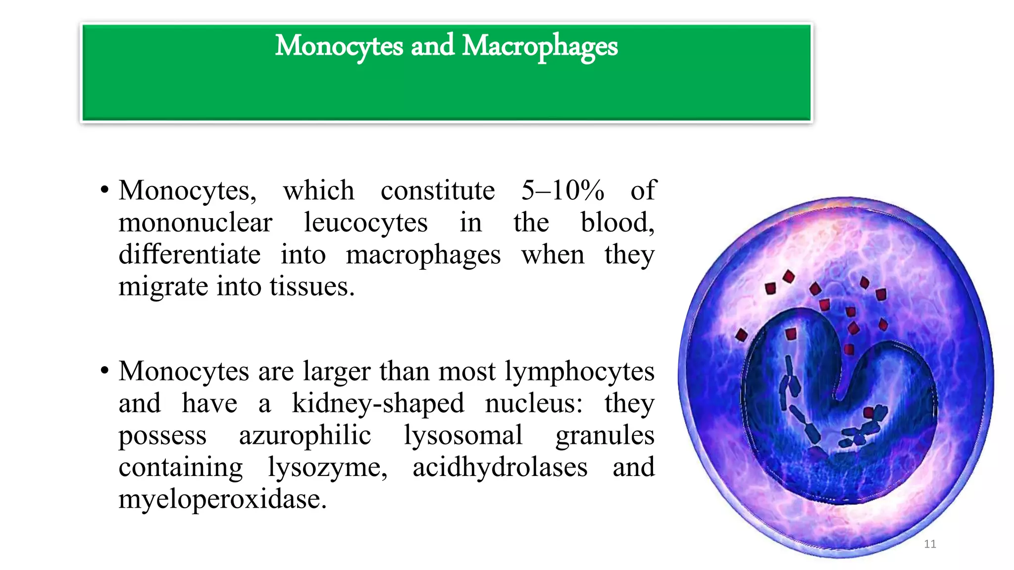
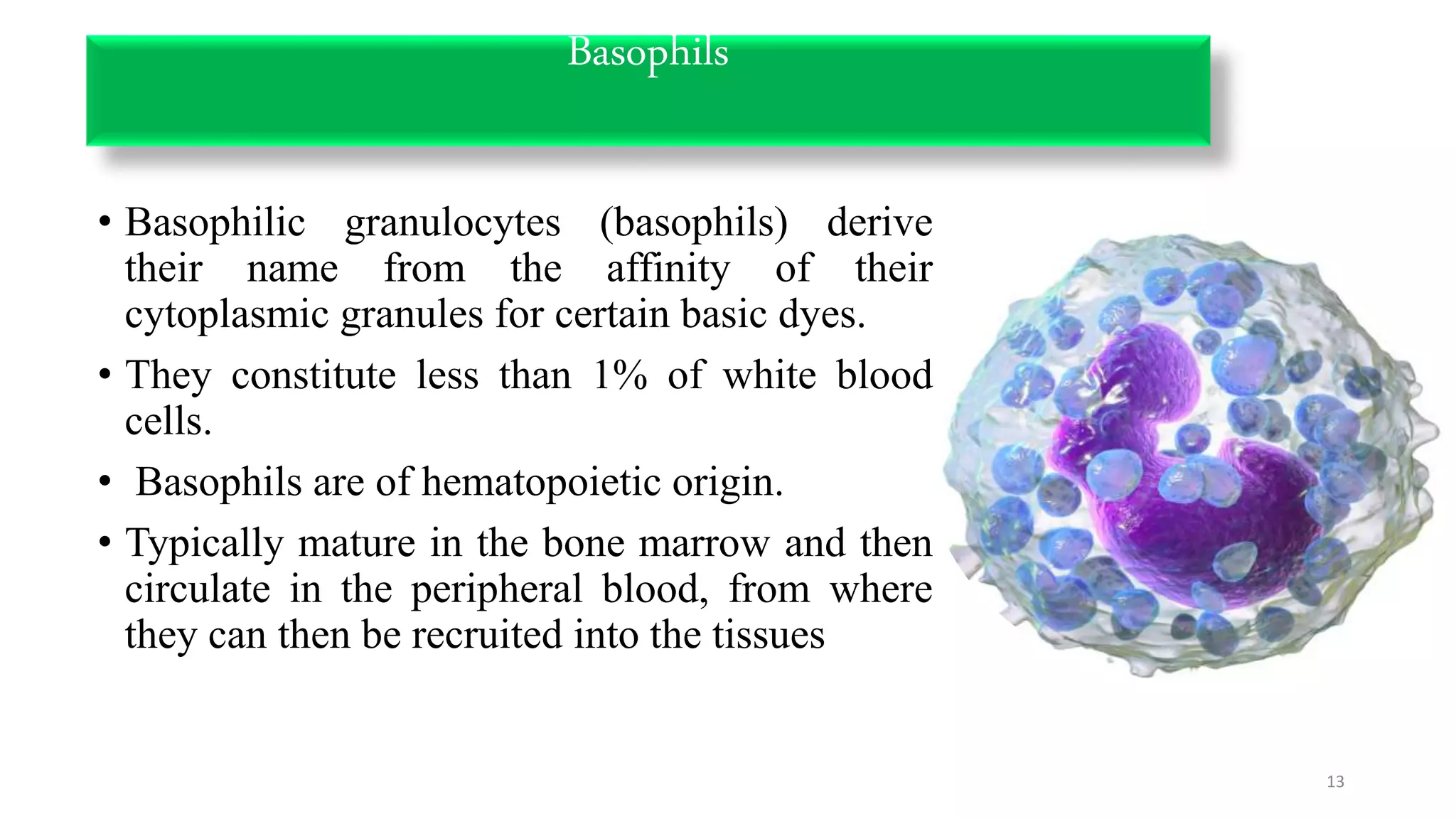
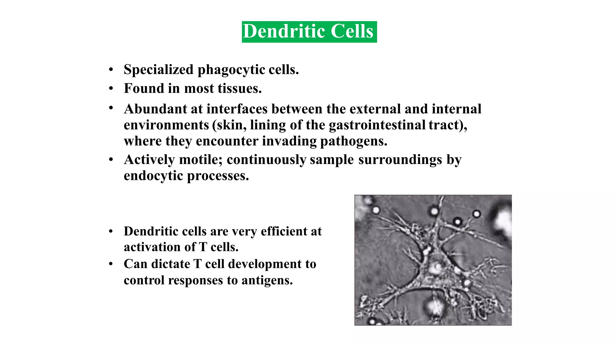
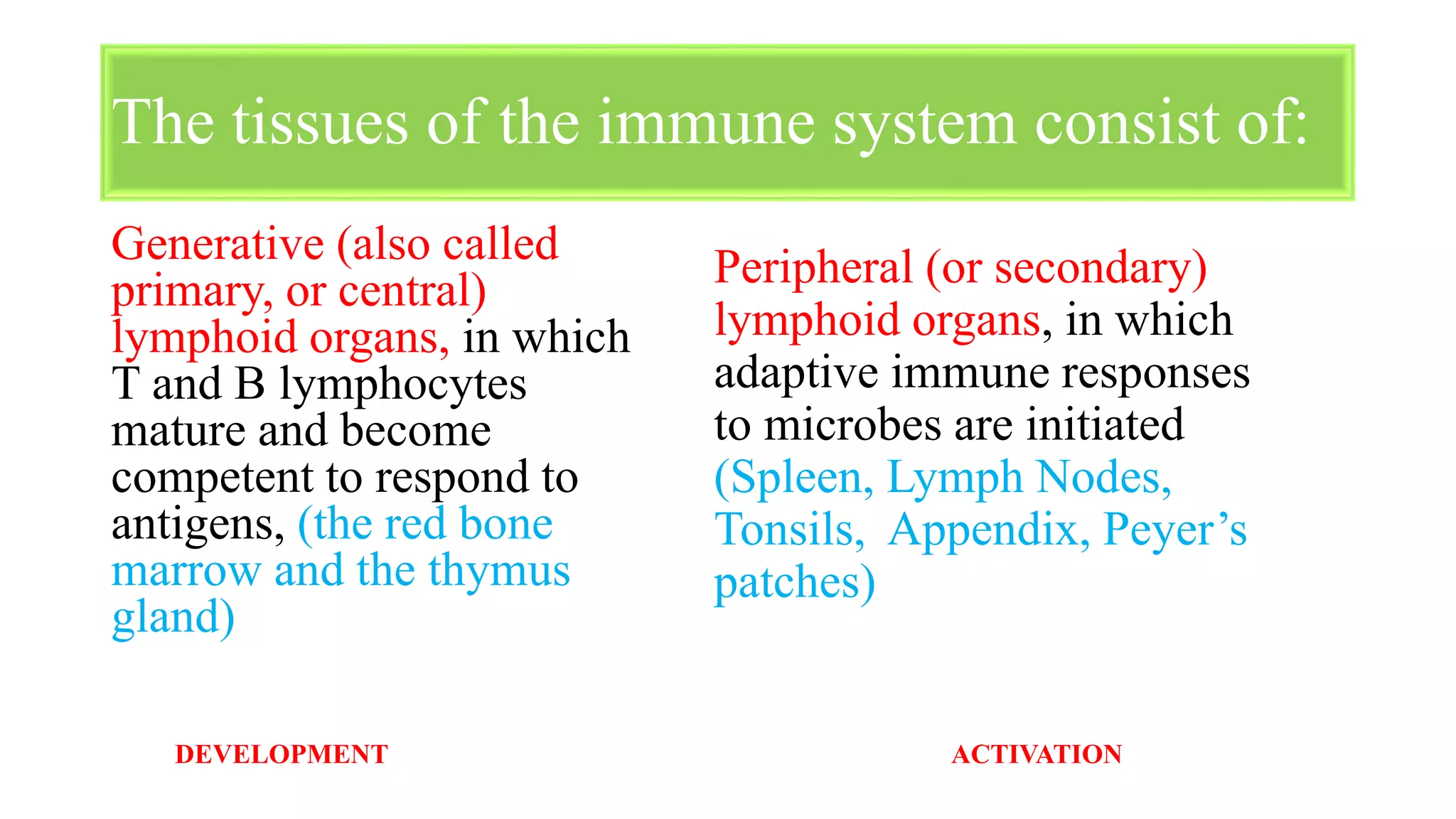
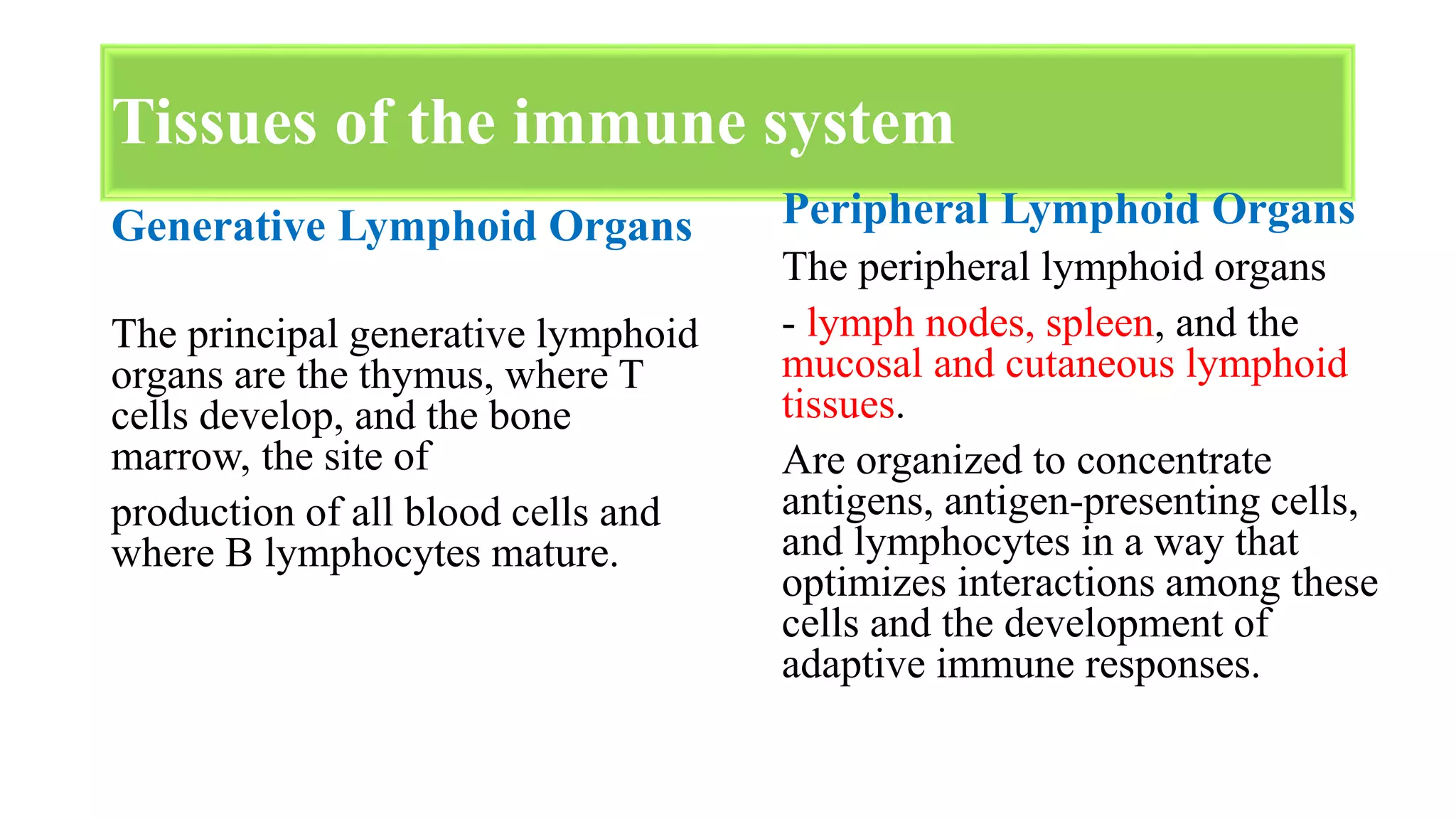
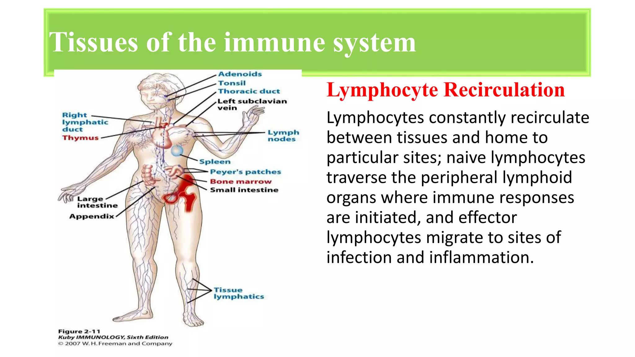
CELLS AND TISSUES OF
THE IMMUNES SYSTEM
J. Nutai Kolleh
Year III Student
A.M. Dogliotti College of Medicine
OBJECTIVES
At the end of this presentation, students should know
the following:
• The cells and tissues of the immune system
• The origin of immune cells
• The functions of immunes cells
• Primary lymphoid and secondary lymphoid tissues
INTRODUCTION
• The immune system comprises multiple cell types
(leukocytes) organized in lymphoid tissues & organs
together with other cell types where they interact to
perform their function.
• All cells in the immune system are derived from stem
cells and either myeloid or lymphoid precursors under
the control of cytokines
• Immunity is the ability of the body to defend itself against
specific invading agents such as bacteria, toxins, viruses, and
foreign tissue is called immunity.
• The main function of the immune system is to defend the
body against a wide variety of pathogenic infectious agents
with vastly differing natures, i.e. viruses, bacteria, fungi,
protozoa and parasitic worms.
• The immune system also play a key role in limiting the
infections.
The Immune System
Cells of the Immune System
Lymphocytes B cells T cells
Natural killer
cell
Dendritic Cells Monocytes
Macrophages Neutrophils Eosinophils
Basophils Mast Cells
Cells Of The Immune System
• The cells of the immune system arise
• from pluripotent hematopoeitic stem cells (HSC)
through two main lines of differentiation
• Myeloid lineage produces phagocytes (neutrophils..)
and other cells
• Lymphoid lineage produces lymphocytes
Development Of Cells Involved In The
Immune Response
NEUTROPHIL
• Comprise the majority of white blood cells (60–70%).
• Often called polymorphonuclear cells (PMN's) because of their multi
lobed nuclei and are larger than most mononuclear blood cells.
• The neutrophil's main role is in inflammation.
– First to arrive at inflammation site
• Neutrophils are attracted into the tissue by chemotactic factors
stimulated by tissue damage
They destroy ingested microorganisms by the phagocytosis and
intracellular digestion of particulate antigens (e.g. bacteria) and to
produce reactive oxygen intermediates (ROI) with antimicrobial
potential.
EOSINOPHIL
• Granulocytes stain intensely with 'eosin’;
bilobed nucleus.
• They comprise 2–5% of white blood cells
• Contain basic crystal granules in cytoplasm to
mediate toxic reactions to large parasites.
• Eosinophils are motile, sometimes phagocytic,
and are particularly active in parasitic
infection.
Eosinophils also produce cytokines,
prostaglandins and leucotrienes
• Unlike neutrophil which normally display
phagocytosis and intracellular digestion, eosinophils
secrete their granule contents for extracellular
digestion of infectious pathogens which are too large
to be engulfed.
• They then secrete their antibiotic granule contents
(including major basic protein and eosinophil cationic
protein) and reactive oxygen species to bring about
damage to the target.
EOSINOPHIL
Monocytes and Macrophages
• Monocytes, which constitute 5–10% of
mononuclear leucocytes in the blood,
differentiate into macrophages when they
migrate into tissues.
• Monocytes are larger than most lymphocytes
and have a kidney-shaped nucleus: they
possess azurophilic lysosomal granules
containing lysozyme, acidhydrolases and
myeloperoxidase.
11
• The blood monocytes arise from myeloid progenitors in the bone
marrow.
• Macrophages can be resident in tissues for prolonged periods of time
where they take on various morphologies, and are known by
different names, depending on their tissues of residence, e.g. Kupffer
cells in the liver, mesangial cells in the kidney and microglial cells
in the brain.
12
Monocytes and Macrophages
Basophils
• Basophilic granulocytes (basophils) derive
their name from the affinity of their
cytoplasmic granules for certain basic dyes.
• They constitute less than 1% of white blood
cells.
• Basophils are of hematopoietic origin.
• Typically mature in the bone marrow and then
circulate in the peripheral blood, from where
they can then be recruited into the tissues
13
Mast Cells
• Formed in tissue from undifferentiated bone marrow
precursors.
Similar importance in allergic reactions as basophils, but
only found in tissues.
•
• Contain granules with preformed mediators to be released
after stimulation
– histamine, prostaglandins
– leukotrienes
Stimulation of mast cells occurs by the anaphylatoxins
(complement proteins C3a and C5a) or by cross-linking of
surface immunoglobulin (IgE).
•
Dendritic Cells
•
•
•
Specialized phagocytic cells.
Found in most tissues.
Abundant at interfaces between the external and internal
environments (skin, lining of the gastrointestinal tract),
where they encounter invading pathogens.
• Actively motile; continuously sample
endocytic processes.
surroundings by
• Dendritic cells are very efficient at
activation of T cells.
Can dictate T cell development to
control responses to antigens.
•
LYMPHOCYTE
• Lymphocytes are 20-40% of WBC, 99% of cells in lymph.
• Lymphocytes includes three types of cells.
• T-lymphocytes or T cells, which are derived from the thymus
and play a role in cell-mediated immunity.
• B-lymphocytes or B-cells which are derived from liver, spleen
and bone marrow are the precursors of plasma cells and play a
role in humoral immunity.
• Natural killer (NK) and killer (K) cells.
16
LYMPHOCYTES ACTIVATION
Mature lymphocytes that have not encountered the antigen for
which they are specific are said to be naive (immunologically
inexperienced).
After they are activated by recognition of antigens and other
signals, lymphocytes differentiate into Effector Cells, which
perform the function of eliminating microbes,
and Memory Cells, which live in a state of heightened
awareness and are able to react rapidly and strongly to combat
the microbe in case it returns.
• The main functions of T
lymphocytes are to exert
effects on other cells, either
regulating the activity of cells of
the immune system or killing
cells that are infected or
malignant.
• T cells have surface antigen
receptors, but there is no
secreted form of these
equivalent to antibodies.
18
The T lymphocytes are associated with
two types of immunological functions:
• The effector functions include
activities such as killing of virally
infected cells and tumors.
• The regulatory function are
represented by their ability to
amplify or suppress through
cytokines or other effector
lymphocytes including B and T
cells.
T Cells
B cells
• B lymphocytes represents 3 to 15% of circulating lymphoid cells and
are primarily defined by surface immunoglobulins (Ig).
• The B lymphocytes are common in areas of antibody production, such
as the germinal centers of the lymph nodes and diffuse lymphoid
tissue of mucosal systems.
• The main function of a B cell is to secrete soluble recognition
molecules called antibodies which specifically bind to an antigen
recognized by that B cell.
• Throughout the life, bone marrow remains the major repository of
stem cells for B lymphocytes.
19
Natural Killer Cells
• Functionally cytotoxic representing an innate
population that kill viral infected or tumor
target cells.
• they do not need to recognize foreign
antigens presented on the target cell.
– NK cells do not have a specific cell receptor. Target
recognition occurs by a Killer Inhibitory Receptor,
KIR, which is lacking on infected and tumor
targets.
• Kill targets by releasing perforin which
damages target
• cell membranes. Can also induce apoptosis in
the target.
 Heterogeneous group of T cells
 Share properties of both T cells and natural
 (NK) cells.
 Recognizes self- and foreign lipids
and glycolipids.
 Constitute only 0.2% of peripheral blood T
cells
Natural Killer T Cells
HELPER T CELLS
• Helper T cells are the major
driving force and the main
regulators of the immune
defense.
• Their primary task is to
activate B cells and killer T
cells.
• However, the helper T cells
themselves must be activated.
21
• APCs are found primarily in
the skin, lymph nodes,
• spleen and thymus.
• Main role is present antigens to
antigen sensitive
• lymphocytes.
• • APCs are classified according
to their ability to
• phagocytose antigens, location
in the body, and expression of
MHC related molecules.
Antigen Presenting Cells
TISSUES OF THE IMMUNE
SYSTEM
Generative (also called
primary, or central)
lymphoid organs, in which
T and B lymphocytes
mature and become
competent to respond to
antigens, (the red bone
marrow and the thymus
gland)
The tissues of the immune system consist of:
Peripheral (or secondary)
lymphoid organs, in which
adaptive immune responses
to microbes are initiated
(Spleen, Lymph Nodes,
Tonsils, Appendix, Peyer’s
patches)
DEVELOPMENT ACTIVATION
Tissues of the immune system
Generative Lymphoid Organs
The principal generative lymphoid
organs are the thymus, where T
cells develop, and the bone
marrow, the site of
production of all blood cells and
where B lymphocytes mature.
Peripheral Lymphoid Organs
The peripheral lymphoid organs
- lymph nodes, spleen, and the
mucosal and cutaneous lymphoid
tissues.
Are organized to concentrate
antigens, antigen-presenting cells,
and lymphocytes in a way that
optimizes interactions among these
cells and the development of
adaptive immune responses.
• Lymph nodes are nodular aggregates of lymphoid tissues located along
lymphatic channels throughout the body
• The spleen is an abdominal organ that serves the same role in immune
responses to blood borne antigen as the lymph nodes do in responses
to lymph-borne antigens.
• Bone marrow – Most defense cells are produced and then also
multiply here. They then migrate from the bone marrow into the
bloodstream and reach other organs and tissues
• Thymus - Certain defense cells are differentiated in the thymus: the
T lymphocytes, or T Cells for short, among other things, are
responsible for coordinating the innate and the adaptive immune
system.
Tissues of the immune system
Lymphatic tissue in the bowel and in other mucous
membranes in the body
• The Bowel plays a central role in defending the body
against pathogens: More than half of all cells that
produce antibodies are found in the bowel wall, especially in the last
part of the small bowel and in the Appendix.
• Other parts of the body where pathogens may enter also
contain lymphatic tissue in the mucous membranes. All this tissue
together is also called Mucosa-associated lymphoid tissue (MALT).
Pathogens might enter the body through the airways or the urinary
tract, with the defense cells being directly beneath the mucous
membrane, they prevent bacteria and viruses from attaching.
Lymphocyte Recirculation
Lymphocytes constantly recirculate
between tissues and home to
particular sites; naive lymphocytes
traverse the peripheral lymphoid
organs where immune responses
are initiated, and effector
lymphocytes migrate to sites of
infection and inflammation.
Tissues of the immune system
Conclusion
• Leukocytes (white blood cells) are immune system cells
involved in defending the body against infectious diseases and
foreign materials. Different types of leukocytes exist, all
produced and derived from a multipotent cell in the bone
marrow known as a hematopoietic stem cell
• The key primary lymphoid organs of the immune system
include the thymus and bone marrow, as well as secondary
lymphatic tissues including spleen, tonsils, lymph vessels,
lymph nodes, adenoids, skin, and liver.
References
• Jeffrey K. Actor, Ph.D.Pathology and Laboratory Medicine, cells and
organ of the immune system
• “Cells and Organs of the Immune System.” Boundless
Microbiology Boundless, 26 May. 2016.
• Dr. Vartika Srivastava, Cells of the immune system
• Robbins and Cotran Pathologic Basis of Disease, 9th Edition
• Bonnie Hylander, Ph.D. Immune Cells and Organs
• https://www.ncbi.nlm.nih.gov/pubmedhealth/PMH0072579/
Cells and tissues of the immune system

Cells and tissues of the immune system

  • 1.
    CELLS AND TISSUESOF THE IMMUNES SYSTEM J. Nutai Kolleh Year III Student A.M. Dogliotti College of Medicine
  • 2.
    OBJECTIVES At the endof this presentation, students should know the following: • The cells and tissues of the immune system • The origin of immune cells • The functions of immunes cells • Primary lymphoid and secondary lymphoid tissues
  • 3.
    INTRODUCTION • The immunesystem comprises multiple cell types (leukocytes) organized in lymphoid tissues & organs together with other cell types where they interact to perform their function. • All cells in the immune system are derived from stem cells and either myeloid or lymphoid precursors under the control of cytokines
  • 4.
    • Immunity isthe ability of the body to defend itself against specific invading agents such as bacteria, toxins, viruses, and foreign tissue is called immunity. • The main function of the immune system is to defend the body against a wide variety of pathogenic infectious agents with vastly differing natures, i.e. viruses, bacteria, fungi, protozoa and parasitic worms. • The immune system also play a key role in limiting the infections. The Immune System
  • 5.
    Cells of theImmune System Lymphocytes B cells T cells Natural killer cell Dendritic Cells Monocytes Macrophages Neutrophils Eosinophils Basophils Mast Cells
  • 6.
    Cells Of TheImmune System • The cells of the immune system arise • from pluripotent hematopoeitic stem cells (HSC) through two main lines of differentiation • Myeloid lineage produces phagocytes (neutrophils..) and other cells • Lymphoid lineage produces lymphocytes
  • 7.
    Development Of CellsInvolved In The Immune Response
  • 8.
    NEUTROPHIL • Comprise themajority of white blood cells (60–70%). • Often called polymorphonuclear cells (PMN's) because of their multi lobed nuclei and are larger than most mononuclear blood cells. • The neutrophil's main role is in inflammation. – First to arrive at inflammation site • Neutrophils are attracted into the tissue by chemotactic factors stimulated by tissue damage They destroy ingested microorganisms by the phagocytosis and intracellular digestion of particulate antigens (e.g. bacteria) and to produce reactive oxygen intermediates (ROI) with antimicrobial potential.
  • 9.
    EOSINOPHIL • Granulocytes stainintensely with 'eosin’; bilobed nucleus. • They comprise 2–5% of white blood cells • Contain basic crystal granules in cytoplasm to mediate toxic reactions to large parasites. • Eosinophils are motile, sometimes phagocytic, and are particularly active in parasitic infection. Eosinophils also produce cytokines, prostaglandins and leucotrienes
  • 10.
    • Unlike neutrophilwhich normally display phagocytosis and intracellular digestion, eosinophils secrete their granule contents for extracellular digestion of infectious pathogens which are too large to be engulfed. • They then secrete their antibiotic granule contents (including major basic protein and eosinophil cationic protein) and reactive oxygen species to bring about damage to the target. EOSINOPHIL
  • 11.
    Monocytes and Macrophages •Monocytes, which constitute 5–10% of mononuclear leucocytes in the blood, differentiate into macrophages when they migrate into tissues. • Monocytes are larger than most lymphocytes and have a kidney-shaped nucleus: they possess azurophilic lysosomal granules containing lysozyme, acidhydrolases and myeloperoxidase. 11
  • 12.
    • The bloodmonocytes arise from myeloid progenitors in the bone marrow. • Macrophages can be resident in tissues for prolonged periods of time where they take on various morphologies, and are known by different names, depending on their tissues of residence, e.g. Kupffer cells in the liver, mesangial cells in the kidney and microglial cells in the brain. 12 Monocytes and Macrophages
  • 13.
    Basophils • Basophilic granulocytes(basophils) derive their name from the affinity of their cytoplasmic granules for certain basic dyes. • They constitute less than 1% of white blood cells. • Basophils are of hematopoietic origin. • Typically mature in the bone marrow and then circulate in the peripheral blood, from where they can then be recruited into the tissues 13
  • 14.
    Mast Cells • Formedin tissue from undifferentiated bone marrow precursors. Similar importance in allergic reactions as basophils, but only found in tissues. • • Contain granules with preformed mediators to be released after stimulation – histamine, prostaglandins – leukotrienes Stimulation of mast cells occurs by the anaphylatoxins (complement proteins C3a and C5a) or by cross-linking of surface immunoglobulin (IgE). •
  • 15.
    Dendritic Cells • • • Specialized phagocyticcells. Found in most tissues. Abundant at interfaces between the external and internal environments (skin, lining of the gastrointestinal tract), where they encounter invading pathogens. • Actively motile; continuously sample endocytic processes. surroundings by • Dendritic cells are very efficient at activation of T cells. Can dictate T cell development to control responses to antigens. •
  • 16.
    LYMPHOCYTE • Lymphocytes are20-40% of WBC, 99% of cells in lymph. • Lymphocytes includes three types of cells. • T-lymphocytes or T cells, which are derived from the thymus and play a role in cell-mediated immunity. • B-lymphocytes or B-cells which are derived from liver, spleen and bone marrow are the precursors of plasma cells and play a role in humoral immunity. • Natural killer (NK) and killer (K) cells. 16
  • 17.
    LYMPHOCYTES ACTIVATION Mature lymphocytesthat have not encountered the antigen for which they are specific are said to be naive (immunologically inexperienced). After they are activated by recognition of antigens and other signals, lymphocytes differentiate into Effector Cells, which perform the function of eliminating microbes, and Memory Cells, which live in a state of heightened awareness and are able to react rapidly and strongly to combat the microbe in case it returns.
  • 18.
    • The mainfunctions of T lymphocytes are to exert effects on other cells, either regulating the activity of cells of the immune system or killing cells that are infected or malignant. • T cells have surface antigen receptors, but there is no secreted form of these equivalent to antibodies. 18 The T lymphocytes are associated with two types of immunological functions: • The effector functions include activities such as killing of virally infected cells and tumors. • The regulatory function are represented by their ability to amplify or suppress through cytokines or other effector lymphocytes including B and T cells. T Cells
  • 19.
    B cells • Blymphocytes represents 3 to 15% of circulating lymphoid cells and are primarily defined by surface immunoglobulins (Ig). • The B lymphocytes are common in areas of antibody production, such as the germinal centers of the lymph nodes and diffuse lymphoid tissue of mucosal systems. • The main function of a B cell is to secrete soluble recognition molecules called antibodies which specifically bind to an antigen recognized by that B cell. • Throughout the life, bone marrow remains the major repository of stem cells for B lymphocytes. 19
  • 20.
    Natural Killer Cells •Functionally cytotoxic representing an innate population that kill viral infected or tumor target cells. • they do not need to recognize foreign antigens presented on the target cell. – NK cells do not have a specific cell receptor. Target recognition occurs by a Killer Inhibitory Receptor, KIR, which is lacking on infected and tumor targets. • Kill targets by releasing perforin which damages target • cell membranes. Can also induce apoptosis in the target.  Heterogeneous group of T cells  Share properties of both T cells and natural  (NK) cells.  Recognizes self- and foreign lipids and glycolipids.  Constitute only 0.2% of peripheral blood T cells Natural Killer T Cells
  • 21.
    HELPER T CELLS •Helper T cells are the major driving force and the main regulators of the immune defense. • Their primary task is to activate B cells and killer T cells. • However, the helper T cells themselves must be activated. 21 • APCs are found primarily in the skin, lymph nodes, • spleen and thymus. • Main role is present antigens to antigen sensitive • lymphocytes. • • APCs are classified according to their ability to • phagocytose antigens, location in the body, and expression of MHC related molecules. Antigen Presenting Cells
  • 22.
    TISSUES OF THEIMMUNE SYSTEM
  • 23.
    Generative (also called primary,or central) lymphoid organs, in which T and B lymphocytes mature and become competent to respond to antigens, (the red bone marrow and the thymus gland) The tissues of the immune system consist of: Peripheral (or secondary) lymphoid organs, in which adaptive immune responses to microbes are initiated (Spleen, Lymph Nodes, Tonsils, Appendix, Peyer’s patches) DEVELOPMENT ACTIVATION
  • 24.
    Tissues of theimmune system Generative Lymphoid Organs The principal generative lymphoid organs are the thymus, where T cells develop, and the bone marrow, the site of production of all blood cells and where B lymphocytes mature. Peripheral Lymphoid Organs The peripheral lymphoid organs - lymph nodes, spleen, and the mucosal and cutaneous lymphoid tissues. Are organized to concentrate antigens, antigen-presenting cells, and lymphocytes in a way that optimizes interactions among these cells and the development of adaptive immune responses.
  • 25.
    • Lymph nodesare nodular aggregates of lymphoid tissues located along lymphatic channels throughout the body • The spleen is an abdominal organ that serves the same role in immune responses to blood borne antigen as the lymph nodes do in responses to lymph-borne antigens. • Bone marrow – Most defense cells are produced and then also multiply here. They then migrate from the bone marrow into the bloodstream and reach other organs and tissues • Thymus - Certain defense cells are differentiated in the thymus: the T lymphocytes, or T Cells for short, among other things, are responsible for coordinating the innate and the adaptive immune system. Tissues of the immune system
  • 26.
    Lymphatic tissue inthe bowel and in other mucous membranes in the body • The Bowel plays a central role in defending the body against pathogens: More than half of all cells that produce antibodies are found in the bowel wall, especially in the last part of the small bowel and in the Appendix. • Other parts of the body where pathogens may enter also contain lymphatic tissue in the mucous membranes. All this tissue together is also called Mucosa-associated lymphoid tissue (MALT). Pathogens might enter the body through the airways or the urinary tract, with the defense cells being directly beneath the mucous membrane, they prevent bacteria and viruses from attaching.
  • 27.
    Lymphocyte Recirculation Lymphocytes constantlyrecirculate between tissues and home to particular sites; naive lymphocytes traverse the peripheral lymphoid organs where immune responses are initiated, and effector lymphocytes migrate to sites of infection and inflammation. Tissues of the immune system
  • 28.
    Conclusion • Leukocytes (whiteblood cells) are immune system cells involved in defending the body against infectious diseases and foreign materials. Different types of leukocytes exist, all produced and derived from a multipotent cell in the bone marrow known as a hematopoietic stem cell • The key primary lymphoid organs of the immune system include the thymus and bone marrow, as well as secondary lymphatic tissues including spleen, tonsils, lymph vessels, lymph nodes, adenoids, skin, and liver.
  • 29.
    References • Jeffrey K.Actor, Ph.D.Pathology and Laboratory Medicine, cells and organ of the immune system • “Cells and Organs of the Immune System.” Boundless Microbiology Boundless, 26 May. 2016. • Dr. Vartika Srivastava, Cells of the immune system • Robbins and Cotran Pathologic Basis of Disease, 9th Edition • Bonnie Hylander, Ph.D. Immune Cells and Organs • https://www.ncbi.nlm.nih.gov/pubmedhealth/PMH0072579/