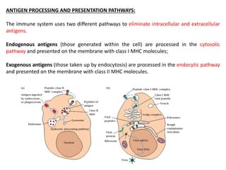
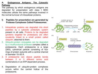
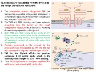
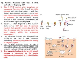
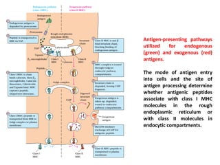
ANTIGEN PROCESSING AND PRESENTATION PATHWAYS
Dr. Manikandan Kathirvel M.Sc., Ph.D., (NET)
Assistant Professor,
Department of Life Sciences,
Kristu Jayanti College (Autonomous),
(Reaccredited with "A" Grade by NAAC)
Affiliated to Bengaluru North University,
K. Narayanapura, Kothanur (PO)
Bengaluru
Email: manikandan@kristujayanti.com
ORCID ID: 0000000270066334
ANTIGEN PROCESSING AND PRESENTATION:
a. The recognition of foreign antigen by a T cell requires that peptides derived from the
antigen be displayed within the cleft of an MHC molecule on the membrane of a cell.
b. The formation of these peptide-MHC complexes requires that a protein antigen be
degraded into peptides by a sequence of events called antigen processing.
c. The degraded peptides then associate with MHC molecules within the cell interior, and
the peptide MHC complexes are transported to the membrane, where they are
displayed to T cells (antigen presentation) for T cell activation and differentiation.
•Class I and class II MHC molecules associate with peptides that have been processed in
different intracellular compartments.
•Class I MHC molecules bind peptides derived from endogenous antigens that have been
processed within the cytoplasm of the cell – Cytosolic Pathway (e.g., normal cellular
proteins, tumor proteins, or viral and bacterial proteins produced within infected cells).
• Class II MHC molecules bind peptides derived from exogenous antigens that are
internalized by phagocytosis or endocytosis and processed within the endocytic pathway.
“All nucleated Cells can present antigen with Class I MHC;
Presentation with Class II MHC is restricted only to antigen presenting cells (APCs)”
•Cells expressing either class I or class II MHC molecules can present antigenic peptides to TC
and TH cells.
•Cells that display peptides associated with class I MHC molecules to CD8+ TC cells and
•Cells that display peptides associated with class II MHC molecules to CD4+ TH cells are
called antigen-presenting cells (APCs).
Three cell types are classified as professional antigen-presenting cells: dendritic cells,
macrophages, and B lymphocytes. These cells differ from each other in their mechanisms
of antigen uptake, in whether they constitutively express class II MHC molecules, and in
their co-stimulatory activity.
ANTIGEN PROCESSING AND PRESENTATION PATHWAYS:
The immune system uses two different pathways to eliminate intracellular and extracellular
antigens.
Endogenous antigens (those generated within the cell) are processed in the cytosolic
pathway and presented on the membrane with class I MHC molecules;
Exogenous antigens (those taken up by endocytosis) are processed in the endocytic pathway
and presented on the membrane with class II MHC molecules.
A. Endogenous Antigens: The
Cytosolic Pathway
B. Exogenous Antigens: The
Endocytic Pathway
A. Endogenous Antigens: The
Cytosolic Pathway
A. Endogenous Antigens: The Cytosolic
Pathway
The pathway by which endogenous antigens are
degraded for presentation with class I MHC
molecules utilizes the same pathways involved in
the normal turnover of intracellular proteins.
I) Peptides for presentation are generated by
Protease Complexes Called Proteasomes:
1. Intracellular proteins are degraded into short
peptides by a cytosolic proteolytic system
present in all cells. Proteins to be degraded
(proteins targeted for proteolysis) are often
covalently linked to a small protein called
ubiquitin, attached to them.
2. Ubiquitin-protein conjugates can be degraded
by a multifunctional protease complex called a
proteasome. Each proteasome is a large
(26S), cylindrical particle consisting of four
rings of protein subunits with a central channel
of diameter 10–50 Å.
3. A proteasome can cleave peptide bonds
between 2 or 3 different amino acid
combinations in an ATP-dependent process.
4. Degradation of ubiquitin-protein complexes
occurs within the central hollow of the
proteasome.
II). Peptides Are Transported from the Cytosol to
the Rough Endoplasmic Reticulum:
1. The transporter protein, designated TAP (for
transporter associated with antigen processing) is
a membrane-spanning heterodimer consisting of
two proteins: TAP1 and TAP2.
2. The TAP1 and TAP2 proteins, each have a domain
projecting into the lumen of the rough
endoplasmic reticulum (RER) and an ATP-binding
domain that projects into the cytosol.
3. (Both TAP1 and TAP2 belong to the family of ATP-
binding cassette proteins found in the membranes of
many cells, including bacteria; these proteins mediate
ATP-dependent transport of amino acids, sugars, ions,
and peptides).
4. Peptides generated in the cytosol by the
proteasome are translocated by TAP into the RER
by a process that requires the hydrolysis of ATP.
5. TAP has the highest affinity for peptides
containing 8–10 amino acids, which is the
optimal peptide length for class I MHC binding.
6. Thus, TAP is optimized to transport peptides that
will interact with class I MHC molecules.
Generation of antigenic peptide–class I MHC complexes in the
cytosolic pathway
III). Peptides Assemble with Class I MHC Aided by
Chaperone Molecules
1. The α chain and β2-microglobulin components of the
class I MHC molecule are synthesized on polysomes
along the rough endoplasmic reticulum.
2. Assembly of these components into a stable class I MHC
molecular complex involves several steps which includes
the participation of molecular chaperones, which
facilitate the folding of polypeptides.
3. The first molecular chaperone involved in class I MHC
assembly is calnexin, a resident membrane protein of
the endoplasmic reticulum.
4. Calnexin associates with the free class I α chain and
promotes its folding. When β2-microglobulin binds to
the α chain, calnexin is released and the class I molecule
associates with the chaperone calreticulin and with
tapasin.
5. Tapasin (TAP-associated protein) brings the TAP
transporter into proximity with the class I molecule and
allows it to acquire an antigenic peptide. The physical
association of the α chain and β2-microglobulin
heterodimer with the TAP protein promotes peptide
capture by the class I molecule. Peptides that are not
bound by class I molecules are rapidly degraded.
6. Once a peptide has bound, the peptide–class I complex
is transported to the plasma membrane and presents it
to TC cells (cytotoxic T cells).
B. Exogenous Antigens: The Endocytic
Pathway:
B. Exogenous Antigens: The Endocytic Pathway:
1. Antigen-presenting cells can internalize antigen by
phagocytosis, endocytosis, or both.
2. Macrophages internalize antigen by both processes,
whereas most other APCs are not phagocytic or are
poorly phagocytic and therefore internalize
exogenous antigen only by endocytosis (either
receptor mediated endocytosis or pinocytosis).
3. B cells, for example, internalize antigen very
effectively by receptor-mediated endocytosis using
antigen-specific membrane antibody as the
receptor.
I). Peptides Are Generated from Internalized
Molecules in Endocytic Vesicles
1. Once an antigen is internalized, it is
degraded into peptides within
compartments of the endocytic processing
pathway.
2. The endocytic pathway appears to involve
three increasingly acidic compartments:
early endosomes (pH 6.0–6.5); late
endosomes, or endolysosomes (pH 5.0–
6.0) and lysosomes (pH 4.5–5.0).
Internalized antigen moves from early to
late endosomes and finally to lysosomes,
encountering hydrolytic enzymes and a
lower pH in each compartment.
3. (Lysosomes contain a unique collection of
more than 40 acid-dependent hydrolases,
including proteases, nucleases,
glycosidases, lipases, phospholipases, and
phosphatases).
4. Within the compartments of the endocytic
pathway, antigen is degraded into
oligopeptides of about 13-18 residues,
which bind to class II MHC molecules.
Generation of antigenic peptides in the
endocytic processing pathway
II). The Invariant Chain Guides
Transport of Class II MHC Molecules
to Endocytic Vesicles
1. When class II MHC molecule are
synthesized within the RER, three
pairs of class II αβ chains associate
with a preassembled trimer of a
protein called invariant chain (Ii,
CD74).
2. This trimeric protein interacts with
the peptide-binding cleft of the class
II molecules, preventing any
endogenously derived peptides from
binding to the cleft while the class II
molecule is within the RER.
III). Peptides Assemble with Class II MHC
Molecules by Displacing CLIP
1. Class II MHC–invariant chain complexes are
transported from the RER, through the Golgi
complex and trans-Golgi network, and then
through the endocytic pathway, moving from
early endosomes to late endosomes, and finally
to lysosomes. As the proteolytic activity
increases in each successive compartment, the
invariant chain is gradually degraded.
2. However, a short fragment of the invariant
chain termed CLIP (for class II–associated
invariant chain peptide) remains bound to the
class II molecule after the invariant chain has
been cleaved within the endosomal
compartment.
3. CLIP physically occupies the peptide-binding
groove of the class II MHC molecule,
presumably preventing any premature binding
of antigenic peptide.
4. Class II MHC molecule called HLA-DM is
required to catalyze the exchange of CLIP with
antigenic peptides, in the presence of HLA-DO.
5. Once a peptide has bound, the peptide–class II
complex is transported to the plasma
membrane and presents it to TH cells (T-helper
cells).
Antigen-presenting pathways
utilized for endogenous
(green) and exogenous (red)
antigens.
The mode of antigen entry
into cells and the site of
antigen processing determine
whether antigenic peptides
associate with class I MHC
molecules in the rough
endoplasmic reticulum or
with class II molecules in
endocytic compartments.

Lecture on Antigen processing and presentation pathways

  • 1.
    ANTIGEN PROCESSING ANDPRESENTATION PATHWAYS Dr. Manikandan Kathirvel M.Sc., Ph.D., (NET) Assistant Professor, Department of Life Sciences, Kristu Jayanti College (Autonomous), (Reaccredited with "A" Grade by NAAC) Affiliated to Bengaluru North University, K. Narayanapura, Kothanur (PO) Bengaluru Email: manikandan@kristujayanti.com ORCID ID: 0000000270066334
  • 2.
    ANTIGEN PROCESSING ANDPRESENTATION: a. The recognition of foreign antigen by a T cell requires that peptides derived from the antigen be displayed within the cleft of an MHC molecule on the membrane of a cell. b. The formation of these peptide-MHC complexes requires that a protein antigen be degraded into peptides by a sequence of events called antigen processing. c. The degraded peptides then associate with MHC molecules within the cell interior, and the peptide MHC complexes are transported to the membrane, where they are displayed to T cells (antigen presentation) for T cell activation and differentiation.
  • 3.
    •Class I andclass II MHC molecules associate with peptides that have been processed in different intracellular compartments. •Class I MHC molecules bind peptides derived from endogenous antigens that have been processed within the cytoplasm of the cell – Cytosolic Pathway (e.g., normal cellular proteins, tumor proteins, or viral and bacterial proteins produced within infected cells). • Class II MHC molecules bind peptides derived from exogenous antigens that are internalized by phagocytosis or endocytosis and processed within the endocytic pathway. “All nucleated Cells can present antigen with Class I MHC; Presentation with Class II MHC is restricted only to antigen presenting cells (APCs)” •Cells expressing either class I or class II MHC molecules can present antigenic peptides to TC and TH cells. •Cells that display peptides associated with class I MHC molecules to CD8+ TC cells and •Cells that display peptides associated with class II MHC molecules to CD4+ TH cells are called antigen-presenting cells (APCs).
  • 4.
    Three cell typesare classified as professional antigen-presenting cells: dendritic cells, macrophages, and B lymphocytes. These cells differ from each other in their mechanisms of antigen uptake, in whether they constitutively express class II MHC molecules, and in their co-stimulatory activity.
  • 5.
    ANTIGEN PROCESSING ANDPRESENTATION PATHWAYS: The immune system uses two different pathways to eliminate intracellular and extracellular antigens. Endogenous antigens (those generated within the cell) are processed in the cytosolic pathway and presented on the membrane with class I MHC molecules; Exogenous antigens (those taken up by endocytosis) are processed in the endocytic pathway and presented on the membrane with class II MHC molecules.
  • 6.
    A. Endogenous Antigens:The Cytosolic Pathway B. Exogenous Antigens: The Endocytic Pathway
  • 7.
    A. Endogenous Antigens:The Cytosolic Pathway
  • 8.
    A. Endogenous Antigens:The Cytosolic Pathway The pathway by which endogenous antigens are degraded for presentation with class I MHC molecules utilizes the same pathways involved in the normal turnover of intracellular proteins. I) Peptides for presentation are generated by Protease Complexes Called Proteasomes: 1. Intracellular proteins are degraded into short peptides by a cytosolic proteolytic system present in all cells. Proteins to be degraded (proteins targeted for proteolysis) are often covalently linked to a small protein called ubiquitin, attached to them. 2. Ubiquitin-protein conjugates can be degraded by a multifunctional protease complex called a proteasome. Each proteasome is a large (26S), cylindrical particle consisting of four rings of protein subunits with a central channel of diameter 10–50 Å. 3. A proteasome can cleave peptide bonds between 2 or 3 different amino acid combinations in an ATP-dependent process. 4. Degradation of ubiquitin-protein complexes occurs within the central hollow of the proteasome.
  • 9.
    II). Peptides AreTransported from the Cytosol to the Rough Endoplasmic Reticulum: 1. The transporter protein, designated TAP (for transporter associated with antigen processing) is a membrane-spanning heterodimer consisting of two proteins: TAP1 and TAP2. 2. The TAP1 and TAP2 proteins, each have a domain projecting into the lumen of the rough endoplasmic reticulum (RER) and an ATP-binding domain that projects into the cytosol. 3. (Both TAP1 and TAP2 belong to the family of ATP- binding cassette proteins found in the membranes of many cells, including bacteria; these proteins mediate ATP-dependent transport of amino acids, sugars, ions, and peptides). 4. Peptides generated in the cytosol by the proteasome are translocated by TAP into the RER by a process that requires the hydrolysis of ATP. 5. TAP has the highest affinity for peptides containing 8–10 amino acids, which is the optimal peptide length for class I MHC binding. 6. Thus, TAP is optimized to transport peptides that will interact with class I MHC molecules. Generation of antigenic peptide–class I MHC complexes in the cytosolic pathway
  • 10.
    III). Peptides Assemblewith Class I MHC Aided by Chaperone Molecules 1. The α chain and β2-microglobulin components of the class I MHC molecule are synthesized on polysomes along the rough endoplasmic reticulum. 2. Assembly of these components into a stable class I MHC molecular complex involves several steps which includes the participation of molecular chaperones, which facilitate the folding of polypeptides. 3. The first molecular chaperone involved in class I MHC assembly is calnexin, a resident membrane protein of the endoplasmic reticulum. 4. Calnexin associates with the free class I α chain and promotes its folding. When β2-microglobulin binds to the α chain, calnexin is released and the class I molecule associates with the chaperone calreticulin and with tapasin. 5. Tapasin (TAP-associated protein) brings the TAP transporter into proximity with the class I molecule and allows it to acquire an antigenic peptide. The physical association of the α chain and β2-microglobulin heterodimer with the TAP protein promotes peptide capture by the class I molecule. Peptides that are not bound by class I molecules are rapidly degraded. 6. Once a peptide has bound, the peptide–class I complex is transported to the plasma membrane and presents it to TC cells (cytotoxic T cells).
  • 11.
    B. Exogenous Antigens:The Endocytic Pathway:
  • 12.
    B. Exogenous Antigens:The Endocytic Pathway: 1. Antigen-presenting cells can internalize antigen by phagocytosis, endocytosis, or both. 2. Macrophages internalize antigen by both processes, whereas most other APCs are not phagocytic or are poorly phagocytic and therefore internalize exogenous antigen only by endocytosis (either receptor mediated endocytosis or pinocytosis). 3. B cells, for example, internalize antigen very effectively by receptor-mediated endocytosis using antigen-specific membrane antibody as the receptor.
  • 13.
    I). Peptides AreGenerated from Internalized Molecules in Endocytic Vesicles 1. Once an antigen is internalized, it is degraded into peptides within compartments of the endocytic processing pathway. 2. The endocytic pathway appears to involve three increasingly acidic compartments: early endosomes (pH 6.0–6.5); late endosomes, or endolysosomes (pH 5.0– 6.0) and lysosomes (pH 4.5–5.0). Internalized antigen moves from early to late endosomes and finally to lysosomes, encountering hydrolytic enzymes and a lower pH in each compartment. 3. (Lysosomes contain a unique collection of more than 40 acid-dependent hydrolases, including proteases, nucleases, glycosidases, lipases, phospholipases, and phosphatases). 4. Within the compartments of the endocytic pathway, antigen is degraded into oligopeptides of about 13-18 residues, which bind to class II MHC molecules. Generation of antigenic peptides in the endocytic processing pathway
  • 14.
    II). The InvariantChain Guides Transport of Class II MHC Molecules to Endocytic Vesicles 1. When class II MHC molecule are synthesized within the RER, three pairs of class II αβ chains associate with a preassembled trimer of a protein called invariant chain (Ii, CD74). 2. This trimeric protein interacts with the peptide-binding cleft of the class II molecules, preventing any endogenously derived peptides from binding to the cleft while the class II molecule is within the RER.
  • 15.
    III). Peptides Assemblewith Class II MHC Molecules by Displacing CLIP 1. Class II MHC–invariant chain complexes are transported from the RER, through the Golgi complex and trans-Golgi network, and then through the endocytic pathway, moving from early endosomes to late endosomes, and finally to lysosomes. As the proteolytic activity increases in each successive compartment, the invariant chain is gradually degraded. 2. However, a short fragment of the invariant chain termed CLIP (for class II–associated invariant chain peptide) remains bound to the class II molecule after the invariant chain has been cleaved within the endosomal compartment. 3. CLIP physically occupies the peptide-binding groove of the class II MHC molecule, presumably preventing any premature binding of antigenic peptide. 4. Class II MHC molecule called HLA-DM is required to catalyze the exchange of CLIP with antigenic peptides, in the presence of HLA-DO. 5. Once a peptide has bound, the peptide–class II complex is transported to the plasma membrane and presents it to TH cells (T-helper cells).
  • 16.
    Antigen-presenting pathways utilized forendogenous (green) and exogenous (red) antigens. The mode of antigen entry into cells and the site of antigen processing determine whether antigenic peptides associate with class I MHC molecules in the rough endoplasmic reticulum or with class II molecules in endocytic compartments.