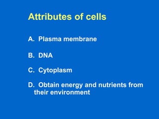
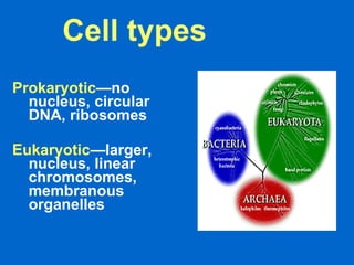
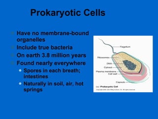
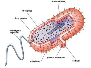
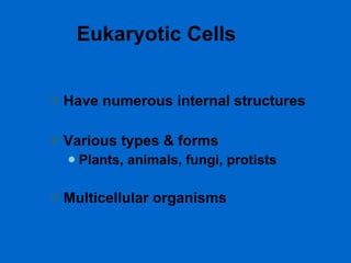
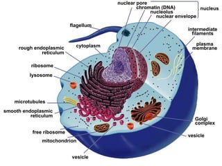
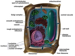
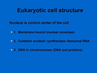
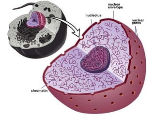
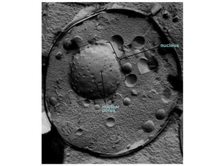
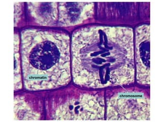
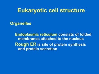
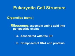
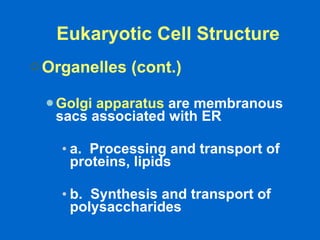
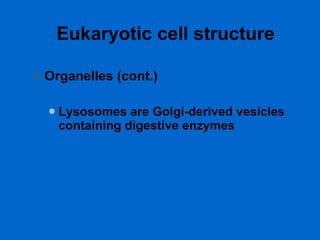
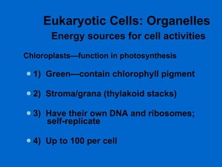
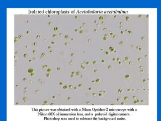
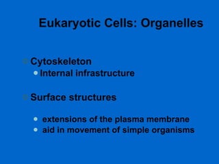
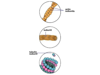
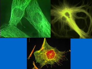
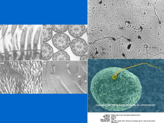
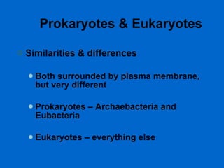
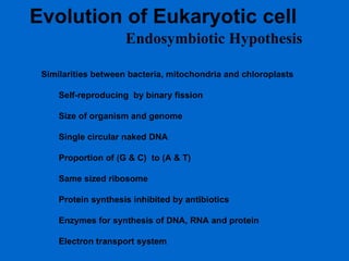
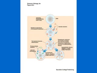
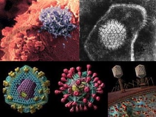
Cell Structure and Function
Attributes of cells   A.  Plasma membrane  B.  DNA  C.  Cytoplasm  D.  Obtain energy and nutrients from their environment
1.  1–100µm 2.  Why is there a limit to cell size?  a.  Surface-to-volume ratio  b.  Distance from surface to center  Cell Size
adult human tallest trees chicken egg frog embryo most eukaryotic cells mitochondrion most bacteria virus proteins atoms diameter of DNA double helix
 
Prokaryotic —no nucleus, circular DNA, ribosomes   Eukaryotic —larger, nucleus, linear chromosomes, membranous organelles  Cell types
Prokaryotic Cells Have no membrane-bound organelles Include true bacteria On earth 3.8 million years Found nearly everywhere Spores in each breath; intestines Naturally in soil, air, hot springs
ribosomes cell wall plasma membrane food granule prokaryotic flagellum cytoplasm nucleoid (DNA)
Eukaryotic Cells Have numerous internal structures Various types & forms Plants, animals, fungi, protists Multicellular organisms
flagellum cytoplasm rough endoplasmic reticulum ribosome microtubules lysosome mitochondrion Golgi  complex  plasma membrane intermediate filaments vesicle smooth endoplasmic reticulum free ribosome vesicle nuclear pore chromatin (DNA) nucleolus nuclear envelope  nucleus
chloroplast central vacuole rough endoplasmic reticulum ribosomes microtubules (part of cytoskeleton) cell wall mitochondrion Golgi complex  plasma membrane intermediate filaments smooth endoplasmic reticulum free ribosome vesicle nuclear pore chromatin nucleolus nuclear envelope  nucleus
Eukaryotic cell structure   Nucleus is control center of the cell  1.  Membrane bound (nuclear envelope)  2.  Contains nucleoli; synthesizes ribosomal RNA  3.  DNA in chromosomes (DNA and proteins)
nuclear  pores chromatin nucleolus nuclear  envelope
nuclear  pores nucleus
chromosome chromatin
0.05 micrometers
Eukaryotic cell structure Organelles  Endoplasmic reticulum   consists of folded membranes attached to the nucleus  Rough ER   is site of protein synthesis and protein secretion
0.5 micrometers smooth endoplasmic reticulum vesicles ribosomes rough endoplasmic reticulum 0.5 micrometers
Eukaryotic Cell Structure Organelles (cont.) Ribosomes   assemble amino acid into polypeptide chains  a.  Associated with the ER  b.  Composed of RNA and proteins
0.5 micrometers ribosomes rough endoplasmic reticulum
0.5 micrometers smooth endoplasmic reticulum vesicles
Eukaryotic Cell Structure Organelles (cont.) Golgi apparatus  are membranous sacs associated with ER a.  Processing and transport of proteins, lipids  b.  Synthesis and transport of polysaccharides
Golgi  complex  vesicles from ER vesicles leaving Golgi  complex
Eukaryotic cell structure Organelles (cont.) Lysosomes are Golgi-derived vesicles containing digestive enzymes
Golgi  complex  vesicles from ER vesicles leaving Golgi  complex
Eukaryotic Cells: Organelles   Energy sources for cell activities Mitochondria provide energy for cellular functions (respiration)  a.  Membrane bound, numerous  b.  Matrix/cristae  c.  Have their own DNA and ribosomes; self-replicate
 
5 micrometers
Eukaryotic Cells: Organelles   Energy sources for cell activities Chloroplasts—function in photosynthesis 1)  Green—contain chlorophyll pigment  2)  Stroma/grana (thylakoid stacks)  3)  Have their own DNA and ribosomes;    self-replicate  4)  Up to 100 per cell
 
 
Eukaryotic Cells: Organelles Cytoskeleton Internal infrastructure Surface structures extensions of the plasma membrane  aid in movement of simple organisms
tubulin subunit actin subunits subunit
 
 
Prokaryotes & Eukaryotes Similarities & differences Both surrounded by plasma membrane, but very different Prokaryotes – Archaebacteria and Eubacteria Eukaryotes – everything else
Evolution of Eukaryotic cell Endosymbiotic Hypothesis Similarities between bacteria, mitochondria and chloroplasts Self-reproducing  by binary fission Size of organism and genome Single circular naked DNA Proportion of (G & C)  to (A & T) Same sized ribosome Protein synthesis inhibited by antibiotics Enzymes for synthesis of DNA, RNA and protein Electron transport system
 
Plant & Animal Cells Similarities Both constructed from eukaryotic cells Both contain similar organelles Both surrounded by cell membrane
Plant & Animal Cells Differences Plants have Cell wall – provides strength & rigidity Have chloroplasts, photosynthetic Animals have Other organelle not found in plants (lysosomes formed from Golgi) Centrioles, important in cell division
Brief Look at Viruses Viruses are acellular Not considered to be living Cause serious diseases in most organisms
 

Cell structure-function

  • 1.
  • 2.
    Attributes of cells A. Plasma membrane B. DNA C. Cytoplasm D. Obtain energy and nutrients from their environment
  • 3.
    1. 1–100µm2. Why is there a limit to cell size? a. Surface-to-volume ratio b. Distance from surface to center Cell Size
  • 4.
    adult human tallesttrees chicken egg frog embryo most eukaryotic cells mitochondrion most bacteria virus proteins atoms diameter of DNA double helix
  • 5.
  • 6.
    Prokaryotic —no nucleus,circular DNA, ribosomes Eukaryotic —larger, nucleus, linear chromosomes, membranous organelles Cell types
  • 7.
    Prokaryotic Cells Haveno membrane-bound organelles Include true bacteria On earth 3.8 million years Found nearly everywhere Spores in each breath; intestines Naturally in soil, air, hot springs
  • 8.
    ribosomes cell wallplasma membrane food granule prokaryotic flagellum cytoplasm nucleoid (DNA)
  • 9.
    Eukaryotic Cells Havenumerous internal structures Various types & forms Plants, animals, fungi, protists Multicellular organisms
  • 10.
    flagellum cytoplasm roughendoplasmic reticulum ribosome microtubules lysosome mitochondrion Golgi complex plasma membrane intermediate filaments vesicle smooth endoplasmic reticulum free ribosome vesicle nuclear pore chromatin (DNA) nucleolus nuclear envelope nucleus
  • 11.
    chloroplast central vacuolerough endoplasmic reticulum ribosomes microtubules (part of cytoskeleton) cell wall mitochondrion Golgi complex plasma membrane intermediate filaments smooth endoplasmic reticulum free ribosome vesicle nuclear pore chromatin nucleolus nuclear envelope nucleus
  • 12.
    Eukaryotic cell structure Nucleus is control center of the cell 1. Membrane bound (nuclear envelope) 2. Contains nucleoli; synthesizes ribosomal RNA 3. DNA in chromosomes (DNA and proteins)
  • 13.
    nuclear poreschromatin nucleolus nuclear envelope
  • 14.
  • 15.
  • 16.
  • 17.
    Eukaryotic cell structureOrganelles Endoplasmic reticulum consists of folded membranes attached to the nucleus Rough ER is site of protein synthesis and protein secretion
  • 18.
    0.5 micrometers smoothendoplasmic reticulum vesicles ribosomes rough endoplasmic reticulum 0.5 micrometers
  • 19.
    Eukaryotic Cell StructureOrganelles (cont.) Ribosomes assemble amino acid into polypeptide chains a. Associated with the ER b. Composed of RNA and proteins
  • 20.
    0.5 micrometers ribosomesrough endoplasmic reticulum
  • 21.
    0.5 micrometers smoothendoplasmic reticulum vesicles
  • 22.
    Eukaryotic Cell StructureOrganelles (cont.) Golgi apparatus are membranous sacs associated with ER a. Processing and transport of proteins, lipids b. Synthesis and transport of polysaccharides
  • 23.
    Golgi complex vesicles from ER vesicles leaving Golgi complex
  • 24.
    Eukaryotic cell structureOrganelles (cont.) Lysosomes are Golgi-derived vesicles containing digestive enzymes
  • 25.
    Golgi complex vesicles from ER vesicles leaving Golgi complex
  • 26.
    Eukaryotic Cells: Organelles Energy sources for cell activities Mitochondria provide energy for cellular functions (respiration) a. Membrane bound, numerous b. Matrix/cristae c. Have their own DNA and ribosomes; self-replicate
  • 27.
  • 28.
  • 29.
    Eukaryotic Cells: Organelles Energy sources for cell activities Chloroplasts—function in photosynthesis 1) Green—contain chlorophyll pigment 2) Stroma/grana (thylakoid stacks) 3) Have their own DNA and ribosomes; self-replicate 4) Up to 100 per cell
  • 30.
  • 31.
  • 32.
    Eukaryotic Cells: OrganellesCytoskeleton Internal infrastructure Surface structures extensions of the plasma membrane aid in movement of simple organisms
  • 33.
    tubulin subunit actinsubunits subunit
  • 34.
  • 35.
  • 36.
    Prokaryotes & EukaryotesSimilarities & differences Both surrounded by plasma membrane, but very different Prokaryotes – Archaebacteria and Eubacteria Eukaryotes – everything else
  • 37.
    Evolution of Eukaryoticcell Endosymbiotic Hypothesis Similarities between bacteria, mitochondria and chloroplasts Self-reproducing by binary fission Size of organism and genome Single circular naked DNA Proportion of (G & C) to (A & T) Same sized ribosome Protein synthesis inhibited by antibiotics Enzymes for synthesis of DNA, RNA and protein Electron transport system
  • 38.
  • 39.
    Plant & AnimalCells Similarities Both constructed from eukaryotic cells Both contain similar organelles Both surrounded by cell membrane
  • 40.
    Plant & AnimalCells Differences Plants have Cell wall – provides strength & rigidity Have chloroplasts, photosynthetic Animals have Other organelle not found in plants (lysosomes formed from Golgi) Centrioles, important in cell division
  • 41.
    Brief Look atViruses Viruses are acellular Not considered to be living Cause serious diseases in most organisms
  • 42.

Editor's Notes

  • #5 Figure: 04-01 Title: Relative sizes. Caption: Dimensions commonly encountered in biology range from about 100 meters (the height of the tallest redwoods) through a few micrometers (the diameter of most cells) to a few nanometers (the diameter of many large molecules).
  • #6 Figure: 04-00UN01 Title: Geometric considerations. Caption: Geometric considerations.
  • #9 Figure: 04-10 Title: A generalized prokaryotic cell. Caption: A generalized prokaryotic cell.
  • #11 Figure: 04-02 Title: A generalized animal cell. Caption: A generalized animal cell.
  • #12 Figure: 04-03 Title: A generalized plant cell. Caption: A generalized plant cell.
  • #14 Figure: 04-04a Title: The nucleus. Caption: (a) The nucleus is bounded by a nuclear envelope. Inside are chromatin (DNA and associated proteins) and a nucleolus.
  • #15 Figure: 04-04b Title: The nucleus. Caption: (b) An electron micrograph of a yeast cell that was frozen and broken open to reveal its internal structures. The large nucleus, with nuclear pores penetrating its nuclear envelope, is clearly visible.
  • #16 Figure: 04-05 Title: Chromosomes. Caption: Chromosomes, seen here in a light micrograph of a dividing cell (on the right) in an onion root tip, are the same material (DNA and proteins) as the chromatin seen in nondividing cells adjacent to it, but in a more compact state.
  • #17 Figure: 04-06 Title: Ribosomes. Caption: Ribosomes.
  • #19 Figure: 04-07 Title: Endoplasmic reticulum. Caption: There are two types of endoplasmic reticulum: rough ER, coated with ribosomes, and smooth ER, without ribosomes. Although in electron micrographs the ER looks like a series of tubes and sacs, it is actually a maze of folded sheets and interlocking channels.
  • #21 Figure: 04-07R-1 Title: Rough endoplasmic reticulum. Caption: Rough endoplasmic reticulum.
  • #22 Figure: 04-07R-2 Title: Smooth endoplasmic reticulum. Caption: Smooth endoplasmic reticulum.
  • #24 Figure: 04-08 Title: The Golgi complex. Caption: The Golgi complex is a stack of flat membranous sacs. Vesicles transport material from the ER to the Golgi (and vice versa) and from the Golgi to plasma membrane, lysosomes, and vesicles. Departing vesicles bud off from the Golgi on one face; arriving vesicles join it on the opposite face.
  • #26 Figure: 04-08 Title: The Golgi complex. Caption: The Golgi complex is a stack of flat membranous sacs. Vesicles transport material from the ER to the Golgi (and vice versa) and from the Golgi to plasma membrane, lysosomes, and vesicles. Departing vesicles bud off from the Golgi on one face; arriving vesicles join it on the opposite face.
  • #29 Figure: 04-E4-2d Title: An SEM photo. Caption: (d) An SEM photo at much higher magnification, showing mitochondria, many of which are sliced open.
  • #34 Figure: 04-2 Title: Cytoskeleton components. Caption: Cytoskeleton components.