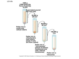
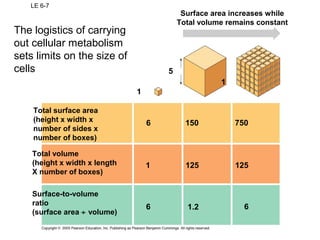
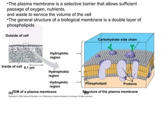
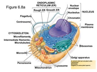
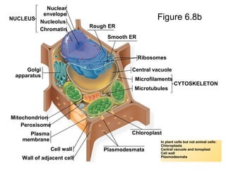
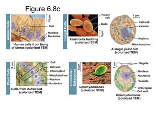
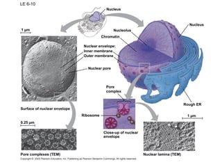
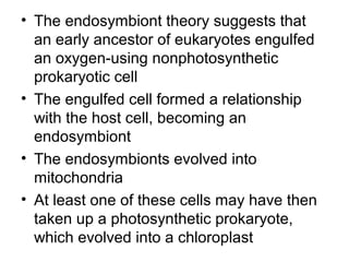
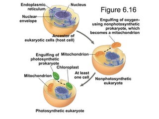
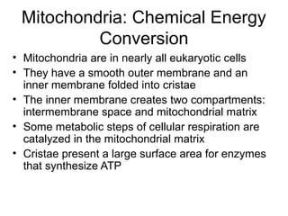
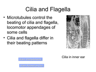
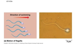
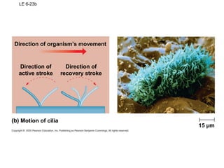
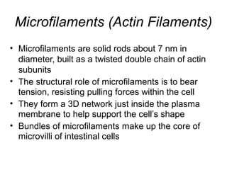
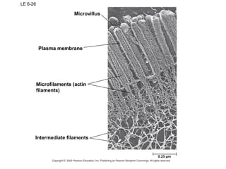
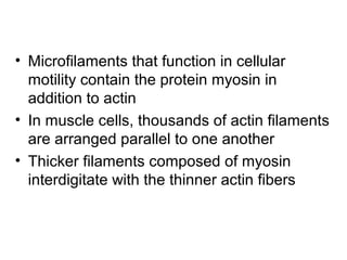
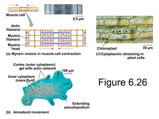
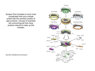
Chapter 6 discusses the basic unit of life, the cell, highlighting the correlation between cell structure and function, and the distinctions between prokaryotic and eukaryotic cells. It covers microscopy techniques for visualizing cells, the cellular components including organelles like the nucleus, endoplasmic reticulum, and mitochondria, and the endomembrane system's role in protein transportation and metabolic functions. Additionally, the chapter explores the cytoskeleton's structure and function, as well as the mechanics of cilia and flagella movement.
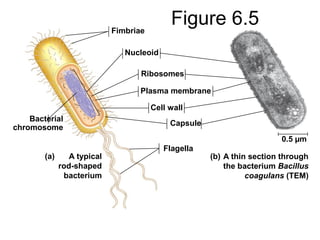
![• Prokaryotic cells have no nucleus
• In a prokaryotic cell, DNA is in an unbound
region called the nucleoid
• Prokaryotic cells lack membrane-bound
organelles
Cell wall of the gram-negative bacterium
Leptothrix discophora. Visible are the inner
and outer membranes and the peptidoglycan
cell wall. (Transmission electron micrograph.)
[© T. J. Beveridge/BPS.]](https://image.slidesharecdn.com/biochapter6notes-151125141544-lva1-app6892/85/Bio-chapter-6-notes-11-320.jpg)


![06atourofthecell-130311053323-phpapp01 [Autoguardado].ppt](https://cdn.slidesharecdn.com/ss_thumbnails/06atourofthecell-130311053323-phpapp01autoguardado-250807125330-0210e941-thumbnail.jpg?width=640&height=640&fit=bounds)