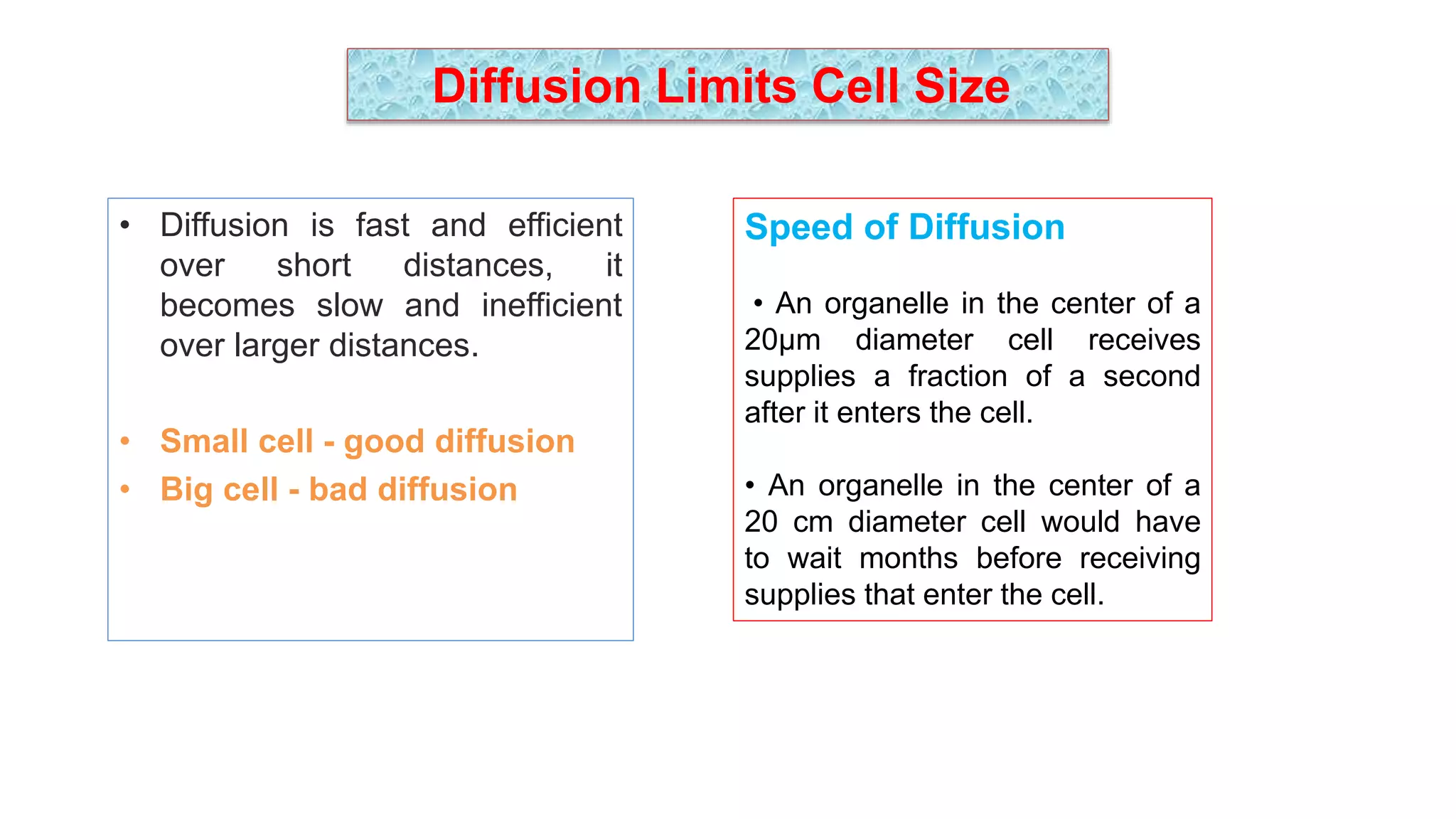
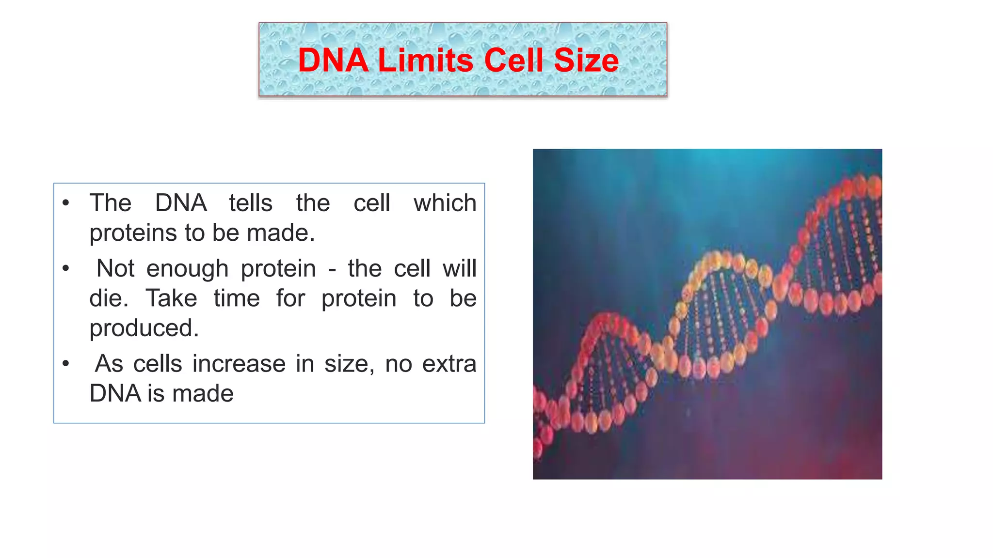
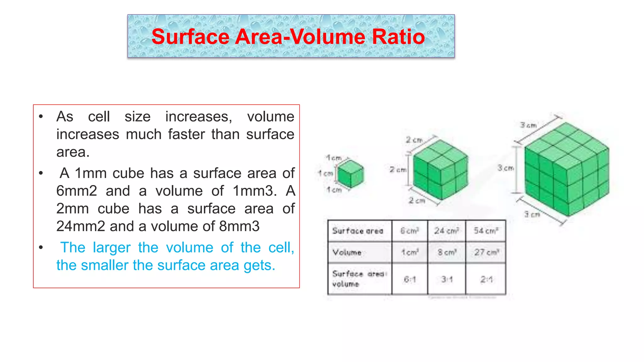
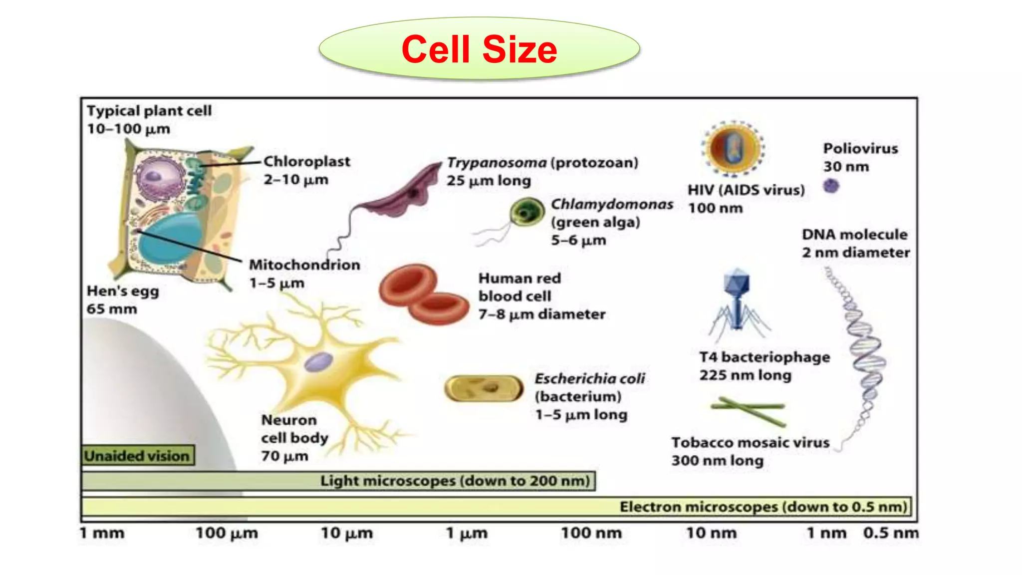
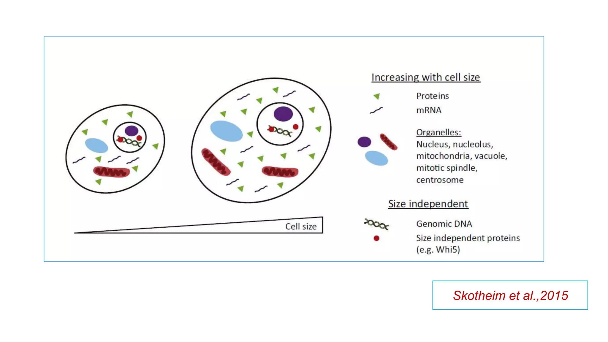
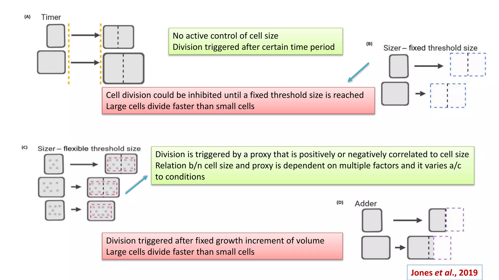
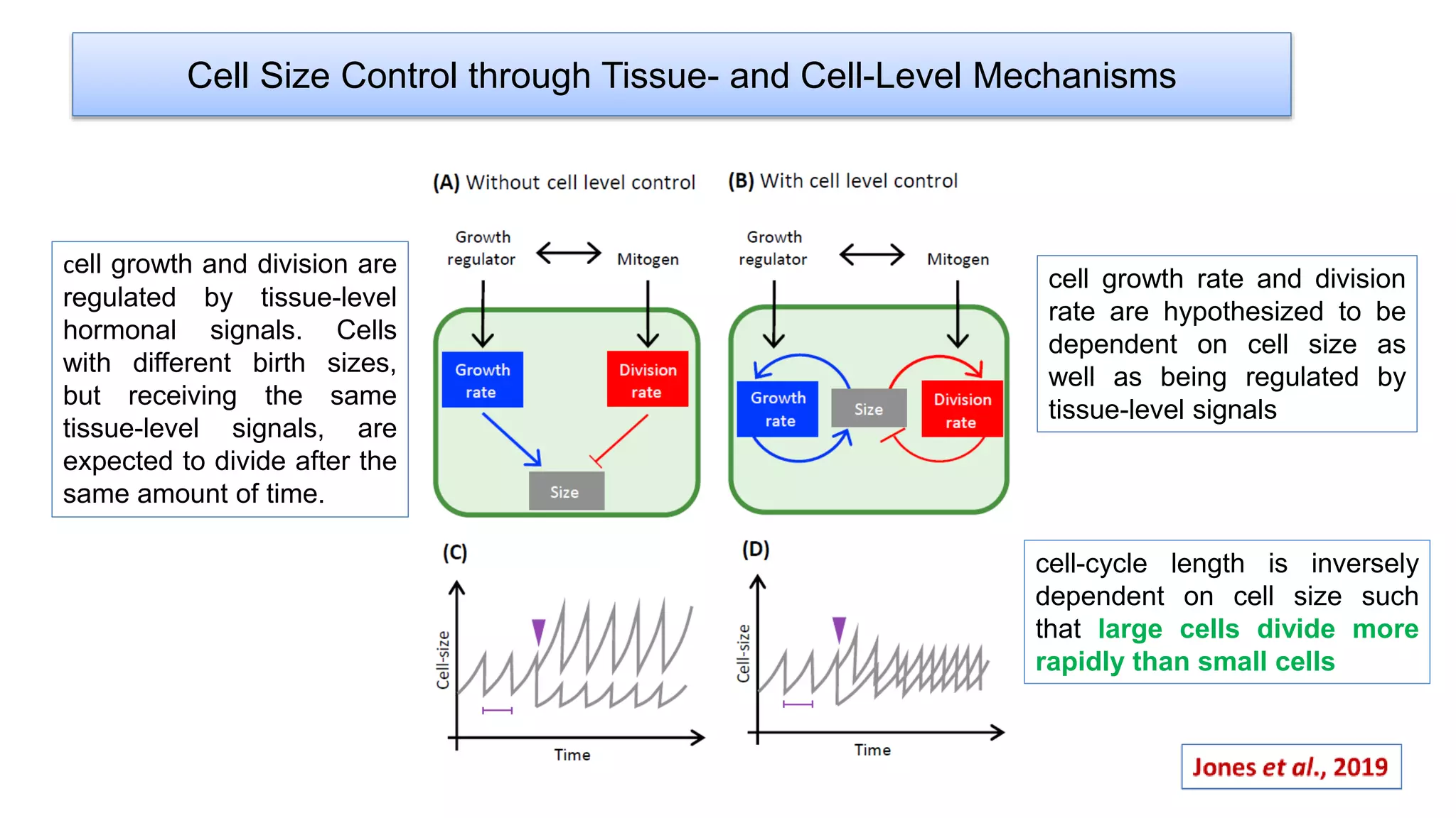
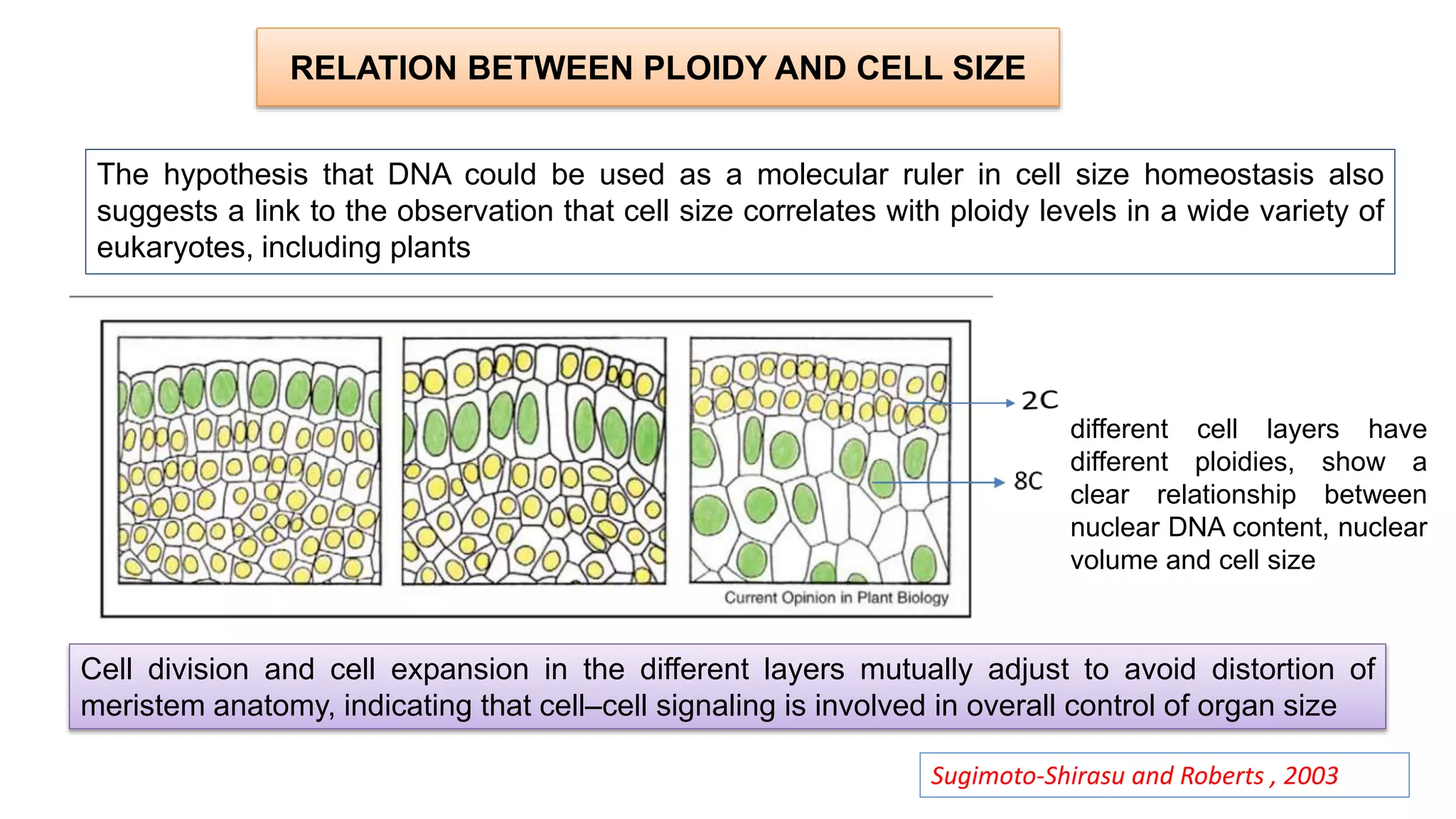
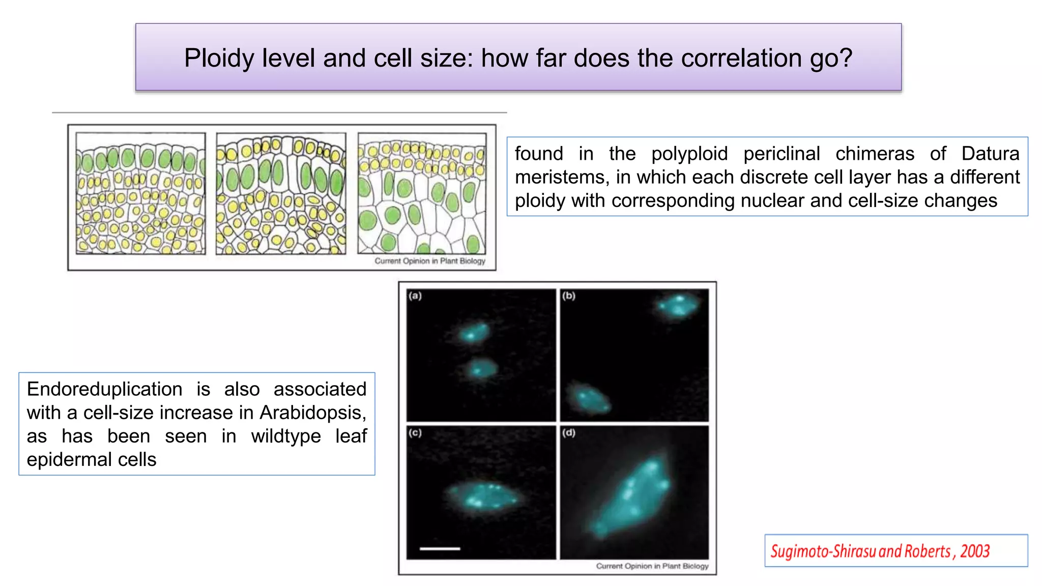
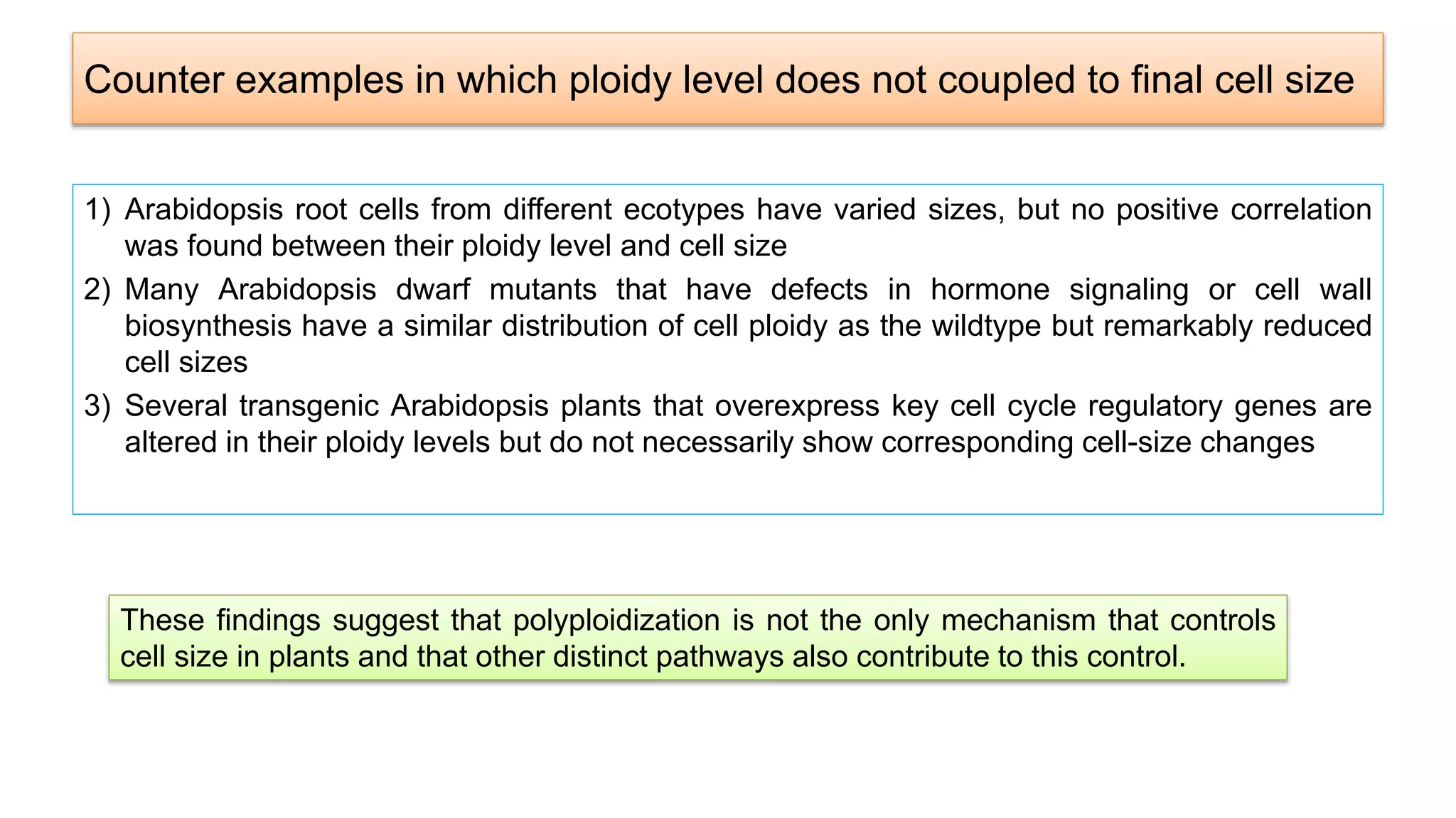
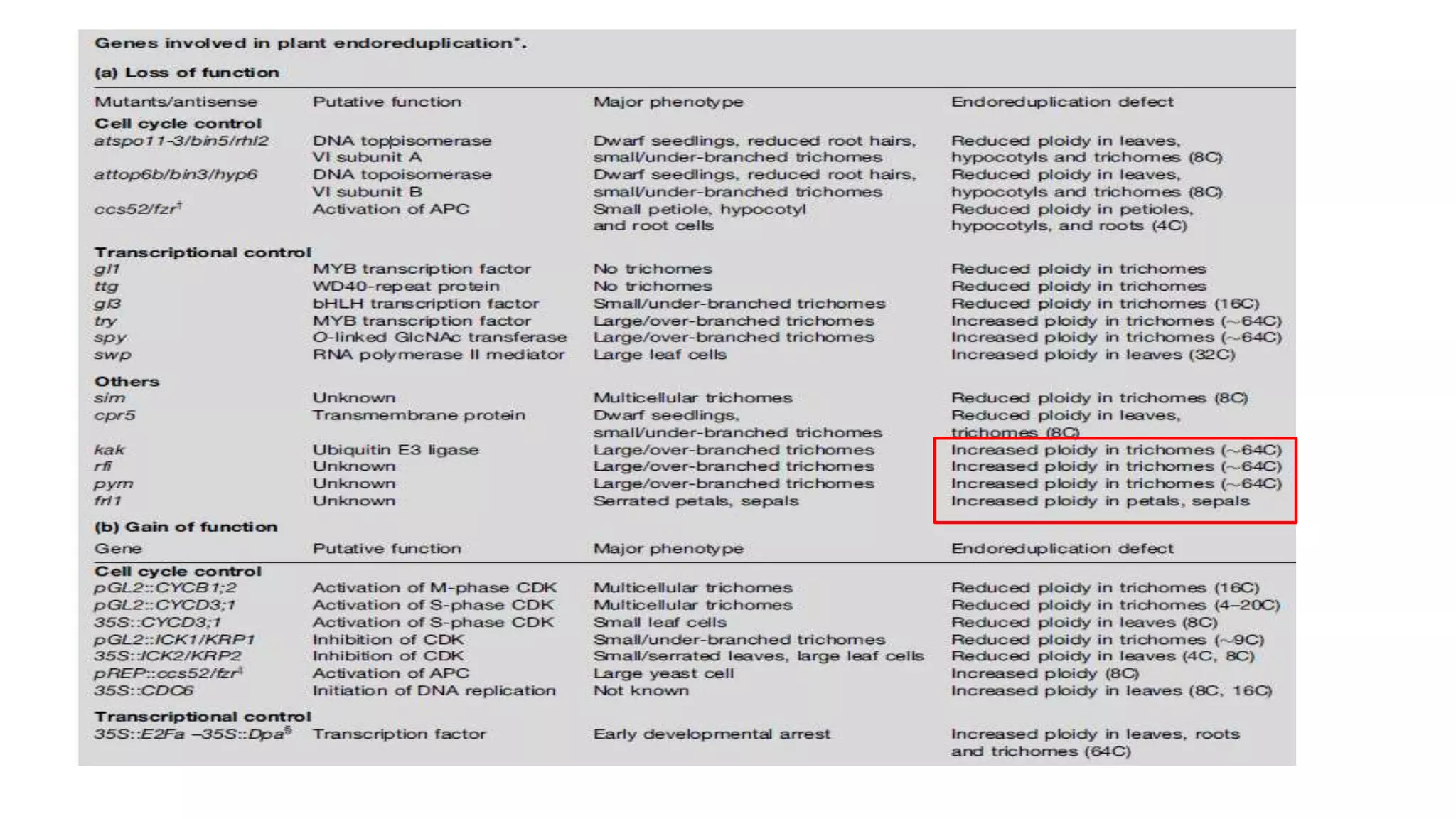
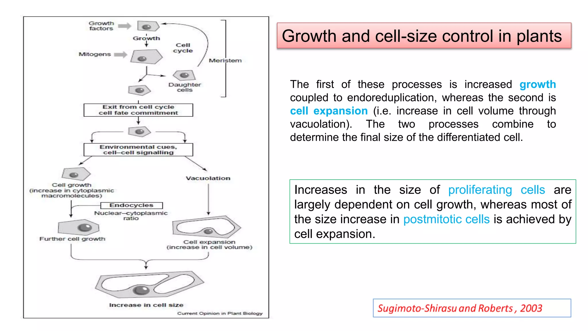
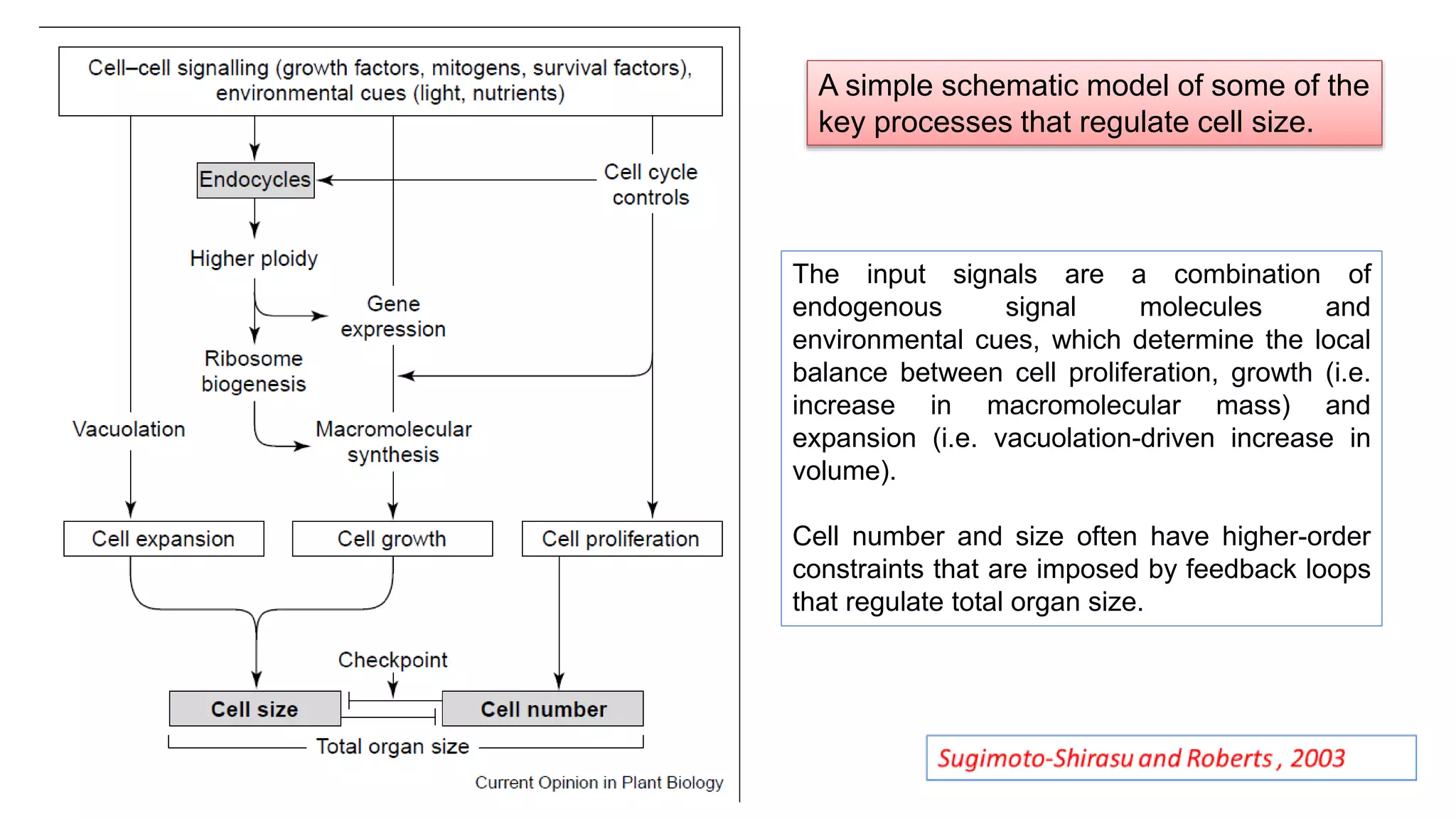
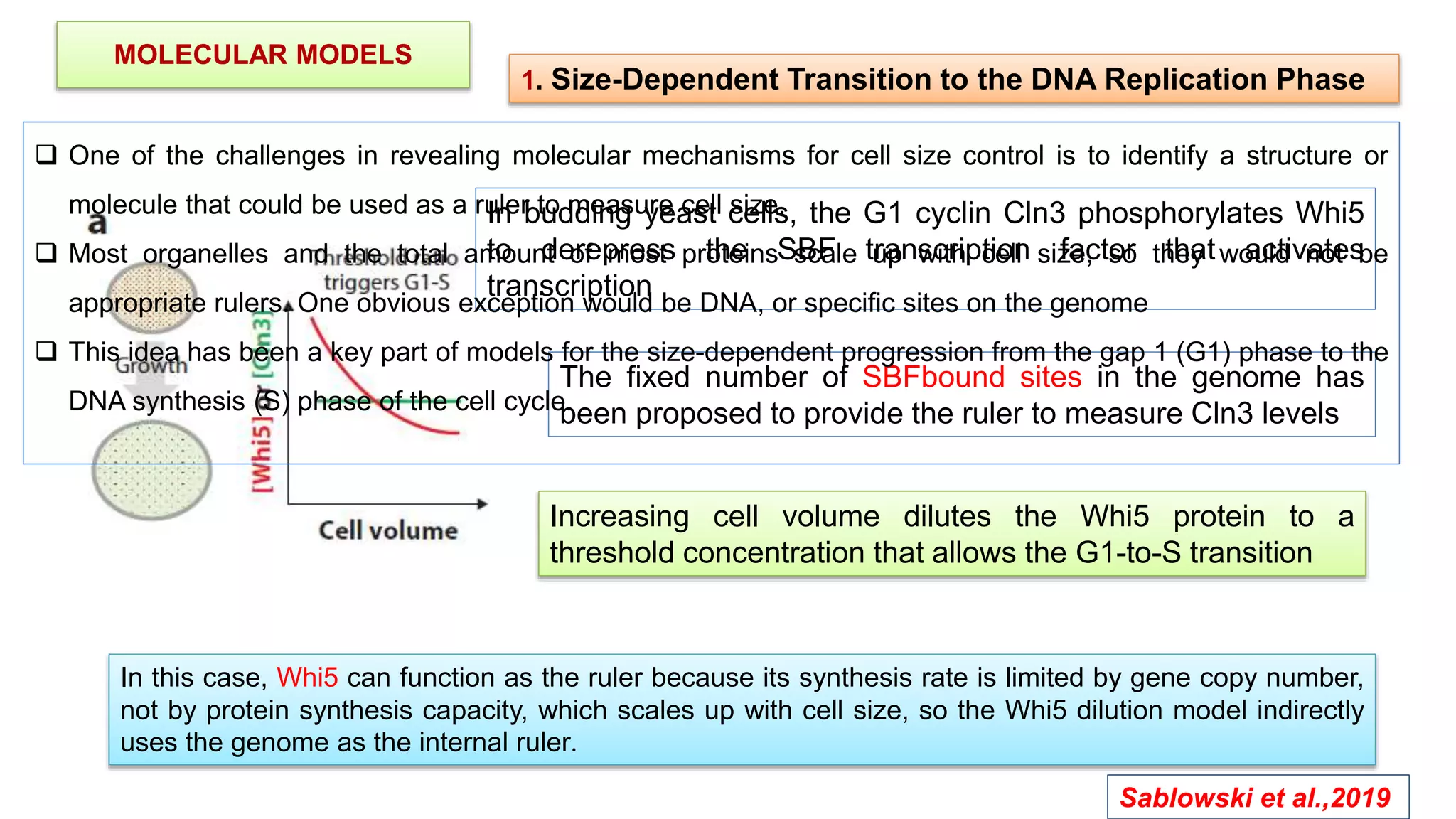
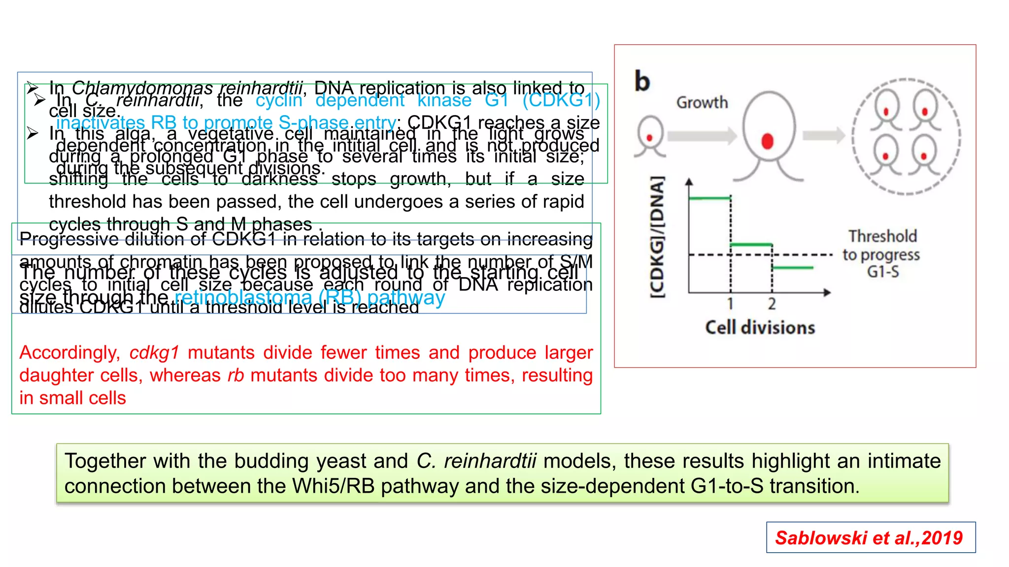
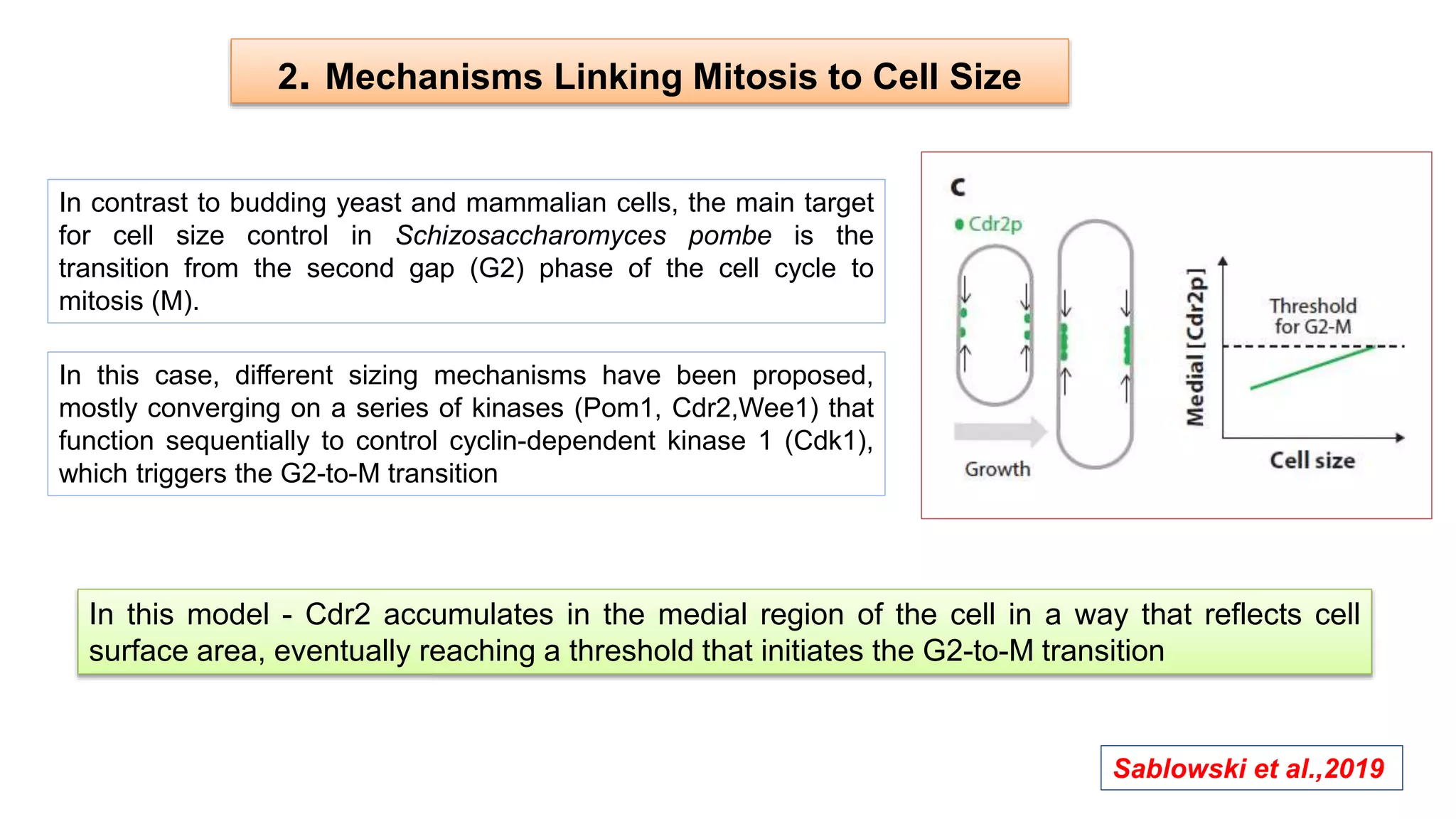
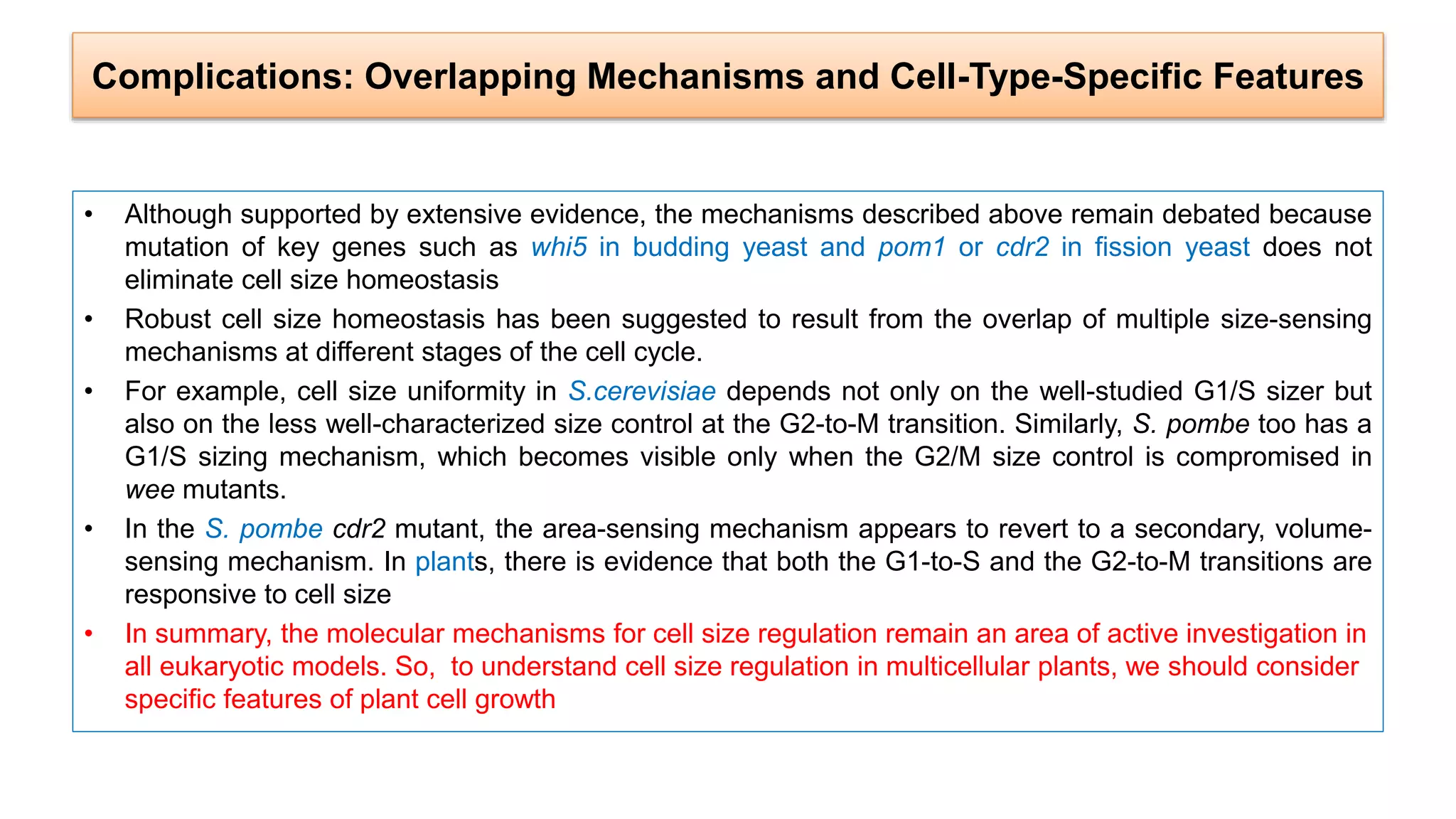
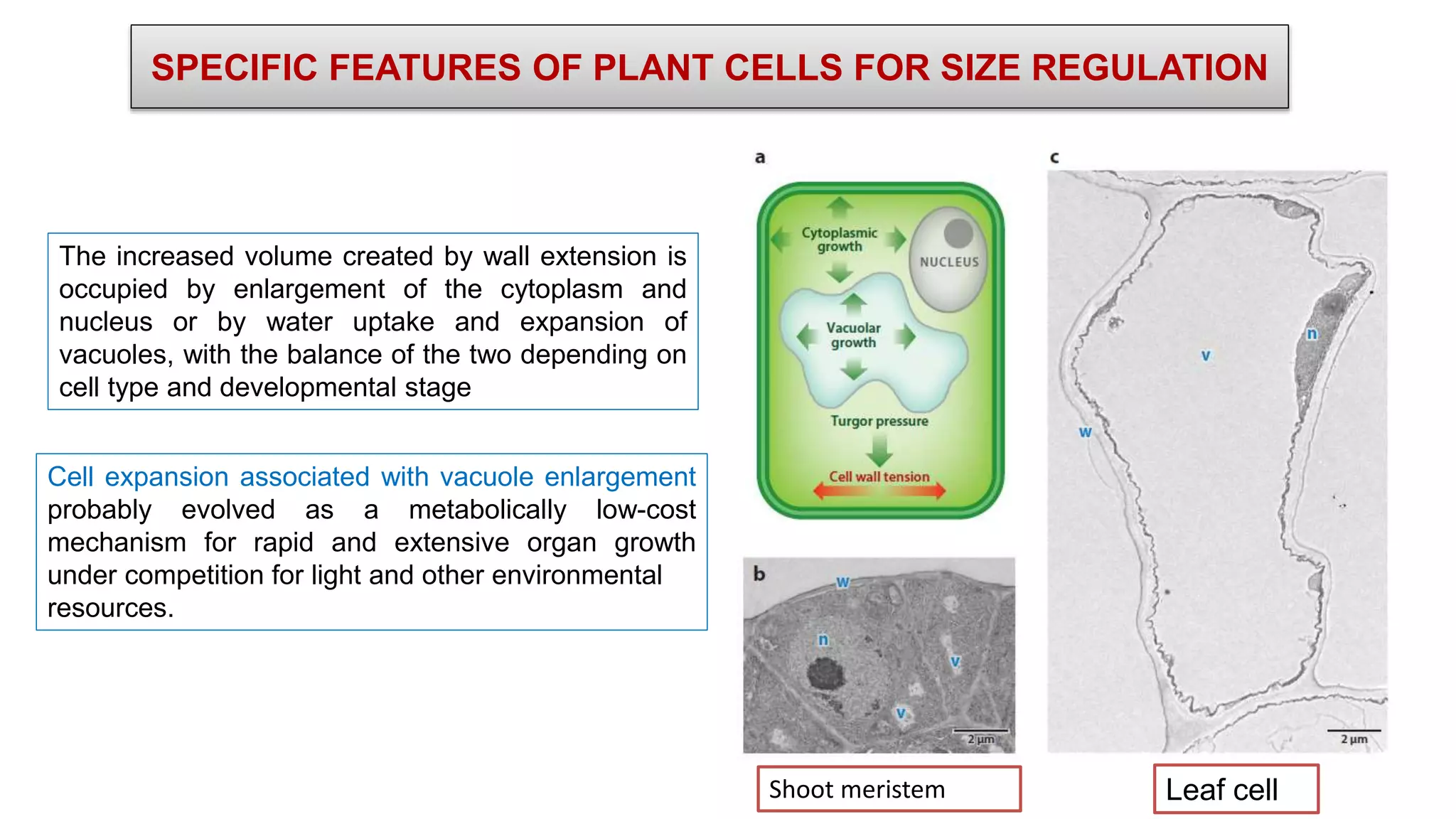
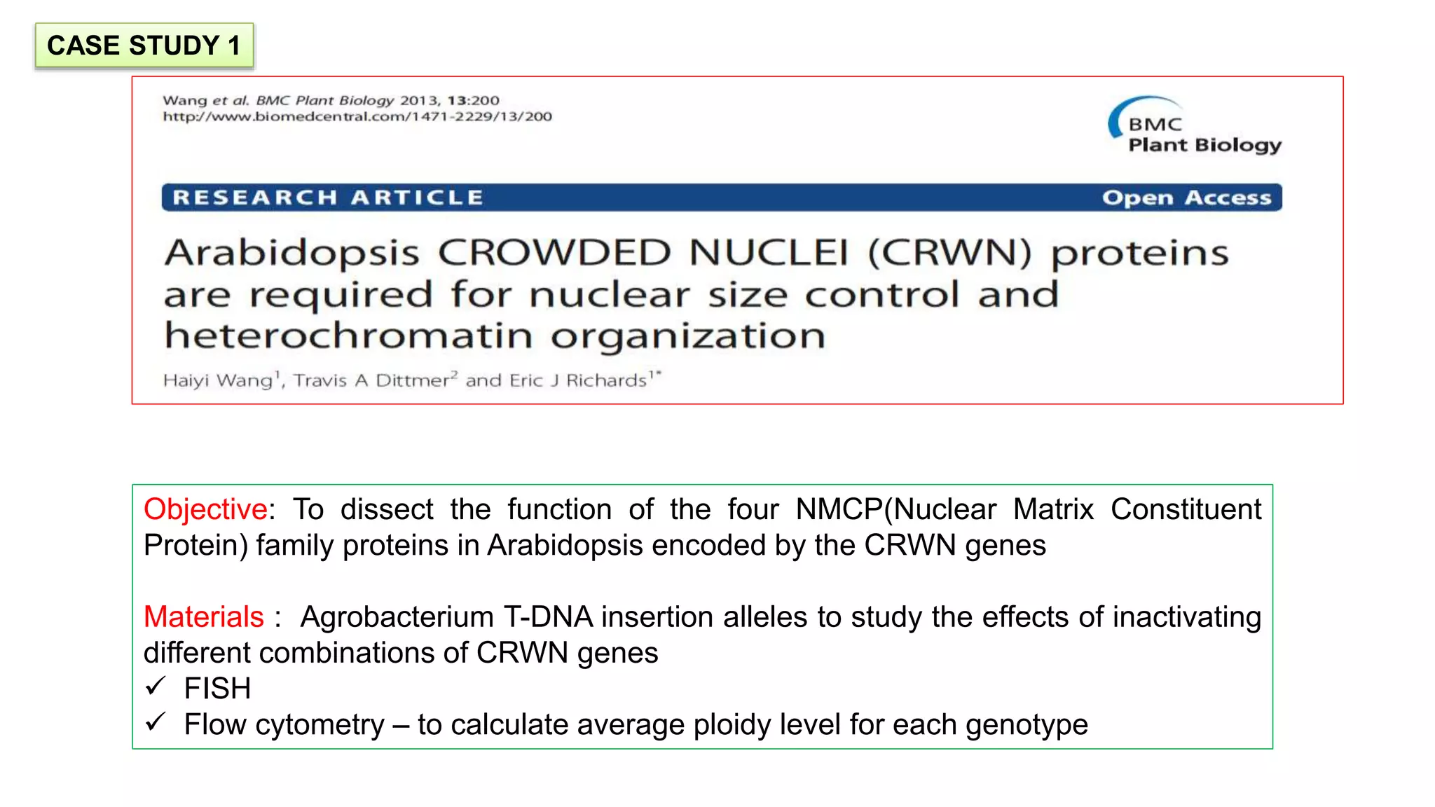
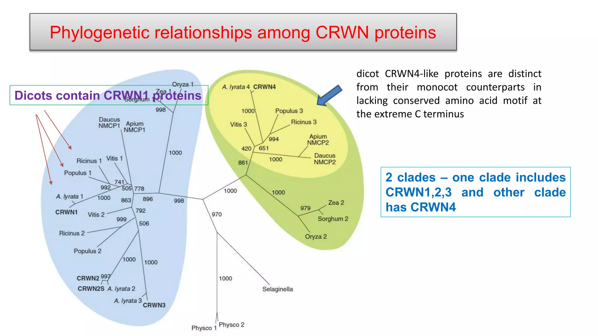
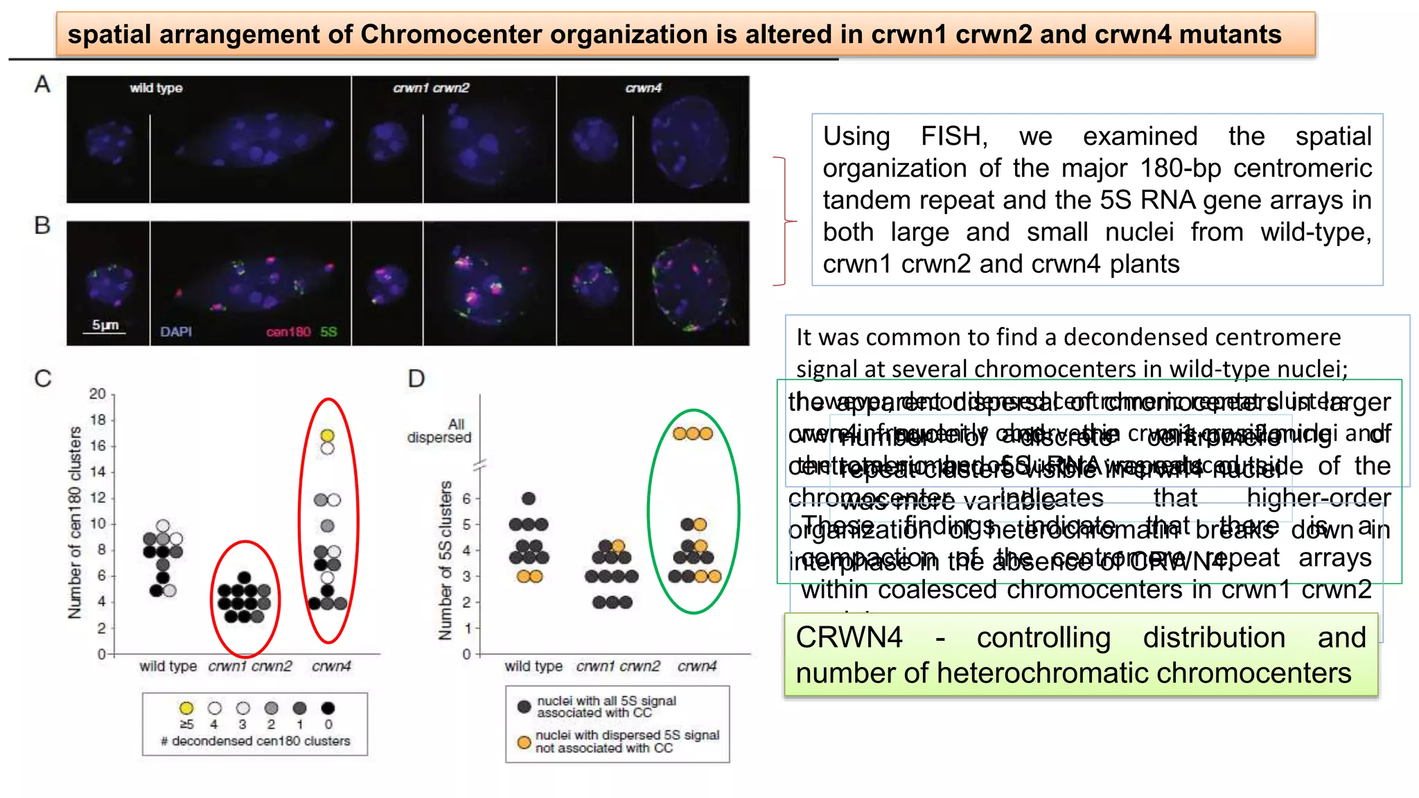
Size is important for cell function and is characteristic of different species and of different cell types in the same organism. How cell size is specified has intrigued researchers ever since cells became visible to biologists. Cell size determines the geometry of all intracellular compartments and sets the scale of biosynthetic processes. Biosynthesis increases in larger cells, which have proportionally more protein, total RNA, total mRNA, and mRNA for specific investigated genes.Plants are a good multicellular system for studying the regulation of cell size. As plant cells cannot migrate because they are encased in a cell wall matrix, their growth and division can be readily tracked and measured by live cell imaging and three-dimensional (3D) image analysis. Plants are also particularly relevant for studying how cell size relates to ploidy and to organ size because of the prominent role of polyploidy in plant evolution, plant development, and crop domestication.
The document discusses transport across cell membranes. It begins by describing the structure and function of cell membranes, including their semipermeable nature. It then explains various transport mechanisms like diffusion, osmosis, facilitated diffusion, active transport, and endocytosis/exocytosis that allow materials to move across membranes. Specific examples are given of how these transport mechanisms function in cells, lungs, and other organisms and systems to maintain homeostasis.
This document discusses different types of cell and organ culture. It describes four main types of culture media: natural media which are obtained from natural sources but have unknown compositions; and three types of artificial media - serum containing media which typically contain 2-10% fetal bovine serum, serum-free defined media which have consistent compositions and reduce contamination risk, and protein-free and chemically defined media. It also outlines techniques for primary cell culture, establishing cell lines, and organ culture methods like plasma clot, agar gel, and raft methods.
This document discusses stem cell niches and the microenvironment that supports stem cells. It outlines various cell types that make up stem cell niches in the bone marrow including mesenchymal stem cells, endothelial cells, and osteoblasts. It describes markers that characterize these cell types and factors they secrete like cytokines, growth factors, and extracellular matrix proteins that regulate stem cell self-renewal and differentiation. Pathways involved in maintaining stem cells like the Wnt signaling pathway are also summarized.
Cell adhesion molecules and matrix proteinsUSmile Ï Ṩṃïlệ
Cell adhesion molecules are proteins located on cell surfaces that are involved in binding between cells or between cells and the extracellular matrix. The three main types are cadherins, integrins, and selectins. Cadherins are calcium-dependent proteins that mediate cell-cell adhesion. Integrins are transmembrane receptors that bind to components of the extracellular matrix and mediate cell-matrix adhesion as well as cell signaling. Selectins mediate the initial capture and rolling of leukocytes along vascular surfaces. Cell adhesion molecules play important physiological roles in processes like leukocyte trafficking, blood coagulation, and morphogenesis. They also have applications as therapeutic targets in areas such as cancer, osteoporosis, and inflammatory diseases.
- The document discusses the structure of the cell membrane and cellular junctions.
- It describes the fluid mosaic model of the cell membrane, which proposes that the membrane is composed of a lipid bilayer with proteins embedded and floating within it, giving it a fluid and mosaic-like structure.
- There are two main types of cellular junctions - anchoring junctions, which attach the cell to other cells or the extracellular matrix, and tight junctions, which form a seal between adjacent cell membranes to control what can pass through the space between them.
wo-dimensional gel electrophoresis, abbreviated as 2-DE or 2-D electrophoresis, is a form of gel electrophoresis commonly used to analyze proteins. Mixtures of proteins are separated by two properties in two dimensions on 2D gels. 2-DE was first independently introduced by O'Farrell and Klose in 1975.
The document summarizes the nutritional requirements of cultured cells. It discusses that cells need nutrients like amino acids, vitamins, inorganic ions, trace elements, monosaccharides, and growth factors to survive and grow in culture. Specific requirements are outlined, including essential amino acids, glutamine, salts like sodium, potassium and calcium, and glucose as an energy source. Serum provides additional growth factors, minerals, lipids and hormones to support cell growth. Characteristics of cell culture and applications are also briefly mentioned.
The cell proliferation will generally decrease with the differentiation of cells, and most cells in adult animals are blocked in the G 0 phase of the cell cycle.
https://www.creative-bioarray.com/Services/cell-proliferation-assay-services.htm
Specialized Tissues, Stem Cells and Tissue RenewalGarry D. Lasaga
This report will talk about the physiology of tissue renewal among different tissues and the important role played by stem cells in such mechanism as wells as the various key signaling mechanism involved.
This document provides an outline for Chapter 4 of the biology textbook "Cell Structure and Function" by Sylvia S. Mader and Michael Windelspecht. The outline covers topics including the cellular level of organization, prokaryotic and eukaryotic cells, organelles such as the nucleus, endomembrane system, cytoskeleton, and energy-related organelles. Diagrams are included to illustrate cell structures like plant and animal cells as well as different types of microscopes used to study cells.
The document discusses the composition and functions of the extracellular matrix (ECM) and cell-cell junctions. The ECM provides structural support to cells and regulates cell behavior. It is composed of fibrous proteins like collagen, polysaccharides like glycosaminoglycans, and adhesion proteins like fibronectin and laminin. Cells interact with the ECM through integrin receptors. Cell-cell junctions allow communication between cells and include adherens junctions, desmosomes, tight junctions, and gap junctions. The ECM and cell-cell junctions are essential for tissue structure and function.
This document provides an overview of cell biology, covering key topics such as:
1. The different types of microscopes used to study cells and their relative capabilities.
2. The main differences between prokaryotic and eukaryotic cells, including their genetic material and internal structures.
3. The structures and functions of eukaryotic cell organelles such as the nucleus, mitochondria, chloroplasts, Golgi apparatus, endoplasmic reticulum, and cytoskeleton. These organelles allow for compartmentalization and specialized functions within eukaryotic cells.
Membrane Dynamics:Properties of biological membrane (plasma membrane)Manju Chhetri
The document discusses membrane dynamics, including:
- Membrane fluidity is affected by temperature, fatty acid composition, and sterol content and allows for lipid movement within bilayers.
- Transbilayer lipid movement between inner and outer leaflets is slow but can be catalyzed by proteins.
- Lipids and proteins can diffuse laterally but this is limited by interactions with structures like lipid rafts and cytoskeleton.
- Caveolin associates with the inner plasma membrane leaflet and forms caveolae, playing roles in transport and signaling.
- Specific proteins cause local membrane curvature and mediate fusion between membranes during processes like endocytosis and viral invasion.
- Integrins attach cells to each other
Electroporation is a method that uses electric pulses to create temporary pores in cell membranes, allowing molecules like DNA to enter cells. It can be used to introduce foreign genes into host cells for transformation or transfection. The electric pulses temporarily permeabilize the membrane, and the DNA enters through newly formed pores and incorporates into the host cell genome. Electroporation has applications in biotech for bacterial, yeast, and plant transformation, as well as gene therapy, cell therapy, and tumor treatment. It allows efficient delivery of DNA vaccines and other molecules into cells with minimal amounts of material.
Vertical Gel Electrophoresis (SDS-PAGE)Srikanth H N
Vertical gel electrophoresis has several advantages over horizontal gel electrophoresis. It allows for the use of a discontinuous buffer system to separate proteins, which is not possible with horizontal gels. The technique involves pouring an acrylamide gel between glass plates to a thickness of less than 2 mm. Samples are loaded and subjected to an electric current, with cations moving toward the cathode and anions toward the anode. Proteins are separated based on their size and charge using techniques like SDS-PAGE, which involves denaturing proteins to impart a uniform charge.
Subculture involves transferring cells from an existing culture to fresh growth medium in order to prolong the life and expand the number of cells. This is necessary because over time toxic metabolites build up and nutrients are depleted in the original culture. Subculture produces a new culture with lower cell density and fresh nutrients. It allows for long-term maintenance of cell lines and increasing cell numbers for industrial or research purposes. Different techniques are used to subculture adherent versus non-adherent cells.
This document discusses cell culture in animal biotechnology. It provides an overview of the history of cell culture from the late 1800s to modern applications. Some key points covered include:
1) Cell culture refers to growing cells in a controlled artificial environment outside of their natural environment. Examples of cell types grown include fibroblasts, lymphocytes, and cells from various tissues.
2) Important milestones in the history of cell culture include Roux maintaining embryonic chick cells in 1885 and Carrel introducing strict aseptic techniques allowing long-term cell culture in 1913.
3) Cell culture has various applications including use as model systems, in toxicity testing, cancer research, virology, drug development, and gene therapy.
The document summarizes four basic mechanisms of transport across cell membranes: diffusion, facilitated diffusion, osmosis, and active transport. Diffusion is the passive movement of molecules from an area of high concentration to low concentration down a gradient. Facilitated diffusion uses protein channels to transport molecules that cannot diffuse directly through the membrane such as glucose. Osmosis is the diffusion of water across a partially permeable membrane to equalize water concentration. Active transport requires energy and transports molecules against a concentration gradient using carrier proteins.
Mitochondria are organelles found in most cells that produce energy through aerobic respiration. They convert nutrients into ATP, which fuels cellular activities, earning mitochondria their nickname as the "powerhouse of the cell." A mitochondrion contains inner and outer membranes that create distinct regions, including the matrix where ATP production occurs through redox reactions and oxidative phosphorylation. Mitochondria play a dominant role in cellular energy conversion by producing ATP using enzymes in the inner membrane.
Bioreactors for animal cell suspension cultureGrace Felciya
This document discusses bioreactors for animal cell suspension culture. It begins by introducing animal cell culture and some key developments that enabled it. There are two main types of culture: primary culture using explants or enzymes, and secondary culture which is derived from primary culture. Cells can be anchorage-dependent, growing in monolayers, or non-anchorage dependent, growing in suspension. Bioreactors provide conditions for mass cultivation of suspension cells. Properties of animal cells require gentle mixing and aeration in bioreactors. Common bioreactor types for suspension culture include stirred tank, continuous flow, and airlift fermentors. Perfusion culture allows continuous medium exchange to achieve high cell densities and productivity.
The plasma membrane is a selectively permeable membrane that surrounds the cell. It is composed of a phospholipid bilayer with embedded proteins. The fluid mosaic model describes the plasma membrane structure, with integral and peripheral proteins embedded within or attached to the fluid phospholipid bilayer. The plasma membrane regulates what enters and exits the cell through passive diffusion, facilitated diffusion, active transport, osmosis, and bulk transport mechanisms like endocytosis and exocytosis.
General principles of cell communicationGoutam Mallik
Cell signaling allows cells to communicate and coordinate their functions. There are several forms of cell signaling, including endocrine signaling where hormones travel through the bloodstream, paracrine signaling between neighboring cells, contact-dependent signaling through cell junctions, and synaptic signaling across nerve cell junctions. In intracellular signaling, receptor activation leads to the production of second messengers that transmit signals within the cell by activating intracellular signaling pathways, ultimately triggering responses like transcription, survival, movement, or metabolic changes.
The cell membrane is a phospholipid bilayer that forms a barrier around cells. The fluidity of the membrane must be maintained within a certain range for proper cell functioning. Several factors influence membrane fluidity, including the length of fatty acid tails, temperature, cholesterol content, and degree of saturation. Longer fatty acid tails, lower temperatures, more cholesterol, and more saturated fatty acids decrease fluidity by increasing interactions between phospholipid molecules.
The cell membrane is composed mainly of lipids and proteins that form a fluid bilayer. Phospholipids are the major lipids and form a bilayer with their hydrophobic tails facing inward and hydrophilic heads facing outward. Membrane proteins can be integral or peripheral. The fluid mosaic model describes membranes as a fluid bilayer with proteins diffusing laterally. Membranes exhibit asymmetry, fluidity, and formation of microdomains. Specific proteins mediate membrane fusion and curvature essential for cellular processes.
The document discusses apoptosis (programmed cell death) through three parts:
1) An introduction to apoptosis, its history, and how it is important in development and physiology.
2) The mechanisms and pathways of apoptosis, including caspases, the intrinsic mitochondrial pathway, extrinsic death receptor pathway, and Bcl-2 family of proteins.
3) The importance of apoptosis in normal development and physiology through tissue sculpting, but that defects can lead to diseases like cancer, autoimmunity, and neurodegeneration when there is too much or too little apoptosis.
Diversity of cell size & shape By KK Sahu SirKAUSHAL SAHU
Cells show tremendous diversity in size, shape, structure and function. Robert Hooke first observed cells in 1665 when examining a thin slice of cork under a microscope. Cells can be prokaryotic or eukaryotic, and range enormously in size from 0.1um to over 2m in length. Cell shape also varies greatly between species, with spherical, flat, elongated and branched shapes that often correlate to a cell's specialized function. This diversity arises from cells differentiating and specializing during development to perform distinct roles in multicellular organisms.
Bacterial Growth and structure in book of bacteriologysubeersomali
This document summarizes bacterial growth and methods for measuring it. It describes the stages of bacterial growth (lag phase, exponential phase, stationary phase, death phase) and the process of binary fission that bacteria use to replicate. It also discusses two common ways to measure bacterial growth: microscopic cell counts, which involve direct observation of cells under a microscope, and turbidity measurements, which measure cell mass indirectly. Microscopic counts have limitations like difficulty distinguishing live from dead cells and achieving precision.
Homecell divisionCell division
Cell division
Miller November 05, 2022
Every living organism depends on the growth and multiplication of its cells for growth and development because a multicellular organism begins as a single cell and undergoes repeated division. The characteristic trait of all living things is an increase in cell size brought on by growth. The cell starts to divide once its growth has reached its maximum. An organism grows vegetatively when its number of cells increases through cell divisions that follow a geometric progression. The three stages of cell division, which is a continuous and dynamic process, are as follows:
Replicating the genome or DNA
Karyokinesis, or nuclear division
Cytokinesis, also known as cell division
Based on the number of genomes present in the daughter cells in comparison to the dividing parent cell, there are two types of cell division: mitosis and meiosis.
1. Mitosis- W. Flemming first used the word mitosis in 1882. Mitosis, also known as somatic division, is the process by which a body cell divides into two daughter cells, each of equal size and with the same number of chromosomes as the parent cell.
2. Meiosis- J. Meiosis was the first to use the term. B. Farmer and J. Smith in 1905 Moore, E. Only the gonads (germ mother cells) undergo meiosis during the development of gametes like sperm and ovum. Meiosis is the process by which chromosomes go from having two copies, or 2N or diploid, to having only one copy, or N or haploid. Additionally known as the reduction process. Every cell that is able to divide undergoes a regular cycle of alterations known as the cell cycle. A cell is diploid when it begins its cycle.
Phases of cell cycle
The cell cycle has two phases: the long interphase, also known as Iphase, and the short mitotic, also known as M-phase, phases. 1. Interphase-
The interphase is the period of time between telophase's conclusion and the start of the following Mphase. The stage is long and complicated, lasting between 10 and 30 hours. The cell develops during this phase by producing biological molecules like lipids, proteins, carbohydrates, and nucleic acids.
First gap, also known as the G1 phase, second gap, also known as the G2 phase, and synthetic phase make up the interphase.
(i) G1 phase- The G1 phase represents the duration between the previous mitosis and the start of DNA synthesis. During this phase, a newly formed cell begins to grow. During this stage, a wide range of biological molecules—including RNAs, proteins, lipids, and some non-histones—are created.
In order to prepare for the DNA replication that will occur next to it, normal metabolism is carried out. This phase does not involve DNA synthesis. (ii) S Phase- Each chromosome is duplicated during this phase by replicating new DNA molecules using the existing DNA as a template. Only in S-phase do histone protein and their mRNA, some non-histone protein, and new nucleosome formation take place. Most eukary
B.sc. Microbiology II Bacteriology Unit 4.1 Bacterial GrowthRai University
This document discusses bacterial growth and the requirements for bacterial growth. It covers the following key points:
1. Bacterial growth occurs through binary fission where a single bacterium divides into two daughter cells. The number of bacterial cells increases exponentially in the exponential growth phase until resources are depleted.
2. Bacteria require carbon, nitrogen, phosphorus, sulfur, water, and trace elements as nutrients for growth. Carbon is obtained from organic sources or carbon dioxide. Nitrogen sources include amino acids, ammonia, and nitrates.
3. In addition to macro nutrients, bacteria may also require small amounts of organic growth factors like vitamins, amino acids, and nucleic acid precursors to support their metabolism and growth
B.Sc. Microbiology IV Bacteriology Unit 4.1 Bacterial GrowthRai University
This document discusses bacterial growth and the requirements for bacterial growth. It covers the following key points:
1. Bacterial growth occurs through binary fission where a parent cell divides into two identical daughter cells. Exponential growth occurs when each new cell divides.
2. Bacteria require nutrients like carbon, nitrogen, sulfur, phosphorus and micronutrients for growth. Carbon sources include CO2, organic molecules, and reduced inorganic molecules. Nitrogen sources include amino acids, ammonia, nitrates and nitrogen gas.
3. In addition to macro/micronutrients, some bacteria also require small amounts of organic growth factors like vitamins, amino acids and nucleic acid precursors to support their metabolism.
B.Sc. Biotech Biochem II BM Unit-2.1 Microbial GrowthRai University
Microbial growth involves an increase in cell number through binary fission. Bacteria require carbon, nitrogen, phosphorus, sulfur, and trace elements for growth, as well as an energy source. Carbon sources include CO2, organic molecules, or reduced inorganic compounds. Nitrogen is obtained from ammonia, nitrate, or nitrogen gas. Growth is also influenced by environmental conditions like temperature and pH. Growth occurs in lag, exponential, stationary, and death phases as measured by cell counts or turbidity over time.
8. Biology and characterization of cultured cellsShailendra shera
Immediate environment and environment of surrounding medium governs the various properties of cell. The in vitro condition markedly affects the cellular property of cultured cells. For e.g. Reduction in Cell–cell and cell-material interaction. Therefore, it is imperative to develop understanding of biology of cells in response to various environmental conditions. Characterization of cells helps to identify the origin, purity and authenticity of cells and cell lines.
Explore the intricate dance of cell division, from the significance of mitosis and meiosis to molecular choreography and implications for health. Unravel the mysteries shaping life's fundamental processes.
The document discusses cell theory and evidence that supports it. It states that all living things are made of cells, cells come from other cells, tissues are made of individual cells, and organelles cannot live independently of cells. It provides examples of unicellular organisms carrying out life functions through metabolism, response, homeostasis, growth, reproduction, and nutrition. It also discusses exceptions like muscle cells and the debates around viruses.
The document provides information about cytology and cell physiology. It begins by defining cytology as the study of cells and cell physiology as the study of cellular mechanisms and interactions. It then discusses the key differences between prokaryotic and eukaryotic cells. Prokaryotic cells like bacteria are simpler, lacking membrane-bound organelles and a nucleus. Eukaryotic cells are more complex with organelles that carry out specialized functions within membrane compartments. The document also summarizes the cell theory that cells are the basic unit of life and arise from preexisting cells.
Infer the significance of cell division.
Differentiate a DNA molecule, a chromosome, and a chromatid.
Characterize the phases of the cell cycle and their control points.
Describe the major events associated with stages of mitosis.
Explain the process of cytokinesis.
Learning Objectives
Describe the role of apoptosis in the life cycle of a cell.
Relate cancer as a result of the malfunction of the cell during the cell cycle.
This document provides information about cell differentiation from a lecture presented by Dr. SHWETA SINGH. It defines cell differentiation as the process by which a cell undergoes changes in gene expression to become a more specialized cell type. This allows multicellular organisms to develop uniquely functional cell types and body plans. The document discusses how all organisms begin as a single cell and must undergo cell division and differentiation to form the complex tissues and organ systems of the adult body. It provides examples of cell differentiation in animal development and plant growth from a single cell.
Microbial growth refers to an increase in number of cells rather than an incr...Smitha Vijayan
Microbial growth refers to an increase in the number of cells rather than size. Most bacteria reproduce through binary fission, where the cell elongates, replicates its DNA, and divides into two daughter cells through septum formation. Some bacteria use other mechanisms like budding or multiple fission. The bacterial cell cycle consists of a growth phase, DNA replication/partitioning phase, and cytokinesis phase to form daughter cells. Environmental factors like temperature, nutrients, and waste accumulation influence the growth curve phases of lag, exponential, stationary, and death phases. Microbes have adapted mechanisms to grow at different temperatures ranges through modifications to enzymes, membranes, and other cellular components.
Cells are the basic building blocks of all living things. They have intricate structures like cell membranes, nuclei that contain DNA, and organelles that perform specialized functions. Cells communicate through signaling pathways and work together to keep the body functioning. Researchers study cellular biology to understand life at its most fundamental level and apply that knowledge to fields like medicine and biotechnology. Continued exploration of cellular structures and processes is providing insights into health and disease while also fueling innovations.
This document discusses several key concepts related to plant development:
1. Plant development includes processes like seed germination, growth of roots and shoots, flowering, fruit formation, and dormancy.
2. Development is influenced by both genetic and environmental factors and involves cell division, growth, differentiation, pattern formation, and morphogenesis.
3. Key stages of development include embryogenesis, organ expansion and maturation, and the arrangement of structures like leaves.
Cell proliferation is the process by which the number of cells increases through cell division and differentiation. It plays a key role in development and tissue homeostasis. There are several techniques to measure cell proliferation, including assays that detect proliferation markers, measure ATP or new RNA synthesis, track membrane integrity, or analyze DNA or metabolic activity. Creative Bioarray offers flexible and accurate cell proliferation assays using various techniques such as DNA synthesis assays with thymidine or BrdU labeling or metabolic assays measuring tetrazolium salts or Alamar Blue, which provide dose response curves and IC50 values with a fast turnaround time.
Method for physiologic phenotype characterization at the single-cell level in...Shashaanka Ashili
Intercellular heterogeneity is a key factor in a variety of core cellular processes including proliferation, stimulus response, carcinogenesis, and drug resistance. However, cell-to-cell variability studies at the single-cell level have been hampered by the lack of enabling experimental techniques. We present a measurement platform that features the capability to quantify oxygen consumption rates of individual, non-interacting and interacting cells
under normoxic and hypoxic conditions. It is based on real-time concentration measurements of metabolites of
interest by means of extracellular optical sensors in cell-isolating microwells of subnanoliter volume. We present the results of a series of measurements of oxygen consumption rates (OCRs) of individual non-interacting and interacting human epithelial cells. We measured the effects of cell-to-cell interactions by using the system’s capability to isolate two and three cells in a single well. The major advantages of the approach are: 1. ratiometric, intensity-based characterization of the metabolic phenotype at the single-cell level, 2. minimal invasiveness due to the distant positioning of sensors, and 3. ability to study the effects of cell-cell interactions on cellular respiration rates.
Similar to cell size control [Autosaved].pptx (20)
Gene trapping is a type of insertional mutagenesis that disrupts gene function by the integration of a vector in the intergeneric sequences. It provides an important and unique method for studying the relationship between gene expression and function and to characterize novel genes and analyze their importance in biological phenomena. It is insertional based gene discovery that utilizes random integration of reporter gene construct into genome and produce dominant expression phenotype.
It is performed with gene trap vectors that simultaneously mutate and report the expression of the endogenous gene at the site of insertion. The transformed cells are selected on the basis of selectable markers. Insertion events are detected and the trapped lines are established. This technique has been used to identify tissue specific and temporally regulated genes in plants and mice. It proves to be a powerful tool of functional genomics.
ROLE OF CYTOKININS IN RETARDING LEAF SENESCENCEsukruthaa
Cytokinins are involved in the control of numerous and important processes associated with plant growth and development. They take part in the control of cell division, chloroplast development, bud differentiation, shoot initiation and growth or leaf senescence.
This document discusses plant epigenetics and its potential for crop improvement. It begins by defining epigenetics as heritable changes in gene expression that do not involve changes to the underlying DNA sequence. It then discusses several epigenetic mechanisms including DNA methylation, histone modifications, and RNA interference. DNA methylation and histone modifications can alter gene expression patterns without changing the DNA sequence. RNA interference is a post-transcriptional gene silencing mechanism. Understanding these epigenetic processes may help improve crops through epigenetic breeding or biotechnology approaches.
The document discusses chasmogamy and cleistogamy in plants. It defines chasmogamy as the production of open, showy flowers that expose reproductive structures for cross-pollination. Cleistogamy refers to small, unopened, self-pollinating flowers. A case study on Viola pubescens found that it produces more chasmogamous flowers in spring when light levels are high, and more cleistogamous flowers in summer when the canopy closes, reducing light levels. Environmental factors like light, temperature, soil moisture, and nutrients can influence the ratio of chasmogamous to cleistogamous flowers produced in various plant species. Cleistogamous flowers provide reproductive assurance when
Groundnut is an important crop grown for food, feed, and oil. The document discusses various quality traits that are important in groundnut breeding programs, including morphological, physical, organoleptic, biological, and biochemical traits. It provides details on the genetics and breeding methods used to improve traits like seed size, shape, color, oil content, protein content, and resistance to aflatoxin contamination. Conventional breeding methods like selection and hybridization as well as modern techniques like genetic engineering are used to develop new groundnut varieties with enhanced quality.
The document discusses various DNA and RNA sequencing methods and technologies. It begins with an overview of sequencing-based markers like DNA sequencing, RNA sequencing, SNPs, epigenetic markers, and omics. The document then provides more details on the history and development of sequencing technologies, including early methods like Sanger and Maxam-Gilbert sequencing. It discusses next generation sequencing platforms like MPSS, 454 pyrosequencing, Illumina, Ion Torrent, ABI-SOLiD, and their approaches. The document concludes with an overview of third generation long-read sequencing technologies like SMRT and nanopore sequencing.
Current Ms word generated power point presentation covers major details about the micronuclei test. It's significance and assays to conduct it. It is used to detect the micronuclei formation inside the cells of nearly every multicellular organism. It's formation takes place during chromosomal sepration at metaphase.
The binding of cosmological structures by massless topological defectsSérgio Sacani
Assuming spherical symmetry and weak field, it is shown that if one solves the Poisson equation or the Einstein field
equations sourced by a topological defect, i.e. a singularity of a very specific form, the result is a localized gravitational
field capable of driving flat rotation (i.e. Keplerian circular orbits at a constant speed for all radii) of test masses on a thin
spherical shell without any underlying mass. Moreover, a large-scale structure which exploits this solution by assembling
concentrically a number of such topological defects can establish a flat stellar or galactic rotation curve, and can also deflect
light in the same manner as an equipotential (isothermal) sphere. Thus, the need for dark matter or modified gravity theory is
mitigated, at least in part.
Unlocking the mysteries of reproduction: Exploring fecundity and gonadosomati...AbdullaAlAsif1
The pygmy halfbeak Dermogenys colletei, is known for its viviparous nature, this presents an intriguing case of relatively low fecundity, raising questions about potential compensatory reproductive strategies employed by this species. Our study delves into the examination of fecundity and the Gonadosomatic Index (GSI) in the Pygmy Halfbeak, D. colletei (Meisner, 2001), an intriguing viviparous fish indigenous to Sarawak, Borneo. We hypothesize that the Pygmy halfbeak, D. colletei, may exhibit unique reproductive adaptations to offset its low fecundity, thus enhancing its survival and fitness. To address this, we conducted a comprehensive study utilizing 28 mature female specimens of D. colletei, carefully measuring fecundity and GSI to shed light on the reproductive adaptations of this species. Our findings reveal that D. colletei indeed exhibits low fecundity, with a mean of 16.76 ± 2.01, and a mean GSI of 12.83 ± 1.27, providing crucial insights into the reproductive mechanisms at play in this species. These results underscore the existence of unique reproductive strategies in D. colletei, enabling its adaptation and persistence in Borneo's diverse aquatic ecosystems, and call for further ecological research to elucidate these mechanisms. This study lends to a better understanding of viviparous fish in Borneo and contributes to the broader field of aquatic ecology, enhancing our knowledge of species adaptations to unique ecological challenges.
Remote Sensing and Computational, Evolutionary, Supercomputing, and Intellige...University of Maribor
Slides from talk:
Aleš Zamuda: Remote Sensing and Computational, Evolutionary, Supercomputing, and Intelligent Systems.
11th International Conference on Electrical, Electronics and Computer Engineering (IcETRAN), Niš, 3-6 June 2024
Inter-Society Networking Panel GRSS/MTT-S/CIS Panel Session: Promoting Connection and Cooperation
https://www.etran.rs/2024/en/home-english/
When I was asked to give a companion lecture in support of ‘The Philosophy of Science’ (https://shorturl.at/4pUXz) I decided not to walk through the detail of the many methodologies in order of use. Instead, I chose to employ a long standing, and ongoing, scientific development as an exemplar. And so, I chose the ever evolving story of Thermodynamics as a scientific investigation at its best.
Conducted over a period of >200 years, Thermodynamics R&D, and application, benefitted from the highest levels of professionalism, collaboration, and technical thoroughness. New layers of application, methodology, and practice were made possible by the progressive advance of technology. In turn, this has seen measurement and modelling accuracy continually improved at a micro and macro level.
Perhaps most importantly, Thermodynamics rapidly became a primary tool in the advance of applied science/engineering/technology, spanning micro-tech, to aerospace and cosmology. I can think of no better a story to illustrate the breadth of scientific methodologies and applications at their best.
This presentation explores a brief idea about the structural and functional attributes of nucleotides, the structure and function of genetic materials along with the impact of UV rays and pH upon them.
Travis Hills' Endeavors in Minnesota: Fostering Environmental and Economic Pr...Travis Hills MN
Travis Hills of Minnesota developed a method to convert waste into high-value dry fertilizer, significantly enriching soil quality. By providing farmers with a valuable resource derived from waste, Travis Hills helps enhance farm profitability while promoting environmental stewardship. Travis Hills' sustainable practices lead to cost savings and increased revenue for farmers by improving resource efficiency and reducing waste.
Phenomics assisted breeding in crop improvementIshaGoswami9
As the population is increasing and will reach about 9 billion upto 2050. Also due to climate change, it is difficult to meet the food requirement of such a large population. Facing the challenges presented by resource shortages, climate
change, and increasing global population, crop yield and quality need to be improved in a sustainable way over the coming decades. Genetic improvement by breeding is the best way to increase crop productivity. With the rapid progression of functional
genomics, an increasing number of crop genomes have been sequenced and dozens of genes influencing key agronomic traits have been identified. However, current genome sequence information has not been adequately exploited for understanding
the complex characteristics of multiple gene, owing to a lack of crop phenotypic data. Efficient, automatic, and accurate technologies and platforms that can capture phenotypic data that can
be linked to genomics information for crop improvement at all growth stages have become as important as genotyping. Thus,
high-throughput phenotyping has become the major bottleneck restricting crop breeding. Plant phenomics has been defined as the high-throughput, accurate acquisition and analysis of multi-dimensional phenotypes
during crop growing stages at the organism level, including the cell, tissue, organ, individual plant, plot, and field levels. With the rapid development of novel sensors, imaging technology,
and analysis methods, numerous infrastructure platforms have been developed for phenotyping.
What is greenhouse gasses and how many gasses are there to affect the Earth.moosaasad1975
What are greenhouse gasses how they affect the earth and its environment what is the future of the environment and earth how the weather and the climate effects.
BREEDING METHODS FOR DISEASE RESISTANCE.pptxRASHMI M G
Plant breeding for disease resistance is a strategy to reduce crop losses caused by disease. Plants have an innate immune system that allows them to recognize pathogens and provide resistance. However, breeding for long-lasting resistance often involves combining multiple resistance genes
Cytokines and their role in immune regulation.pptx
cell size control [Autosaved].pptx
1. CELL SIZE CONTROL IN PLANTS
TAMIL NADU AGRICULTURAL UNIVERSITY
Doctoral Seminar : II
BY
BHIMIREDDY SUKRUTHA
2020608008
II PhD
Department of Plant Breeding and Genetics
2. Advisory Committee
Advisory
Committee
Name
Designation
Chairperson
Dr. S. Rajeswari Professor & Head, Dept. of Cotton
CPBG, TNAU, Coimbatore
Member
Dr. N. Premalatha
Asst. Professor (PBG),Dept. of Cotton,
CPBG, TNAU, Coimbatore
Member Dr. N. Manikanda Boopathi
Professor (Biotech.), DPB,
CPMB&B, TNAU, Coimbatore
Member
Dr. K.Thirukumaran Associate Professor, Dept of Agronomy,
TNAU, Coimbatore.
Additional
Member
Dr. A. Manivannan
Senior Scientist (Genetics)
Central Institute of Cotton Research,
Regional station, Coimbatore
4. Diffusion limits the cell size
DNA limits cell size
Surface area to volume ratio
Why are cells small??
5. Diffusion Limits Cell Size
• Diffusion is fast and efficient
over short distances, it
becomes slow and inefficient
over larger distances.
• Small cell - good diffusion
• Big cell - bad diffusion
Speed of Diffusion
• An organelle in the center of a
20μm diameter cell receives
supplies a fraction of a second
after it enters the cell.
• An organelle in the center of a
20 cm diameter cell would have
to wait months before receiving
supplies that enter the cell.
6. DNA Limits Cell Size
• The DNA tells the cell which
proteins to be made.
• Not enough protein - the cell will
die. Take time for protein to be
produced.
• As cells increase in size, no extra
DNA is made
7. Surface Area-Volume Ratio
• As cell size increases, volume
increases much faster than surface
area.
• A 1mm cube has a surface area of
6mm2 and a volume of 1mm3. A
2mm cube has a surface area of
24mm2 and a volume of 8mm3
• The larger the volume of the cell,
the smaller the surface area gets.
10. As an organism grows, can the cells get bigger?
Before a cell gets too large, it
divides into two “daughter”
cells.
Before cell division takes place,
the cell must replicate all of its
DNA.
The cell then divides and
solves the problem of the cell
becoming too large.
11. 2 Reasons why cells divide
• As the cell grows larger the cells DNA will no longer be able to serve the
increasing needs of the growing cell.
• The larger the cell the more trouble it has moving nutrients such as food,
oxygen and water and wastes across the cell membrane.
12. Possible scenarios in which centrosome centering (a) and spindle
elongation (b) set the upper and lower limit of cell size
If the cell exceeds the upper limit of size,
the centrosome, and consequently the
mitotic spindle, cannot position at the cell
center, leading to nonsymmetrical cell
division.
If the cell falls below the lower limit, the
centrosome may not stably position at the
cell center due to the excess elastic forces
of the microtubules
13. If the cell exceeds the upper limit, astral
microtubules do not reach the cell
cortex, potentially leading to insufficient
spindle elongation.
If the cell falls below the lower limit,
there may not be sufficient space for
accurate chromosome segregation
compared to the size of the cell’s
chromosomes.
Marshall et al., 2012
14. • The genetic control of the characteristic cell sizes of different species and tissues is a long-
standing enigma. Plants are convenient for studying this question in a multicellular context,
as their cells do not move and are easily tracked and measured from organ initiation in the
meristems to subsequent morphogenesis and differentiation.
• Molecular mechanisms for cell size control have implications for how cell size responds to
changes in ploidy, which are particularly important in plant development and evolution.
• Plants are also particularly relevant for studying how cell size relates to ploidy and to organ
size because of the prominent role of polyploidy in plant evolution, plant development, and
crop domestication
Early experiments using sea urchins of different ploidy revealed that cell size is strongly
affected by nuclear contents, while experiments using protozoans showed that
progression through cell division depends on cytoplasmic volume
In multicellular organisms, cell size regulation is particularly complex, partly because cell
autonomous mechanisms for size regulation overlap with developmental control of
growth and cell division by intercellular signals
15. PHENOMENOLOGICAL MODELS FOR CELL SIZE CONTROL
3 models: Timer model
Sizer model
Adder model
Sablowski et al.,2019
Jones et al., 2019
16. No active control of cell size
Division triggered after certain time period
Division triggered after fixed growth increment of volume
Large cells divide faster than small cells
Cell division could be inhibited until a fixed threshold size is reached
Large cells divide faster than small cells
Division is triggered by a proxy that is positively or negatively correlated to cell size
Relation b/n cell size and proxy is dependent on multiple factors and it varies a/c
to conditions
Jones et al., 2019
17. Cell Size Control through Tissue- and Cell-Level Mechanisms
cell growth and division are
regulated by tissue-level
hormonal signals. Cells
with different birth sizes,
but receiving the same
tissue-level signals, are
expected to divide after the
same amount of time.
cell growth rate and division
rate are hypothesized to be
dependent on cell size as
well as being regulated by
tissue-level signals
cell-cycle length is inversely
dependent on cell size such
that large cells divide more
rapidly than small cells
18. RELATION BETWEEN PLOIDY AND CELL SIZE
The hypothesis that DNA could be used as a molecular ruler in cell size homeostasis also
suggests a link to the observation that cell size correlates with ploidy levels in a wide variety of
eukaryotes, including plants
Sugimoto-Shirasu and Roberts , 2003
different cell layers have
different ploidies, show a
clear relationship between
nuclear DNA content, nuclear
volume and cell size
Cell division and cell expansion in the different layers mutually adjust to avoid distortion of
meristem anatomy, indicating that cell–cell signaling is involved in overall control of organ size
19. Endoreduplication
• One common mechanism by which plants achieve this increase in cell size is through
increasing their ploidy level by successive rounds of DNA replication, a process called
endoreduplication.
• Although endoreduplication is widespread in eukaryotes, it is commonplace in plants . Not
only higher plants but also algal and fern cells undergo endoreduplication.
• The entire complement of chromosomes is usually re-replicated during endoreduplication
but, depending on the final configuration of chromosomes, two distinct processes can be
discerned.
• In one of these processes chromosomes go through condensation and de-condensation
stages after replication and sister chromatids separate, resulting in polyploidy; in the second,
the chromosomes replicate without undergoing such condensation stages and sister
chromatids remain closely associated, resulting in polyteny
20. How do cells switch from mitosis to endoreduplication?
• Although this process is still poorly understood, it is clear that it requires important changes
in the cell cycle machinery.
• For instance, cyclins that are active in G2 are no longer expressed, and others have modified
activities as was reported for E cyclins in Drosophila: E cyclin is present at constant levels in
mitotic cells, but fluctuating levels are required for multiple rounds of endocycles.
• One of the components necessary for the transition to endoreduplication is the FIZZY
RELATED protein, first identified in Drosophila. It was proposed that FIZZY RELATED
functions in the degradation and inactivation of mitotic cyclins during interphase, thus
allowing endoreduplication to occur.
22. trichome (32C)
stomatal guard cells (2C) small epidermal cell (4C)
large epidermal cell (8C)
The brightly stained heterochromatic
centromeric regions are few in
number (ten or fewer), strongly
suggesting that the endoreduplicated
chromosomes are polytene in nature
in the 32C trichomes.
The volume of each nucleus is
directly proportional to its ploidy.
Endoreduplication in the Arabidopsis leaf epidermis
23. Ploidy level and cell size: how far does the correlation go?
found in the polyploid periclinal chimeras of Datura
meristems, in which each discrete cell layer has a different
ploidy with corresponding nuclear and cell-size changes
Endoreduplication is also associated
with a cell-size increase in Arabidopsis,
as has been seen in wildtype leaf
epidermal cells
24. Counter examples in which ploidy level does not coupled to final cell size
1) Arabidopsis root cells from different ecotypes have varied sizes, but no positive correlation
was found between their ploidy level and cell size
2) Many Arabidopsis dwarf mutants that have defects in hormone signaling or cell wall
biosynthesis have a similar distribution of cell ploidy as the wildtype but remarkably reduced
cell sizes
3) Several transgenic Arabidopsis plants that overexpress key cell cycle regulatory genes are
altered in their ploidy levels but do not necessarily show corresponding cell-size changes
These findings suggest that polyploidization is not the only mechanism that controls
cell size in plants and that other distinct pathways also contribute to this control.
25.
26. The first of these processes is increased growth
coupled to endoreduplication, whereas the second is
cell expansion (i.e. increase in cell volume through
vacuolation). The two processes combine to
determine the final size of the differentiated cell.
Growth and cell-size control in plants
Increases in the size of proliferating cells are
largely dependent on cell growth, whereas most of
the size increase in postmitotic cells is achieved by
cell expansion.
27. A simple schematic model of some of the
key processes that regulate cell size.
The input signals are a combination of
endogenous signal molecules and
environmental cues, which determine the local
balance between cell proliferation, growth (i.e.
increase in macromolecular mass) and
expansion (i.e. vacuolation-driven increase in
volume).
Cell number and size often have higher-order
constraints that are imposed by feedback loops
that regulate total organ size.
28. MOLECULAR MODELS
1. Size-Dependent Transition to the DNA Replication Phase
In budding yeast cells, the G1 cyclin Cln3 phosphorylates Whi5
to derepress the SBF transcription factor that activates
transcription
The fixed number of SBFbound sites in the genome has
been proposed to provide the ruler to measure Cln3 levels
Increasing cell volume dilutes the Whi5 protein to a
threshold concentration that allows the G1-to-S transition
In this case, Whi5 can function as the ruler because its synthesis rate is limited by gene copy number,
not by protein synthesis capacity, which scales up with cell size, so the Whi5 dilution model indirectly
uses the genome as the internal ruler.
One of the challenges in revealing molecular mechanisms for cell size control is to identify a structure or
molecule that could be used as a ruler to measure cell size.
Most organelles and the total amount of most proteins scale up with cell size, so they would not be
appropriate rulers. One obvious exception would be DNA, or specific sites on the genome
This idea has been a key part of models for the size-dependent progression from the gap 1 (G1) phase to the
DNA synthesis (S) phase of the cell cycle.
Sablowski et al.,2019
29. Progressive dilution of CDKG1 in relation to its targets on increasing
amounts of chromatin has been proposed to link the number of S/M
cycles to initial cell size because each round of DNA replication
dilutes CDKG1 until a threshold level is reached
Accordingly, cdkg1 mutants divide fewer times and produce larger
daughter cells, whereas rb mutants divide too many times, resulting
in small cells
In Chlamydomonas reinhardtii, DNA replication is also linked to
cell size.
In this alga, a vegetative cell maintained in the light grows
during a prolonged G1 phase to several times its initial size;
shifting the cells to darkness stops growth, but if a size
threshold has been passed, the cell undergoes a series of rapid
cycles through S and M phases .
The number of these cycles is adjusted to the starting cell
size through the retinoblastoma (RB) pathway
In C. reinhardtii, the cyclin dependent kinase G1 (CDKG1)
inactivates RB to promote S-phase entry; CDKG1 reaches a size
dependent concentration in the intitial cell and is not produced
during the subsequent divisions.
Together with the budding yeast and C. reinhardtii models, these results highlight an intimate
connection between the Whi5/RB pathway and the size-dependent G1-to-S transition.
Sablowski et al.,2019
30. 2. Mechanisms Linking Mitosis to Cell Size
In contrast to budding yeast and mammalian cells, the main target
for cell size control in Schizosaccharomyces pombe is the
transition from the second gap (G2) phase of the cell cycle to
mitosis (M).
In this case, different sizing mechanisms have been proposed,
mostly converging on a series of kinases (Pom1, Cdr2,Wee1) that
function sequentially to control cyclin-dependent kinase 1 (Cdk1),
which triggers the G2-to-M transition
In this model - Cdr2 accumulates in the medial region of the cell in a way that reflects cell
surface area, eventually reaching a threshold that initiates the G2-to-M transition
Sablowski et al.,2019
31. Complications: Overlapping Mechanisms and Cell-Type-Specific Features
• Although supported by extensive evidence, the mechanisms described above remain debated because
mutation of key genes such as whi5 in budding yeast and pom1 or cdr2 in fission yeast does not
eliminate cell size homeostasis
• Robust cell size homeostasis has been suggested to result from the overlap of multiple size-sensing
mechanisms at different stages of the cell cycle.
• For example, cell size uniformity in S.cerevisiae depends not only on the well-studied G1/S sizer but
also on the less well-characterized size control at the G2-to-M transition. Similarly, S. pombe too has a
G1/S sizing mechanism, which becomes visible only when the G2/M size control is compromised in
wee mutants.
• In the S. pombe cdr2 mutant, the area-sensing mechanism appears to revert to a secondary, volume-
sensing mechanism. In plants, there is evidence that both the G1-to-S and the G2-to-M transitions are
responsive to cell size
• In summary, the molecular mechanisms for cell size regulation remain an area of active investigation in
all eukaryotic models. So, to understand cell size regulation in multicellular plants, we should consider
specific features of plant cell growth
32. SPECIFIC FEATURES OF PLANT CELLS FOR SIZE REGULATION
The increased volume created by wall extension is
occupied by enlargement of the cytoplasm and
nucleus or by water uptake and expansion of
vacuoles, with the balance of the two depending on
cell type and developmental stage
Cell expansion associated with vacuole enlargement
probably evolved as a metabolically low-cost
mechanism for rapid and extensive organ growth
under competition for light and other environmental
resources.
Leaf cell
Shoot meristem
33.
34. Objective: To dissect the function of the four NMCP(Nuclear Matrix Constituent
Protein) family proteins in Arabidopsis encoded by the CRWN genes
Materials : Agrobacterium T-DNA insertion alleles to study the effects of inactivating
different combinations of CRWN genes
FISH
Flow cytometry – to calculate average ploidy level for each genotype
CASE STUDY 1
35. Phylogenetic relationships among CRWN proteins
2 clades – one clade includes
CRWN1,2,3 and other clade
has CRWN4
Dicots contain CRWN1 proteins
dicot CRWN4-like proteins are distinct
from their monocot counterparts in
lacking conserved amino acid motif at
the extreme C terminus
36. Whole plant phenotypes of crwn mutants at rosette stage
Plants carrying a mutation in any
single CRWN gene had
phenotypes similar to wild-type
Columbia plants, as did the
double crwn2 crwn3 and crwn3
crwn4 mutants.
Triple mutant plants carrying
only CRWN2 or CRWN3 were
extremely stunted, but still viable
that suggests CRWN2 or
CRWN3 alone can cover the
minimum requirements for the
entire CRWN protein family.
plants carrying only CRWN1 or only CRWN4 were not recovered, suggesting that CRWN1 and
CRWN4 are specialized and that neither protein alone can express the full range of functions of the
CRWN protein family
37. Nuclear phenotypes of crwn mutants in adult leaf tissue
deficiency of CRWN1 or CRWN4
reduced nuclear size, while loss of
CRWN2 or CRWN3 had no
effect
In contrast, combination of a crwn1 with a
crwn4 mutation had an additive effect on
nuclear size. These findings indicate that
CRWN1 and CRWN4 are the major
determinants of nuclear size among the
CRWN paralogs.
spherical
Combining a crwn1 mutation with a crwn2
or crwn3 mutation had a synergistic effect
on nuclear size, suggesting that CRWN1
function overlaps, at least partially, with
those of CRWN2 and CRWN3.
Double mutant combinations containing
crwn4 and either crwn2 or crwn3 did not
show additive phenotypes but rather
resembled crwn4.
38. Relationship between nuclear size and DNA
content - Average leaf guard cell nuclear
sizes in crwn mutants
Effects of crwn mutations on nuclear size and nuclear DNA density in leaf cells
crwn mutants showed a decrease in
endopolyploidy levels, particularly the crwn
triple mutants and the crwn1 crwn2 double
mutant
Consistent with their effects on nuclear size shown in
Figure 4A Neither the crwn2 nor crwn3 mutation
affected nuclear size in guard cells.
CRWN1 plays the major role in affecting nuclear size in the absence of changes in endopolyploidy
Direct correlation between endopolyploidy
and nuclear size in wild-type Arabidopsis
cells prompted them to examine this
relationship within the crwn mutants
With the exception of crwn2 and crwn3, the crwn
mutations caused a more pronounced reduction
in nuclear size than predicted from the observed
endopolyploidy level. As a consequence, crwn
mutants display a spectrum of nuclear
DNA densities
However, crwn2 crwn3, crwn2 crwn4, and crwn3
crwn4 double mutants had nuclei approximately
20% smaller than those seen in wild-type guard
cells, suggesting some functional redundancy
among CRWN2, CRWN3 and CRWN4 proteins
39. Chromocenter morphology changes in crwn mutants Role of CRWN proteins on the
internal organization of the nucleus
average chromocenter number in crwn1,
crwn2 and crwn3 leaf cell nuclei was
similar to that seen in wild-type leaf cell
nuclei
crwn4 nuclei exhibited a wide range of
chromocenter numbers (2–27)
To explore the chromocenter phenotype in
more detail, aggregation index (AI), to
characterize the distribution of visible
DAPI-bright spots within interphase nuclei
Chromocenter
in
cell
nuclei
Chromocenter number remains fairly constant over a
wide range of nuclear sizes and endopolyploidy levels
(2n to 16n), most likely as a result of lateral association
of sister chromatids after endoreduplication
AI index of wild-type nuclei averaged close to
0.1 nd was not affected significantly by nuclear
size. The absence of a significant correlation
between AI and nuclear size indicates that
chromocenter organization remains constant
across different endopolyploidy levels in wild-
type nuclei
The variability in chromocenter size and
number in crwn mutant nuclei suggests that
CRWN proteins are required for proper
organization of heterochromatin in interphase
nuclei.
40. spatial arrangement of Chromocenter organization is altered in crwn1 crwn2 and crwn4 mutants
It was common to find a decondensed centromere
signal at several chromocenters in wild-type nuclei;
however, decondensed centromeric repeat clusters
were infrequently observed in crwn1 crwn2 nuclei and
the total number of clusters was reduced
number of discrete centromere
repeat clusters visible in crwn4 nuclei
was more variable
the apparent dispersal of chromocenters in larger
crwn4 nuclei and the mis-positioning of
centromeric and 5S RNA repeats outside of the
chromocenter indicates that higher-order
organization of heterochromatin breaks down in
interphase in the absence of CRWN4.
Using FISH, we examined the spatial
organization of the major 180-bp centromeric
tandem repeat and the 5S RNA gene arrays in
both large and small nuclei from wild-type,
crwn1 crwn2 and crwn4 plants
These findings indicate that there is a
compaction of the centromere repeat arrays
within coalesced chromocenters in crwn1 crwn2
nuclei
CRWN4 - controlling distribution and
number of heterochromatic chromocenters
41. Conclusion
• This study addresses fundamental questions about how plant cells specify and control the morphology
of their nuclei and its relationship with internal chromatin organization.
• CRWN proteins are important architectural components of plant nuclei which play diverse roles in both
heterochromatin organization and the control of nuclear morphology.
• CRWN 1 and CRWN4 are major determinants of nuclear size and shape out of which CRWN1- in
controlling nuclear size
CRWN4 - controlling distribution and number of heterochromatic chromocenters.
• The specificity of the nuclear morphological and higher-order chromatin organization defects seen in
crwn mutants reveals the interplay between nuclear morphology and the three-dimensional packaging
of the genome.
42. CASE STUDY 2
Regulation of grain size is crucial for improving crop yield and is also a basic aspect in developmental
biology. However, the genetic and molecular mechanisms underlying grain size control in crops remain
largely unknown despite their central importance
Aim: To reveal a significant genetic and molecular mechanism of the GSK2-OML4 regulatory module
in grain size Control
Materials:
wild type - japonica var Zhonghuajing (ZHJ)
ꞃ-Rays to irradiate the grains of the wild-type ZHJ
RNA Extraction and RT-qPCR Analysis
43. • As grain size is a key component of grain weight, regulation of grain size is a crucial strategy to
increase grain production. Grain growth is restricted by spikelet hulls, which influence final grain size in
rice. The growth of the spikelet hull is determined by cell proliferation and cell expansion processes.
• Several genes that regulate the grain size by influencing cell proliferation in the spikelet hull have been
described in rice- GW2, GW5, GW8, GS5 etc
• SHAGGY-LIKE KINASE2 (GSK2), a homologue of the Arabidopsis (Arabidopsis thaliana)
BRASSINOSTEROID INSENSITIVE2 (BIN2) kinase, has been reported to influence grain size and also
other growth processes in rice.
• Overexpression of GSK2 leads to small grains and short plants, whereas downregulation of GSK2
produces long grains.
• To further reveal the mechanisms of grain size determination, we have identified several grain size
genes whose loss and gain of function lead to opposite effects on grain size in rice - LARGE1, which
encodes MEI2-LIKE PROTEIN4 (OML4) with three RNA recognition motif (RRM) domains, regulates
grain size and weight by restricting cell expansion in spikelet hulls in rice
• The large1-1 mutant forms large and heavy grains, while overexpression of OML4 causes small and
light grains.
44. LARGE1 Influences Grain Size and Plant Morphology
large1-1 mutant displayed large grains and tall
plants. LARGE1 negatively regulates grain
size and weight in rice
LARGE1 also negatively influences
panicle length
The number of grains per panicle in large1-1
was decreased in comparison with that in
ZHJ. These results suggest that LARGE1
influences mainly the grain size in rice.
45. LARGE1 Regulates Cell Expansion in Spikelet Hulls
Scanning electron microscopy analysis of lemma
Indicate that the long and wide grain
phenotypes of large1-1 result from
the long and wide cells in spikelet
hulls. Thus, LARGE1 regulates
grain size by limiting cell
expansion in spikelet hulls.
Grain growth is limited by spikelet hulls,
and spikelet hull growth is determined by
cell proliferation and cell expansion. To
uncover the cellular mechanism for
LARGE1 in grain growth, investigation
carried in cells of ZHJ and large1-1
spikelet hulls.
outer epidermal cells in large1-1
lemmas were longer and wider cells
than those of ZHJ lemmas, while cell
numbers in large1-1 lemmas were
similar to that in the wild-type lemmas in
both longitudinal and transverse
directions
46. LARGE1 Encodes the Mei2-Like Protein OML4
INDEL contained a 4-bp
deletion in large1-1 in the
gene (LOC_Os02g31290)
which leads to a premature
stop codon
confirmed this deletion
in LOC_Os02g31290 by
developing a derived
CAPS marker
A genetic complementation test
was conducted to confirm whether
the deletion in LOC_Os02g31290
was responsible for the large1-1
phenotypes. The genomic
fragment of LOC_ Os02g31290
(gLARGE1) was transformed into
the large1-1 mutant, and 11
transgenic lines were generated.
The Glarge1 construct
complemented the large-grain
phenotypes of the large1-1 mutant
Complementation test
supported that the LARGE1
gene is LOC_Os02g31290
The mutation in large1-1
resulted in a premature
stop codon. The proteins
encoded by large1- 1
(OML4large1-1) lacked
RRM motifs, which
indicated that large1-1 is a
loss-of-function allele.
Expression of OML4 in developing
panicles using RT-qPCR analysis
Subcellular localization of OML4
in rice, they generated gLARGE1-
GFP transgenic plants. the
gLARGE1-GFP construct rescued
the phenotypes of the large1-1
mutant, indicating that the
LARGE1-GFP fusion protein is
functional.
GFP signal in gLARGE1-
GFP; large1-1 roots was
predominantly detected in
nuclei. This indicates that
OML4 is localized in nuclei
in rice
47. Overexpression of OML4
Results in Short Grains Due to
Short Cells in Spikelet Hulls
The average length of proActin:OML4
panicles was significantly decreased
compared with that of ZHJ
As proActin:OML4 transgenic lines
produced short grains, they tested
whether overexpression of OML4
could decrease cell length in spikelet
hulls
Revealed that OML4 affects grain
growth by limiting cell expansion
in spikelet hulls.
48. OML4 Physically Interacts with GSK2 in Vitro and in Vivo
To further understand the
molecular role of OML4 in grain
growth control, they identified its
interacting partners through a
yeast two hybrid Assay. The OML4
full-length protein was used as the
bait. Among several interacting
proteins, six different clones
corresponding to GSK2 were found
in this screen. GSK2 has been
reported to restrict grain growth in
rice, suggesting that GSK2 is a
candidate OML4-interacting
partner. We further confirmed the
interaction of OML4 with the full
length GSK2 in yeast cells
Interaction between OML4 and GSK2
in plant cells using the firefly luciferase
(LUC)
performed a bimolecular
fluorescence complementation
(BiFC) assay to test the
interaction between OML4 and
GSK2 in plant cells
strong YFP fluorescence in nuclei
when coexpressed OML4-cYFP
and GSK2-nYFP in N. benthamiana
leaves which indicate that OML4
associates with GSK2 in plant
cells.
For invitro asay, they expressed
maltose binding protein (MBP)–
fused OML4 (OML4- MBP) and
glutathione S-transferase (GST)
tag–fused GSK2 (GSK2-GST)
proteins in Escherichia coli cells.
The coimmunoprecipitation analyss
were used to examine the
association of GSK2 and OML4 in
N. benthamiana. We coexpressed
GSK2-GFP and OML4-MYC in N.
benthamiana leaves. Total proteins
were isolated and incubated with
MYC beads to immunoprecipitated
OML4-MYC.
GSK2-GFP proteins were detected
in the immunoprecipitated OML4-
MYC complexes, indicating that
GSK2 associated with OML4 in
vivo
49. GSK2 Acts Genetically with OML4 to Regulate Grain Size
GSK2-RNAi lines showed longer and
slightly wider grains than ZHJ,
indicating that GSK2 predominantly
regulates grain length in rice
GSK2-RNAi spikelet hulls
contained longer and slightly
wider epidermal cells than ZHJ
spikelet hulls
cell number in the grain-length and grain-width
directions in GSK2-RNAi lemmas was similar to
that in the wild-type lemmas demonstrates that
GSK2 controls grain growth by limiting cell
elongation in spikelet hulls.
crossed large1-1 with GSK2-RNAi and
isolated large1-1;GSK2-RNAi plants. As
shown in Figure, the length of large1-1
grains was increased by 16.24% in
comparison with that of ZHJ, while the
length of large1-1;GSK2-RNAi grains was
increased by 7.90% compared with GSK2-
RNAi.
To investigate in detail the role of GSK2 in
grain size control, they downregulated the
expression of GSK2 using RNA interference
(RNAi; GSK2-RNAi)
These results suggest that GSK2 acts, at least in part,
in a common genetic pathway with OML4 to control
grain length
50. Conclusion
• MEI2-LIKE PROTEIN4 (OML4) encoded by the LARGE1 gene is phosphorylated by
GLYCOGEN SYNTHASE KINASE2 (GSK2) and negatively controls grain size and weight in
rice.
• Loss of function of OML4 leads to large and heavy grains, while overexpression of
OML4 causes small and light grains
• OML4 regulates grain size by restricting cell expansion in the spikelet hull
• Biochemical analyses showed that the GSK2 physically interacts with OML4 and
phosphorylates it, thereby possibly influencing the stability of OML4. Genetic analyses
support that GSK2 and OML4 act, at least in part, in a common pathway to control grain size
in rice
• Revealed the genetic and molecular mechanism of a GSK2-OML4 regulatory module in grain
size control, suggesting that this pathway is a suitable target for improving seed size and
weight in crops.
51. • Cell-size control in animals appears to be fundamentally different from that of yeast, whereas
plants, with their own unique cellular structures and lifestyle
• Identifying further genetic variants with altered ploidy and/or cell size and characterising
them more systematically will lead to a better understanding of cell-size control in plants.
• A main challenge for the future will be to reveal the molecular mechanisms of cell size
regulation in plants, the extent to which they include conserved strategies and genetic
pathways, and how they relate to plant-specific features such as vacuolar growth and growth
constrained by interconnected cell walls
52. References
• Wang, H., Dittmer, T.A and Richards, E.J. 2013. Arabidopsis CROWDED NUCLEI (CRWN) proteins are
required for nuclear size control and heterochromatin organization. BMC Plant Biology. 13:200.
• Marco D’Ario and Robert Sablowski. 2019. Cell Size Control in Plants. Annual Review of Genetics.
https://doi.org/10.1146/annurev-genet-112618-043602.
• Jones et al., 2019. Double or Nothing? Cell Division and Cell Size Control. Trends in Plant Science.
https://doi.org/10.1016/j.tplants.2019.09.005
• Keiko Sugimoto-Shirasu and Keith Roberts. 2003. ‘‘Big it up’’: endoreduplication and cell-size control in
plants. Current Opinion in Plant Biology. 6:544–553.
• Kurt M.Schmolle and Jan M.Skotheim. 2015. The Biosynthetic Basis of Cell Size Control . Trends in
Cell Biology. http://dx.doi.org/10.1016/j.tcb.2015.10.006
53. • Lyu, J., Wang, D., Duan, P., Liu., Huang, K., Zeng, D., Zhang, L., Dong, G., Li, Y., Xu, R., Zhang, B.,
Huang, X., Li, N., Wang, Y., Qian, Q and Li, Y. 2020. Control of Grain Size and Weight by the GSK2-
LARGE1/OML4 Pathway in Rice. The Plant Cell. Vol. 32: 1905–1918
• Marshall et al., 2012. What determines cell size? BMC Biology. 10:101.
http://www.biomedcentral.com/1741-7007/10/101
• Kondorosi, E., Roudier, F and Gendreau, E. 2000. Plant cell-size control: growing by ploidy? Current
Opinion in Plant Biology. 3:488–492
• Traas, J., Hülskamp, M., Gendreau, E and Höfte,H. 1998. Endoreduplication and development: rule
without dividing? Current Opinion in Plant Biology. 1:498–503
Editor's Notes
show a clear relationship between nuclear DNA content, nuclear volume and cell size. The periclinal chimeras were induced by colchicine treatment
and the green nuclei in each case are 8C, whereas the yellow nuclei are 2C. From the left to right, the apices have 8C nuclei in L1, L2 and L3,
respectively. Cell division and cell expansion in the different layers mutually adjust to avoid distortion of meristem anatomy, indicating that cell–cell
signalling is involved in overall control of organ size.
Such a positive correlation has been documented in many plant species, both at the whole-plant level (e.g. tetraploid plants) and at the level of a single endoreduplicated cell . In fact, many agricultural crops, including coffee, potato and banana, that give higher yield than their native relatives are polyploid, and this is usually linked to their increased cell sizes.
First of all, we need to re-emphasise that cell-size increase in plants is driven by two very distinct processes: cell growth, which involves an increase in total cytoplasmic macromolecular mass (of proteins and nucleic acids for example), and cell expansion, which involves increased cell volume through vacuolation
One clade includes three of the Arabidopsis paralogs, CRWN1, CRWN2 and CRWN3, while CRWN4 belongs to the other clade. Within each clade, the monocot proteins, represented by maize, sorghum and rice, group independently from the dicot proteins. Only two CRWN paralogs exist in these monocots – one CRWN1-like and one CRWN4-like.
used Agrobacterium T-DNA insertion
alleles to study the effects of inactivating different
combinations of CRWN genes
The direct correlation between endopolyploidy and nuclear size in wild-type Arabidopsis cells prompted us to examine this relationship within the crwn mutants.
A conspicuous feature of Arabidopsis interphase nuclei are discrete foci of heterochromatin, or chromocenters, visualized as bright spots after staining with fluorescent DNA-intercalating dyes [22]. A typical interphase nucleus contains approximately ten chromocenters corresponding to the number of diploid chromosomes (2n = 10) [23]. Chromocenter number remains fairly constant over a wide range of nuclear sizes and endopolyploidy levels (2n to 16n), most likely as a result of lateral association of sister chromatids after endoreduplication
Arabidopsis chromocenters are comprised of large segments of repetitive DNA such as the tandemly-arrayed centromeric and 5S RNA repeats located within pericentromeric regions. Using fluorescent in situ hybridization (FISH), we examined the spatial organization of the major 180-bp centromeric tandem repeat and the 5S RNA gene arrays in both large and small nuclei from wild-type, crwn1 crwn2 and crwn4 plants (Figure 7A, B).
Grain growth is limited by spikelet hulls, and spikelet hull growth is determined by cell proliferation and cell expansion processes.
To uncover the cellular mechanism for LARGE1 in grain growth, we investigated cells in ZHJ and large1-1 spikelet hulls.
The MutMap approach was used to identify the large1-1 mutation.We crossed ZHJ with large1-1 and generated an F2 population. In the F2 population, the progeny segregation showed that the single recessive mutation determines the large-grain phenotype of large1-1. The genomic DNAs from F2 plants with the large-grain phenotype were pooled and applied for whole-genome resequencing. The wild-type ZHJ was also sequenced as a control. Single-nucleotide polymorphism (SNP) analyses were performed as described previously. We detected 3913 SNPs and 1280 small insertions and deletions (INDELs) between ZHJ and the pooled F2 plants with large1-1 phenotypes. The SNP:INDEL ratio in the pooled F2 plants was calculated in the whole genome Among them, only one INDEL in the coding region had an SNP:INDEL ratio 5 1. This INDEL contained a 4-bp deletion in large1-1 in the gene (LOC_Os02g31290
To further explore the functions of OML4 in grain growth, we generated the proActin:OML4 construct, transformed it into ZHJ, and isolated 14 transgenic lines. The proActin:OML4 transgenic plants had short grains compared with ZHJ (Figures 4A to 4C), while the width of proActin:OML4 grains was similar to that of ZHJ
(Figure 4D). The grains were also significantly lighter than those of ZHJ (Figure 4E). Grain length of proActin:OML4 transgenic lines was associated with the expression levels of OML4 (Figure 4F). These data reveal that OML4 functions to restrict grain growth in rice.
As proActin:OML4 transgenic lines produced short grains, we tested whether overexpression of OML4 could decrease cell length in spikelet hulls. We examined the size of outer epidermal cells in the wild-type and proActin:OML4spikelet hulls (Figures4N and 4O). Outer epidermal cells in proActin:OML4 spikelet hulls
were shorter than those of ZHJ spikelet hulls (Figures 4P and 4Q). By contrast, the number of epidermal cells in the longitudinal and transverse direction in proActin:OML4spikelet hulls was similar to that in ZHJ spikelet hulls (Figures 4R and 4S). These results further revealed thatOML4 affects grain growth by limiting cell expansion in spikelet hulls.
To investigate in detail the role of GSK2 in grain size
control, we downregulated the expression of GSK2 using RNA
interference (RNAi; GSK2-RNAi)