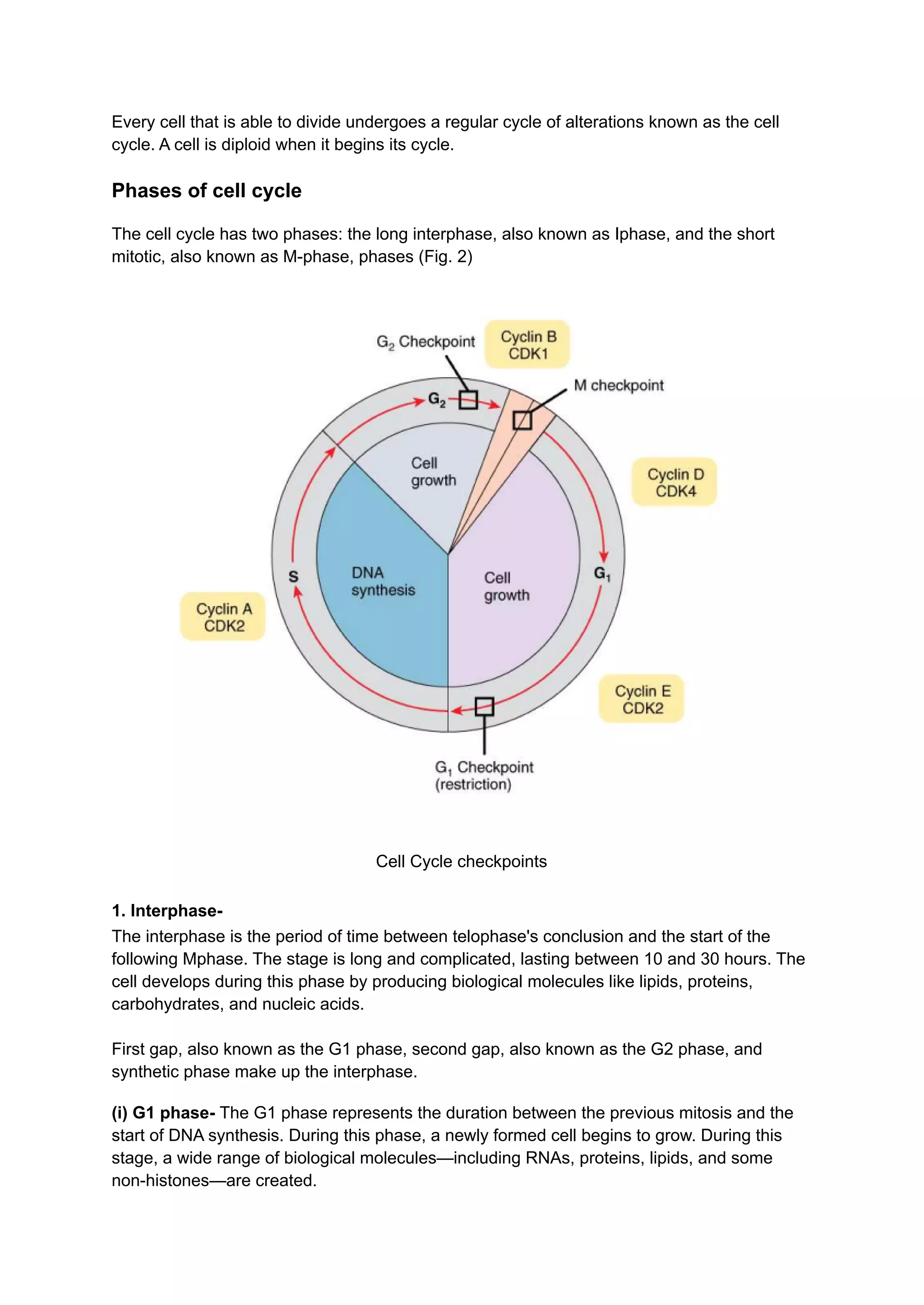
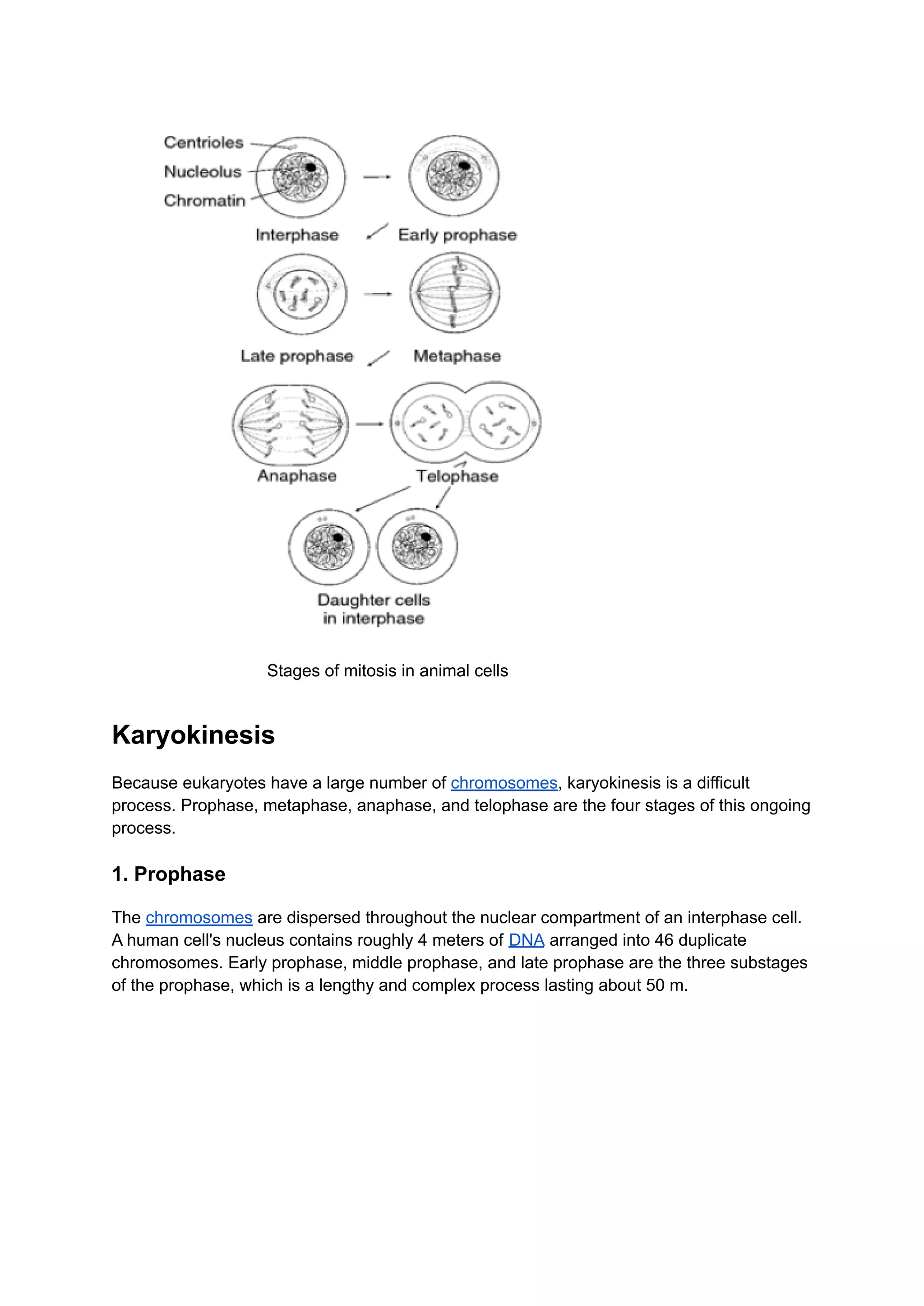
SlideShare a Scribd company logo
Cell division
Every living organism depends on the growth and multiplication of its cells for growth and
development because a multicellular organism begins as a single cell and undergoes
repeated division. The characteristic trait of all living things is an increase in cell size brought
on by growth.
Contents
1. Introduction
2. Cell Cycle
3. Control of Cell Cycle
4. Mitosis
5. Karyokinesis
6. Cytokinesis
7. Significance of Mitosis
8. Meiosis
9. Divisions of Meiosis
10. Cytokinesis
Introduction
The cell starts to divide once its growth has reached its maximum. An organism grows
vegetatively when its number of cells increases through cell divisions that follow a geometric
progression. The three stages of cell division, which is a continuous and dynamic process,
are as follows:
1. Replicating the genome or DNA
2. Karyokinesis, or nuclear division
3. Cytokinesis, also known as cell division
Based on the number of genomes present in the daughter cells in comparison to the dividing
parent cell, there are two types of cell division: mitosis and meiosis.
1. Mitosis- W. Flemming first used the word mitosis in 1882. Mitosis, also known as somatic
division, is the process by which a body cell divides into two daughter cells, each of equal
size and with the same number of chromosomes as the parent cell.
2. Meiosis- J. Meiosis was the first to use the term. B. Farmer and J. Smith in 1905 Moore,
E. Only the gonads (germ mother cells) undergo meiosis during the development of gametes
like sperm and ovum. Meiosis is the process by which chromosomes go from having two
copies, or 2N or diploid, to having only one copy, or N or haploid. Additionally known as the
reduction process.
Cell Cycle
Every cell that is able to divide undergoes a regular cycle of alterations known as the cell
cycle. A cell is diploid when it begins its cycle.
Phases of cell cycle
The cell cycle has two phases: the long interphase, also known as Iphase, and the short
mitotic, also known as M-phase, phases (Fig. 2)
Cell Cycle checkpoints
1. Interphase-
The interphase is the period of time between telophase's conclusion and the start of the
following Mphase. The stage is long and complicated, lasting between 10 and 30 hours. The
cell develops during this phase by producing biological molecules like lipids, proteins,
carbohydrates, and nucleic acids.
First gap, also known as the G1 phase, second gap, also known as the G2 phase, and
synthetic phase make up the interphase.
(i) G1 phase- The G1 phase represents the duration between the previous mitosis and the
start of DNA synthesis. During this phase, a newly formed cell begins to grow. During this
stage, a wide range of biological molecules—including RNAs, proteins, lipids, and some
non-histones—are created.
In order to prepare for the DNA replication that will occur next to it, normal metabolism is
carried out. This phase does not involve DNA synthesis.
● Lampbrush Chromosomes
● Polytene Chromosomes
● Structure of Nucleus
(ii) S Phase- Each chromosome is duplicated during this phase by replicating new DNA
molecules using the existing DNA as a template. Only in S-phase do histone protein and
their mRNA, some non-histone protein, and new nucleosome formation take place. Most
eukaryotes spend 6 to 8 hours in the S-phase.
(iii) G2 Phase- Between DNA synthesis and nuclear division is the G2 phase. During this
phase, protein synthesis and RNA transcription both continue. At this stage, the cell
continues to grow and begins to prepare for division.
The cytoplasmic organelles, including centrioles, mitochondria, and the Golgi apparatus,
double in number during this stage. Proteins for the spindle and asters are also produced,
and metabolic activity stores energy for the upcoming mitosis. Most cells go through the G2
phase between 2 and 5 hours.
2. Mitotic Phase
Mitotic phase comes after the interphase. The already duplicated chromosomes are evenly
distributed to the daughter cells, which have the exact same genetic makeup as the parent
cell, during the mitotic phase. Although not as precise as the DNA, the other parts of the cell
(organelles and molecules) are equally distributed among the daughter cells.
The daughter cells begin the G1 phase of the subsequent cell cycle after mitosis is
complete. Since the chromatin of the nucleus is packed into the visible chromosomes, which
are released by the breakdown of the nuclear envelope, many structural and physiological
changes occur in the cell during mitosis. The cytoskeletal elements and membranous
components undergo a significant reorganization.
The breakdown of the endoplasmic reticulum and the Golgi apparatus into tiny vesicles
stops the movement of the proteins. In order to form the spindle, which takes up the majority
of the cell and aids in the distribution of chromosomes into daughter cells, microtubules
dissociate into tubulin dimers. For the cytoplasmic division, actin filaments are reorganised to
form a contractile ring.
Control of Cell Cycle
1. Nucleo-cytoplasmic Ratio
Hertwig proposed in 1910 that cell division begins when the volume of the nucleus to the
volume of the cytoplasm ratio is disturbed. Proteins, nucleic acids, lipids, and other cellular
constituents are synthesized as the cell expands.
Materials move back and forth through the nuclear and cell membranes as these molecules
are created. The surface of the nucleus becomes inadequate for the exchange of materials
between the nucleus and the cytoplasm necessary for further growth at a critical point when
the volume of the cell grows faster than its surface.
At this point, the cell divides and recaptures the ideal and effective nucleo-cytoplasmic ratio
that permits growth. There are significant exceptions to the rule that cell division occurs after
a cell reaches a certain size.
2. Surface-Volume Ratio
The volume of a cell grows faster than its surface area as it gets bigger. The surface of the
cell is used to draw in all the materials needed for its growth and maintenance. When the
surface area can no longer support the large volume of the cell, a stage will be reached.
It is believed that a critical point exists at which cell division begins, greatly increasing the
surface without increasing the volume. The case of starved cells, which can divide without
doubling in size and produce smaller daughter cells, disproves this theory.
3. Nucleolus
Cell division is stopped when the nucleolus is damaged during a critical phase (telophase or
mid-prophase).
4. Cyclic Nucleotides
cAMP and cGMP concentrations frequently change while cells divide. As the cell enters the
S phase and mitosis, the concentration of cAMP decreases from its high point during the G1
phase.
The variation in cGMP concentration, however, frequently follows the opposite pattern. As a
result, changing the amount of any of these nucleotides has the ability to initiate or halt the
entry of many cells into the S phase and subsequent M phase.
In many cells, the quantity of these cyclic nucleotides remains constant over the course of
the cell cycle. Additionally, cyclic nucleotides are absent from plant cells. Given this
information, it is no longer believed that cyclic AMP and GMP control the cell cycle.
5. Phosphorylation
Phosphate groups are added to the histone groups during the cell cycle, most notably to H1
as the cell enters the S phase. They increase during the M phase and are then taken off
when mitosis is complete, just before G1 begins.
During the cell cycle, phosphate groups are also added to and removed from non-histone
proteins. Due to the fact that these proteins have been discovered to regulate the activity of
genes in RNA transcription during interphase, it is thought that changes in the histones and
non-histones may play a part in the regulation of the cell cycle.
6. Cyclin
Cyclin builds up during interphase and degrades during mitosis, so it appears that cyclin
concentration regulates mitosis.
Mitosis
In 1875, German biologist Eduard Strasburger published the first description of mitosis.
Walther Flemming later described the same in 1879 and later referred to it as "mitosis" in
1882.
It is also known as somatic division because it is the most typical way for eukaryotes to
divide their cells, and it occurs in the body's somatic cells. However, it happens in
undifferentiated germ cells in the gonads. In plants, it occurs in the meristematic tissue cells.
The average time for mitosis is between 30 and 3 hours.
A parent cell divides into two identical daughter cells during mitosis, and each of the
daughter cells has a nucleus with the same amount of DNA, chromosomes, and genetic
material as the parent cell. It also goes by the name "equational division" for this reason.
Karyokinesis, or the division of the nucleus, and cytokinesis, or the division of the cytoplasm,
are the two main processes that take place during mitosis.
Stages of mitosis in animal cells
Karyokinesis
Because eukaryotes have a large number of chromosomes, karyokinesis is a difficult
process. Prophase, metaphase, anaphase, and telophase are the four stages of this ongoing
process.
1. Prophase
The chromosomes are dispersed throughout the nuclear compartment of an interphase cell.
A human cell's nucleus contains roughly 4 meters of DNA arranged into 46 duplicate
chromosomes. Early prophase, middle prophase, and late prophase are the three substages
of the prophase, which is a lengthy and complex process lasting about 50 m.
Mitosis and Cytokinesis Continuum
A) Early prophase
As mitosis enters the early prophase, the following things happen:
(i) The cytoplasm thickens and the cell shape almost becomes rounded.
(ii) The tubulin dimers are polymerized around the centrioles, which are close to the nucleus,
to form the short radiating microtubules. The two centriole pairs, also known as diplosomes,
begin to move to the cell's opposite ends.
Asters are the microtubules that surround each pair of centrioles and resemble the body of a
star. The pericentriolar cloud, an amorphous region of cytoplasm, is what separates the
microtubules, also known as astral rays, from the centrioles and keeps them from coming
into contact. By incorporating more tubulin dimers, the microtubules stretching between the
diplosomes spreading outgrow in number and length.
As a result, asters move the duplicated centrioles to the cell's opposite ends, where they will
eventually split into two daughter cells during cytokinesis. Although the centrioles play no
part in the spindle's formation, they might be concerned with how the spindle is oriented.
(iii) To form a mitotic spindle, long microtubules gather on one side of the nucleus. The
mother-daughter centriole pair is located at each pole of the spindle, where microtubules are
arranged in bundles called spindle fibers.
(iv) By losing water and gradually coiling, the chromosomes that initially look like threads in
the nucleus gradually transform into short, thick rods that are then visible.
Each chromosome appears longitudinally double because of the duplication of DNA and
chromosomal proteins during the interphase. Each chromosome is made up of two sister
chromatids that are identical to one another and are joined at the primary constriction, also
known as the centromere. At the centromere, where the spindle microtubules connect to it,
each chromatid has a structure resembling a disc. The term "kinetochore" refers to this disc.
B) Middle prophase
It includes the following events:
(i) The chromosomes eventually take on their characteristic sizes and can be distinguished
from one another. They continue to get shorter, and thicker, and their chromatids uncoil.
(ii) Nucleoli shrink over time until they eventually simply disappear. Small vesicles that are
separated from the nuclear envelope start to spread throughout the cytoplasm. The lamina
separates into its constituent protein molecules.
C) Late Prophase
This phase involves the following events:
(i) The chromosomes and other nuclear components are released into the cytoplasm as a
result of the nuclear envelope completely rupturing.
(ii) The spindle develops into its proper size and shape.
(iii) The centriole pairs are pushed to the opposite ends of the cell by the expanding
spindles.
2. Metaphase
The brief and simple metaphase lasts for 2 to 10 minutes and includes the following
activities:
A. The spindle resides in the nucleus' region.
B. The chromosomes relocate to the spindle's equatorial plane.
C. Some spindle microtubules reach the chromosomes and connect to them. Chromosome
or kinetochore microtubules are what these are.
D. An equatorial or metaphase plate, made up of the aligned chromosomes, forms in the
middle of the spindle. The kinetochores, with the chromatid arms trailing on the sides, form
this plate. It is perpendicular to the spindle's long axis. The chromosomes are completely
aligned into a plate during metaphase and are waiting for their chromatids to separate.
3. Anaphase
Anaphase only lasts two to three minutes and includes the following activities:
A. Each chromosome's sister chromatids slightly separate at the primary constriction,
causing their kinetochores to stretch in the direction of the spindle's opposing poles.
Chromatid separation occurs almost simultaneously in every chromosome. Because they
are no longer bound to their duplicates, the chromatids are now referred to as
chromosomes.
B. The chromatids completely separate from their former partners and begin to move to the
opposite poles of the spindle after a brief period of time. Each chromosome's attached
microtubules pull it along, leaving its kinetochore leads and arms in their wake. As a result,
depending on where the kinetochore is located, the chromosomes are pulled into the shapes
of V, J, and I. (Either metacentric, sub-metacentric, or telocentric)
C. The two poles distance themselves from one another as the chromosomes move toward
their respective poles through spindle elongation.
When all of the chromatids reach their opposite poles, anaphase is complete. Each
metaphase chromosome sends one chromatid to each pole of the spindle, and both sets of
chromatids carry the exact same genetic material.
4. Telophase
The telophase is a lengthy and complex phase that lasts for about an hour. Each group of
chromosomes is used to reconstruct the nucleus during this phase. It involves the
development progress:
A. At each pole, the chromosomes spread out and grow long and lean. They eventually
blend together like they were in an interphase cell.
B. The nuclear envelope is gradually rebuilt around each group of chromosomes. The
individual unfolding chromosomes first associate with the membrane vesicles, partially
enclosing each chromosome. Then, they combine to create an envelope that encloses each
pole's full complement of chromosomes. The nuclear envelope's reconstruction and the
reassociation of the lamina proteins result in the formation of a full lamina.
C. In the prophase, material from the nucleolar organizer site returns to the nucleolar
organizer site and forms a small nucleolus. This material is made up of partially processed
ribosomal subunits and processing enzymes. Then, the processing of this earlier material
continues.
At this time, new rRNA transcription also starts. It gradually picks up speed until it reaches
the high level required for interphase cells. Additionally, the nucleolus expands until it
reaches its typical size. Old and new rRNA and ribosomal proteins are both present in the
nucleolus, which was reformed at telophase.
Transcription of all three RNA types gradually returns to normal as chromosomes turn into
chromatin and nucleoli are rebuilt.
By depolymerizing microtubules, the spindle starts to disappear, the asters shrink, and the
centrioles move into their characteristic interphase position next to the nucleus on one side.
At the spindle equator, short spindle microtubules persist for a while as a marker for the area
where the cytoplasm will subsequently divide. Read more
Cytokinesis
The division of cytoplasm is known as cytokinesis. It completes cell division by enclosing the
daughter nuclei created by karyokinesis in different cells. At the metaphase, cytoplasmic
movements equalise the distribution of mitochondria and other cell organelles in the two
halves of the cell signal cytokinesis. Animal cells and plant cells divide in different ways.
Significance of Mitosis
Mitosis has manifold significance-
Maintenance of Size- The process of mitosis aids in maintaining cell size. A fully developed
cell divides by the process of mitosis rather than continuing to grow.
Growth- Repeated mitotic cell division transforms a fertilized egg into an embryo, and then
ultimately into an adult.
Maintenance of Chromosome Number- Mitosis ensures that each cell in an individual has
an equal number of chromosomes. Since DNA is duplicated in S phase before mitosis, each
cell receives a complete set of genetic information during mitosis.
Repair- New cells are produced during mitosis to replace old, worn-out, and dying cells.
Healing and Regeneration- For the purpose of wound healing and regeneration, mitosis
generates new cells.
Reproduction- The process of mitosis causes multiplication in acellular organisms. It is
crucial to sexual and asexual reproduction in multicellular organisms as well.
Evidence of Basic Relationship of Organisms-Because mitosis occurs in many different
types of organisms, it supports the fundamental connection between all living things.
Meiosis
Potentially, August Weismann predicted in 1887 that during gamete formation, the number of
chromosomes must be cut in half. Reduction division was demonstrated by Edouard Van
Beneden in 1887. In 1905, J.B. Farmer and Moore coined the term "meiosis."
All eukaryotic cells go through mitosis, whereas meiosis only occurs in specific cells and at a
specific time. Meiosis is a process that only occurs in the cells of sexually reproducing
organisms, and in multicellular organisms, only specific cells undergo the transition from
mitosis to meiosis at a specific point in the life cycle.
Meiosis is the process by which animals, some lower plants, and different protist and fungus
groups produce gametes or gametic nuclei. During meiosis, higher plants produce spores.
Gametophytes, which produce gametes through mitosis, are structures that produce
gametes and are derived from the spores.
Meiosis entails two divisions that happen quickly after one another, with the chromosomes
replicating just once. As a result, a parent cell divides into four daughter cells, each of which
has half the chromosomes and nuclear DNA of the parent cell. Therefore, meiosis is also
referred to as reduction division. The first and second meiotic divisions, also known as
meiosis-I and meiosis-II, are the two stages of meiosis. Read more
Divisions of Meiosis
First meiotic division, also known as Meiosis I: The two homologous chromosomes of each
pair separate from one another and move to different daughter cells during the first meiotic
division. As a result, the condition of the chromosomes changes from diploid to haploid.
Therefore, meiosis-I is also referred to as heterotypic division. Prophase 1, Metaphase 1,
Anaphase 1, and Telophase 1 are the names of the four phases that make up this division.
Cytokinesis
In an animal cell, cytoplasm divides at its center through constriction, whereas in a plant cell,
cell plates form. There are two daughter cells as a result. The latter has half as many nuclear
DNA molecules and chromosomes as the former, respectively. When this is accomplished,
the reduction division is completed. Animals form mature gametes during meiosis II, which
produces cells. They stop dividing at that point.
Before a new being can emerge, a gamete must fuse with another suitable gamete. The
spores are the cells produced during meiosis II in plants. The spores don't have to fuse in
pairs in order to grow into new individuals. In actuality, the spore's capacity to instantly give
rise to a new person is the main distinction between it and a gamete.

More Related Content

Similar to Cell division

Heredity g8
Heredity g8Heredity g8
Mitosis
MitosisMitosis
cell division.pptx
cell division.pptxcell division.pptx
cell division.pptx
JanhaviBurade
 
Cell cycle
Cell cycleCell cycle
Cell cycle
ishita1994
 
Eukaryotic cell division cycle
Eukaryotic cell division cycleEukaryotic cell division cycle
Eukaryotic cell division cycle
ArchanaSoni3
 
Overview of Cell Cycle by Javali.G.
Overview of Cell Cycle by Javali.G.Overview of Cell Cycle by Javali.G.
Overview of Cell Cycle by Javali.G.
javaliG
 
542.ppt
542.ppt542.ppt
542.ppt
HumaTahreem2
 
Cell cycle and division
Cell cycle and divisionCell cycle and division
Cell cycle and division
Rohit Mondal
 
Cell cycles and its regulation presented by dr.sibi p ittiyavirah, professor,...
Cell cycles and its regulation presented by dr.sibi p ittiyavirah, professor,...Cell cycles and its regulation presented by dr.sibi p ittiyavirah, professor,...
Cell cycles and its regulation presented by dr.sibi p ittiyavirah, professor,...
Dr.SIBI P ITTIYAVIRAH
 
Cell cycle and cell death
Cell cycle and cell deathCell cycle and cell death
Cell cycle and cell death
Vamsi kumar
 
Cell cycle
Cell cycleCell cycle
Cell cycle
biOlOgyBINGE
 
Cell cycle ppt.
Cell cycle ppt.Cell cycle ppt.
Cell cycle ppt.
Shabana2428
 
Cell division – mitosis
Cell division – mitosisCell division – mitosis
Cell division – mitosis
Anna Purna
 
Lecture 18 cell cycle and its regulation converted
Lecture 18 cell cycle and its regulation convertedLecture 18 cell cycle and its regulation converted
Lecture 18 cell cycle and its regulation converted
yogeshsharma880
 
Presentation on Cell Cycle
Presentation on Cell CyclePresentation on Cell Cycle
Presentation on Cell Cycle
Dr. Kaushik Kumar Panigrahi
 
Microbial growth refers to an increase in number of cells rather than an incr...
Microbial growth refers to an increase in number of cells rather than an incr...Microbial growth refers to an increase in number of cells rather than an incr...
Microbial growth refers to an increase in number of cells rather than an incr...
Smitha Vijayan
 
1.6 cell division
1.6 cell division1.6 cell division
1.6 cell division
Bob Smullen
 
Cell cycle
Cell cycleCell cycle
Cell cycle
Prasann Saha
 
Cell cycle
Cell cycleCell cycle
Cell cycle
LIFE SCIENCES
 
cell cycel , and its stages
 cell cycel , and its stages  cell cycel , and its stages
cell cycel , and its stages
ajayurs123
 

Similar to Cell division (20)

Heredity g8
Heredity g8Heredity g8
Heredity g8
 
Mitosis
MitosisMitosis
Mitosis
 
cell division.pptx
cell division.pptxcell division.pptx
cell division.pptx
 
Cell cycle
Cell cycleCell cycle
Cell cycle
 
Eukaryotic cell division cycle
Eukaryotic cell division cycleEukaryotic cell division cycle
Eukaryotic cell division cycle
 
Overview of Cell Cycle by Javali.G.
Overview of Cell Cycle by Javali.G.Overview of Cell Cycle by Javali.G.
Overview of Cell Cycle by Javali.G.
 
542.ppt
542.ppt542.ppt
542.ppt
 
Cell cycle and division
Cell cycle and divisionCell cycle and division
Cell cycle and division
 
Cell cycles and its regulation presented by dr.sibi p ittiyavirah, professor,...
Cell cycles and its regulation presented by dr.sibi p ittiyavirah, professor,...Cell cycles and its regulation presented by dr.sibi p ittiyavirah, professor,...
Cell cycles and its regulation presented by dr.sibi p ittiyavirah, professor,...
 
Cell cycle and cell death
Cell cycle and cell deathCell cycle and cell death
Cell cycle and cell death
 
Cell cycle
Cell cycleCell cycle
Cell cycle
 
Cell cycle ppt.
Cell cycle ppt.Cell cycle ppt.
Cell cycle ppt.
 
Cell division – mitosis
Cell division – mitosisCell division – mitosis
Cell division – mitosis
 
Lecture 18 cell cycle and its regulation converted
Lecture 18 cell cycle and its regulation convertedLecture 18 cell cycle and its regulation converted
Lecture 18 cell cycle and its regulation converted
 
Presentation on Cell Cycle
Presentation on Cell CyclePresentation on Cell Cycle
Presentation on Cell Cycle
 
Microbial growth refers to an increase in number of cells rather than an incr...
Microbial growth refers to an increase in number of cells rather than an incr...Microbial growth refers to an increase in number of cells rather than an incr...
Microbial growth refers to an increase in number of cells rather than an incr...
 
1.6 cell division
1.6 cell division1.6 cell division
1.6 cell division
 
Cell cycle
Cell cycleCell cycle
Cell cycle
 
Cell cycle
Cell cycleCell cycle
Cell cycle
 
cell cycel , and its stages
 cell cycel , and its stages  cell cycel , and its stages
cell cycel , and its stages
 

More from Mammalssite

Instinct and Learning Behavior.pdf
Instinct and Learning Behavior.pdfInstinct and Learning Behavior.pdf
Instinct and Learning Behavior.pdf
Mammalssite
 
How young animals grow up?
How young animals grow up?How young animals grow up?
How young animals grow up?
Mammalssite
 
Circulation, Gas Exchange, and Temperature Regulation in Mammals.pdf
Circulation, Gas Exchange, and Temperature Regulation in Mammals.pdfCirculation, Gas Exchange, and Temperature Regulation in Mammals.pdf
Circulation, Gas Exchange, and Temperature Regulation in Mammals.pdf
Mammalssite
 
Structure of Nucleus
Structure of NucleusStructure of Nucleus
Structure of Nucleus
Mammalssite
 
Chromosomes | Polytene Chromosomes | Lampbrush Chromosomes
Chromosomes | Polytene Chromosomes | Lampbrush ChromosomesChromosomes | Polytene Chromosomes | Lampbrush Chromosomes
Chromosomes | Polytene Chromosomes | Lampbrush Chromosomes
Mammalssite
 
External Structure and locomotion.pdf
External Structure and locomotion.pdfExternal Structure and locomotion.pdf
External Structure and locomotion.pdf
Mammalssite
 

More from Mammalssite (6)

Instinct and Learning Behavior.pdf
Instinct and Learning Behavior.pdfInstinct and Learning Behavior.pdf
Instinct and Learning Behavior.pdf
 
How young animals grow up?
How young animals grow up?How young animals grow up?
How young animals grow up?
 
Circulation, Gas Exchange, and Temperature Regulation in Mammals.pdf
Circulation, Gas Exchange, and Temperature Regulation in Mammals.pdfCirculation, Gas Exchange, and Temperature Regulation in Mammals.pdf
Circulation, Gas Exchange, and Temperature Regulation in Mammals.pdf
 
Structure of Nucleus
Structure of NucleusStructure of Nucleus
Structure of Nucleus
 
Chromosomes | Polytene Chromosomes | Lampbrush Chromosomes
Chromosomes | Polytene Chromosomes | Lampbrush ChromosomesChromosomes | Polytene Chromosomes | Lampbrush Chromosomes
Chromosomes | Polytene Chromosomes | Lampbrush Chromosomes
 
External Structure and locomotion.pdf
External Structure and locomotion.pdfExternal Structure and locomotion.pdf
External Structure and locomotion.pdf
 

Recently uploaded

Language Across the Curriculm LAC B.Ed.
Language Across the  Curriculm LAC B.Ed.Language Across the  Curriculm LAC B.Ed.
Language Across the Curriculm LAC B.Ed.
Atul Kumar Singh
 
The approach at University of Liverpool.pptx
The approach at University of Liverpool.pptxThe approach at University of Liverpool.pptx
The approach at University of Liverpool.pptx
Jisc
 
Azure Interview Questions and Answers PDF By ScholarHat
Azure Interview Questions and Answers PDF By ScholarHatAzure Interview Questions and Answers PDF By ScholarHat
Azure Interview Questions and Answers PDF By ScholarHat
Scholarhat
 
BÀI TẬP BỔ TRỢ TIẾNG ANH GLOBAL SUCCESS LỚP 3 - CẢ NĂM (CÓ FILE NGHE VÀ ĐÁP Á...
BÀI TẬP BỔ TRỢ TIẾNG ANH GLOBAL SUCCESS LỚP 3 - CẢ NĂM (CÓ FILE NGHE VÀ ĐÁP Á...BÀI TẬP BỔ TRỢ TIẾNG ANH GLOBAL SUCCESS LỚP 3 - CẢ NĂM (CÓ FILE NGHE VÀ ĐÁP Á...
BÀI TẬP BỔ TRỢ TIẾNG ANH GLOBAL SUCCESS LỚP 3 - CẢ NĂM (CÓ FILE NGHE VÀ ĐÁP Á...
Nguyen Thanh Tu Collection
 
Normal Labour/ Stages of Labour/ Mechanism of Labour
Normal Labour/ Stages of Labour/ Mechanism of LabourNormal Labour/ Stages of Labour/ Mechanism of Labour
Normal Labour/ Stages of Labour/ Mechanism of Labour
Wasim Ak
 
Mule 4.6 & Java 17 Upgrade | MuleSoft Mysore Meetup #46
Mule 4.6 & Java 17 Upgrade | MuleSoft Mysore Meetup #46Mule 4.6 & Java 17 Upgrade | MuleSoft Mysore Meetup #46
Mule 4.6 & Java 17 Upgrade | MuleSoft Mysore Meetup #46
MysoreMuleSoftMeetup
 
The Accursed House by Émile Gaboriau.pptx
The Accursed House by Émile Gaboriau.pptxThe Accursed House by Émile Gaboriau.pptx
The Accursed House by Émile Gaboriau.pptx
DhatriParmar
 
Group Presentation 2 Economics.Ariana Buscigliopptx
Group Presentation 2 Economics.Ariana BuscigliopptxGroup Presentation 2 Economics.Ariana Buscigliopptx
Group Presentation 2 Economics.Ariana Buscigliopptx
ArianaBusciglio
 
S1-Introduction-Biopesticides in ICM.pptx
S1-Introduction-Biopesticides in ICM.pptxS1-Introduction-Biopesticides in ICM.pptx
S1-Introduction-Biopesticides in ICM.pptx
tarandeep35
 
Introduction to AI for Nonprofits with Tapp Network
Introduction to AI for Nonprofits with Tapp NetworkIntroduction to AI for Nonprofits with Tapp Network
Introduction to AI for Nonprofits with Tapp Network
TechSoup
 
A Strategic Approach: GenAI in Education
A Strategic Approach: GenAI in EducationA Strategic Approach: GenAI in Education
A Strategic Approach: GenAI in Education
Peter Windle
 
A Survey of Techniques for Maximizing LLM Performance.pptx
A Survey of Techniques for Maximizing LLM Performance.pptxA Survey of Techniques for Maximizing LLM Performance.pptx
A Survey of Techniques for Maximizing LLM Performance.pptx
thanhdowork
 
special B.ed 2nd year old paper_20240531.pdf
special B.ed 2nd year old paper_20240531.pdfspecial B.ed 2nd year old paper_20240531.pdf
special B.ed 2nd year old paper_20240531.pdf
Special education needs
 
The basics of sentences session 5pptx.pptx
The basics of sentences session 5pptx.pptxThe basics of sentences session 5pptx.pptx
The basics of sentences session 5pptx.pptx
heathfieldcps1
 
STRAND 3 HYGIENIC PRACTICES.pptx GRADE 7 CBC
STRAND 3 HYGIENIC PRACTICES.pptx GRADE 7 CBCSTRAND 3 HYGIENIC PRACTICES.pptx GRADE 7 CBC
STRAND 3 HYGIENIC PRACTICES.pptx GRADE 7 CBC
kimdan468
 
Multithreading_in_C++ - std::thread, race condition
Multithreading_in_C++ - std::thread, race conditionMultithreading_in_C++ - std::thread, race condition
Multithreading_in_C++ - std::thread, race condition
Mohammed Sikander
 
Digital Tools and AI for Teaching Learning and Research
Digital Tools and AI for Teaching Learning and ResearchDigital Tools and AI for Teaching Learning and Research
Digital Tools and AI for Teaching Learning and Research
Vikramjit Singh
 
Unit 8 - Information and Communication Technology (Paper I).pdf
Unit 8 - Information and Communication Technology (Paper I).pdfUnit 8 - Information and Communication Technology (Paper I).pdf
Unit 8 - Information and Communication Technology (Paper I).pdf
Thiyagu K
 
Unit 2- Research Aptitude (UGC NET Paper I).pdf
Unit 2- Research Aptitude (UGC NET Paper I).pdfUnit 2- Research Aptitude (UGC NET Paper I).pdf
Unit 2- Research Aptitude (UGC NET Paper I).pdf
Thiyagu K
 
The Challenger.pdf DNHS Official Publication
The Challenger.pdf DNHS Official PublicationThe Challenger.pdf DNHS Official Publication
The Challenger.pdf DNHS Official Publication
Delapenabediema
 

Recently uploaded (20)

Language Across the Curriculm LAC B.Ed.
Language Across the  Curriculm LAC B.Ed.Language Across the  Curriculm LAC B.Ed.
Language Across the Curriculm LAC B.Ed.
 
The approach at University of Liverpool.pptx
The approach at University of Liverpool.pptxThe approach at University of Liverpool.pptx
The approach at University of Liverpool.pptx
 
Azure Interview Questions and Answers PDF By ScholarHat
Azure Interview Questions and Answers PDF By ScholarHatAzure Interview Questions and Answers PDF By ScholarHat
Azure Interview Questions and Answers PDF By ScholarHat
 
BÀI TẬP BỔ TRỢ TIẾNG ANH GLOBAL SUCCESS LỚP 3 - CẢ NĂM (CÓ FILE NGHE VÀ ĐÁP Á...
BÀI TẬP BỔ TRỢ TIẾNG ANH GLOBAL SUCCESS LỚP 3 - CẢ NĂM (CÓ FILE NGHE VÀ ĐÁP Á...BÀI TẬP BỔ TRỢ TIẾNG ANH GLOBAL SUCCESS LỚP 3 - CẢ NĂM (CÓ FILE NGHE VÀ ĐÁP Á...
BÀI TẬP BỔ TRỢ TIẾNG ANH GLOBAL SUCCESS LỚP 3 - CẢ NĂM (CÓ FILE NGHE VÀ ĐÁP Á...
 
Normal Labour/ Stages of Labour/ Mechanism of Labour
Normal Labour/ Stages of Labour/ Mechanism of LabourNormal Labour/ Stages of Labour/ Mechanism of Labour
Normal Labour/ Stages of Labour/ Mechanism of Labour
 
Mule 4.6 & Java 17 Upgrade | MuleSoft Mysore Meetup #46
Mule 4.6 & Java 17 Upgrade | MuleSoft Mysore Meetup #46Mule 4.6 & Java 17 Upgrade | MuleSoft Mysore Meetup #46
Mule 4.6 & Java 17 Upgrade | MuleSoft Mysore Meetup #46
 
The Accursed House by Émile Gaboriau.pptx
The Accursed House by Émile Gaboriau.pptxThe Accursed House by Émile Gaboriau.pptx
The Accursed House by Émile Gaboriau.pptx
 
Group Presentation 2 Economics.Ariana Buscigliopptx
Group Presentation 2 Economics.Ariana BuscigliopptxGroup Presentation 2 Economics.Ariana Buscigliopptx
Group Presentation 2 Economics.Ariana Buscigliopptx
 
S1-Introduction-Biopesticides in ICM.pptx
S1-Introduction-Biopesticides in ICM.pptxS1-Introduction-Biopesticides in ICM.pptx
S1-Introduction-Biopesticides in ICM.pptx
 
Introduction to AI for Nonprofits with Tapp Network
Introduction to AI for Nonprofits with Tapp NetworkIntroduction to AI for Nonprofits with Tapp Network
Introduction to AI for Nonprofits with Tapp Network
 
A Strategic Approach: GenAI in Education
A Strategic Approach: GenAI in EducationA Strategic Approach: GenAI in Education
A Strategic Approach: GenAI in Education
 
A Survey of Techniques for Maximizing LLM Performance.pptx
A Survey of Techniques for Maximizing LLM Performance.pptxA Survey of Techniques for Maximizing LLM Performance.pptx
A Survey of Techniques for Maximizing LLM Performance.pptx
 
special B.ed 2nd year old paper_20240531.pdf
special B.ed 2nd year old paper_20240531.pdfspecial B.ed 2nd year old paper_20240531.pdf
special B.ed 2nd year old paper_20240531.pdf
 
The basics of sentences session 5pptx.pptx
The basics of sentences session 5pptx.pptxThe basics of sentences session 5pptx.pptx
The basics of sentences session 5pptx.pptx
 
STRAND 3 HYGIENIC PRACTICES.pptx GRADE 7 CBC
STRAND 3 HYGIENIC PRACTICES.pptx GRADE 7 CBCSTRAND 3 HYGIENIC PRACTICES.pptx GRADE 7 CBC
STRAND 3 HYGIENIC PRACTICES.pptx GRADE 7 CBC
 
Multithreading_in_C++ - std::thread, race condition
Multithreading_in_C++ - std::thread, race conditionMultithreading_in_C++ - std::thread, race condition
Multithreading_in_C++ - std::thread, race condition
 
Digital Tools and AI for Teaching Learning and Research
Digital Tools and AI for Teaching Learning and ResearchDigital Tools and AI for Teaching Learning and Research
Digital Tools and AI for Teaching Learning and Research
 
Unit 8 - Information and Communication Technology (Paper I).pdf
Unit 8 - Information and Communication Technology (Paper I).pdfUnit 8 - Information and Communication Technology (Paper I).pdf
Unit 8 - Information and Communication Technology (Paper I).pdf
 
Unit 2- Research Aptitude (UGC NET Paper I).pdf
Unit 2- Research Aptitude (UGC NET Paper I).pdfUnit 2- Research Aptitude (UGC NET Paper I).pdf
Unit 2- Research Aptitude (UGC NET Paper I).pdf
 
The Challenger.pdf DNHS Official Publication
The Challenger.pdf DNHS Official PublicationThe Challenger.pdf DNHS Official Publication
The Challenger.pdf DNHS Official Publication
 

Cell division

  • 1. Cell division Every living organism depends on the growth and multiplication of its cells for growth and development because a multicellular organism begins as a single cell and undergoes repeated division. The characteristic trait of all living things is an increase in cell size brought on by growth. Contents 1. Introduction 2. Cell Cycle 3. Control of Cell Cycle 4. Mitosis 5. Karyokinesis 6. Cytokinesis 7. Significance of Mitosis 8. Meiosis 9. Divisions of Meiosis 10. Cytokinesis Introduction The cell starts to divide once its growth has reached its maximum. An organism grows vegetatively when its number of cells increases through cell divisions that follow a geometric progression. The three stages of cell division, which is a continuous and dynamic process, are as follows: 1. Replicating the genome or DNA 2. Karyokinesis, or nuclear division 3. Cytokinesis, also known as cell division Based on the number of genomes present in the daughter cells in comparison to the dividing parent cell, there are two types of cell division: mitosis and meiosis. 1. Mitosis- W. Flemming first used the word mitosis in 1882. Mitosis, also known as somatic division, is the process by which a body cell divides into two daughter cells, each of equal size and with the same number of chromosomes as the parent cell. 2. Meiosis- J. Meiosis was the first to use the term. B. Farmer and J. Smith in 1905 Moore, E. Only the gonads (germ mother cells) undergo meiosis during the development of gametes like sperm and ovum. Meiosis is the process by which chromosomes go from having two copies, or 2N or diploid, to having only one copy, or N or haploid. Additionally known as the reduction process. Cell Cycle
  • 2. Every cell that is able to divide undergoes a regular cycle of alterations known as the cell cycle. A cell is diploid when it begins its cycle. Phases of cell cycle The cell cycle has two phases: the long interphase, also known as Iphase, and the short mitotic, also known as M-phase, phases (Fig. 2) Cell Cycle checkpoints 1. Interphase- The interphase is the period of time between telophase's conclusion and the start of the following Mphase. The stage is long and complicated, lasting between 10 and 30 hours. The cell develops during this phase by producing biological molecules like lipids, proteins, carbohydrates, and nucleic acids. First gap, also known as the G1 phase, second gap, also known as the G2 phase, and synthetic phase make up the interphase. (i) G1 phase- The G1 phase represents the duration between the previous mitosis and the start of DNA synthesis. During this phase, a newly formed cell begins to grow. During this stage, a wide range of biological molecules—including RNAs, proteins, lipids, and some non-histones—are created.
  • 3. In order to prepare for the DNA replication that will occur next to it, normal metabolism is carried out. This phase does not involve DNA synthesis. ● Lampbrush Chromosomes ● Polytene Chromosomes ● Structure of Nucleus (ii) S Phase- Each chromosome is duplicated during this phase by replicating new DNA molecules using the existing DNA as a template. Only in S-phase do histone protein and their mRNA, some non-histone protein, and new nucleosome formation take place. Most eukaryotes spend 6 to 8 hours in the S-phase. (iii) G2 Phase- Between DNA synthesis and nuclear division is the G2 phase. During this phase, protein synthesis and RNA transcription both continue. At this stage, the cell continues to grow and begins to prepare for division. The cytoplasmic organelles, including centrioles, mitochondria, and the Golgi apparatus, double in number during this stage. Proteins for the spindle and asters are also produced, and metabolic activity stores energy for the upcoming mitosis. Most cells go through the G2 phase between 2 and 5 hours. 2. Mitotic Phase Mitotic phase comes after the interphase. The already duplicated chromosomes are evenly distributed to the daughter cells, which have the exact same genetic makeup as the parent cell, during the mitotic phase. Although not as precise as the DNA, the other parts of the cell (organelles and molecules) are equally distributed among the daughter cells. The daughter cells begin the G1 phase of the subsequent cell cycle after mitosis is complete. Since the chromatin of the nucleus is packed into the visible chromosomes, which are released by the breakdown of the nuclear envelope, many structural and physiological changes occur in the cell during mitosis. The cytoskeletal elements and membranous components undergo a significant reorganization. The breakdown of the endoplasmic reticulum and the Golgi apparatus into tiny vesicles stops the movement of the proteins. In order to form the spindle, which takes up the majority of the cell and aids in the distribution of chromosomes into daughter cells, microtubules dissociate into tubulin dimers. For the cytoplasmic division, actin filaments are reorganised to form a contractile ring. Control of Cell Cycle 1. Nucleo-cytoplasmic Ratio
  • 4. Hertwig proposed in 1910 that cell division begins when the volume of the nucleus to the volume of the cytoplasm ratio is disturbed. Proteins, nucleic acids, lipids, and other cellular constituents are synthesized as the cell expands. Materials move back and forth through the nuclear and cell membranes as these molecules are created. The surface of the nucleus becomes inadequate for the exchange of materials between the nucleus and the cytoplasm necessary for further growth at a critical point when the volume of the cell grows faster than its surface. At this point, the cell divides and recaptures the ideal and effective nucleo-cytoplasmic ratio that permits growth. There are significant exceptions to the rule that cell division occurs after a cell reaches a certain size. 2. Surface-Volume Ratio The volume of a cell grows faster than its surface area as it gets bigger. The surface of the cell is used to draw in all the materials needed for its growth and maintenance. When the surface area can no longer support the large volume of the cell, a stage will be reached. It is believed that a critical point exists at which cell division begins, greatly increasing the surface without increasing the volume. The case of starved cells, which can divide without doubling in size and produce smaller daughter cells, disproves this theory. 3. Nucleolus Cell division is stopped when the nucleolus is damaged during a critical phase (telophase or mid-prophase). 4. Cyclic Nucleotides cAMP and cGMP concentrations frequently change while cells divide. As the cell enters the S phase and mitosis, the concentration of cAMP decreases from its high point during the G1 phase. The variation in cGMP concentration, however, frequently follows the opposite pattern. As a result, changing the amount of any of these nucleotides has the ability to initiate or halt the entry of many cells into the S phase and subsequent M phase. In many cells, the quantity of these cyclic nucleotides remains constant over the course of the cell cycle. Additionally, cyclic nucleotides are absent from plant cells. Given this information, it is no longer believed that cyclic AMP and GMP control the cell cycle. 5. Phosphorylation Phosphate groups are added to the histone groups during the cell cycle, most notably to H1 as the cell enters the S phase. They increase during the M phase and are then taken off when mitosis is complete, just before G1 begins.
  • 5. During the cell cycle, phosphate groups are also added to and removed from non-histone proteins. Due to the fact that these proteins have been discovered to regulate the activity of genes in RNA transcription during interphase, it is thought that changes in the histones and non-histones may play a part in the regulation of the cell cycle. 6. Cyclin Cyclin builds up during interphase and degrades during mitosis, so it appears that cyclin concentration regulates mitosis. Mitosis In 1875, German biologist Eduard Strasburger published the first description of mitosis. Walther Flemming later described the same in 1879 and later referred to it as "mitosis" in 1882. It is also known as somatic division because it is the most typical way for eukaryotes to divide their cells, and it occurs in the body's somatic cells. However, it happens in undifferentiated germ cells in the gonads. In plants, it occurs in the meristematic tissue cells. The average time for mitosis is between 30 and 3 hours. A parent cell divides into two identical daughter cells during mitosis, and each of the daughter cells has a nucleus with the same amount of DNA, chromosomes, and genetic material as the parent cell. It also goes by the name "equational division" for this reason. Karyokinesis, or the division of the nucleus, and cytokinesis, or the division of the cytoplasm, are the two main processes that take place during mitosis.
  • 6. Stages of mitosis in animal cells Karyokinesis Because eukaryotes have a large number of chromosomes, karyokinesis is a difficult process. Prophase, metaphase, anaphase, and telophase are the four stages of this ongoing process. 1. Prophase The chromosomes are dispersed throughout the nuclear compartment of an interphase cell. A human cell's nucleus contains roughly 4 meters of DNA arranged into 46 duplicate chromosomes. Early prophase, middle prophase, and late prophase are the three substages of the prophase, which is a lengthy and complex process lasting about 50 m.
  • 7. Mitosis and Cytokinesis Continuum A) Early prophase As mitosis enters the early prophase, the following things happen: (i) The cytoplasm thickens and the cell shape almost becomes rounded. (ii) The tubulin dimers are polymerized around the centrioles, which are close to the nucleus, to form the short radiating microtubules. The two centriole pairs, also known as diplosomes, begin to move to the cell's opposite ends. Asters are the microtubules that surround each pair of centrioles and resemble the body of a star. The pericentriolar cloud, an amorphous region of cytoplasm, is what separates the microtubules, also known as astral rays, from the centrioles and keeps them from coming into contact. By incorporating more tubulin dimers, the microtubules stretching between the diplosomes spreading outgrow in number and length. As a result, asters move the duplicated centrioles to the cell's opposite ends, where they will eventually split into two daughter cells during cytokinesis. Although the centrioles play no part in the spindle's formation, they might be concerned with how the spindle is oriented.
  • 8. (iii) To form a mitotic spindle, long microtubules gather on one side of the nucleus. The mother-daughter centriole pair is located at each pole of the spindle, where microtubules are arranged in bundles called spindle fibers. (iv) By losing water and gradually coiling, the chromosomes that initially look like threads in the nucleus gradually transform into short, thick rods that are then visible. Each chromosome appears longitudinally double because of the duplication of DNA and chromosomal proteins during the interphase. Each chromosome is made up of two sister chromatids that are identical to one another and are joined at the primary constriction, also known as the centromere. At the centromere, where the spindle microtubules connect to it, each chromatid has a structure resembling a disc. The term "kinetochore" refers to this disc. B) Middle prophase It includes the following events: (i) The chromosomes eventually take on their characteristic sizes and can be distinguished from one another. They continue to get shorter, and thicker, and their chromatids uncoil. (ii) Nucleoli shrink over time until they eventually simply disappear. Small vesicles that are separated from the nuclear envelope start to spread throughout the cytoplasm. The lamina separates into its constituent protein molecules. C) Late Prophase This phase involves the following events: (i) The chromosomes and other nuclear components are released into the cytoplasm as a result of the nuclear envelope completely rupturing. (ii) The spindle develops into its proper size and shape. (iii) The centriole pairs are pushed to the opposite ends of the cell by the expanding spindles. 2. Metaphase The brief and simple metaphase lasts for 2 to 10 minutes and includes the following activities: A. The spindle resides in the nucleus' region. B. The chromosomes relocate to the spindle's equatorial plane.
  • 9. C. Some spindle microtubules reach the chromosomes and connect to them. Chromosome or kinetochore microtubules are what these are. D. An equatorial or metaphase plate, made up of the aligned chromosomes, forms in the middle of the spindle. The kinetochores, with the chromatid arms trailing on the sides, form this plate. It is perpendicular to the spindle's long axis. The chromosomes are completely aligned into a plate during metaphase and are waiting for their chromatids to separate. 3. Anaphase Anaphase only lasts two to three minutes and includes the following activities: A. Each chromosome's sister chromatids slightly separate at the primary constriction, causing their kinetochores to stretch in the direction of the spindle's opposing poles. Chromatid separation occurs almost simultaneously in every chromosome. Because they are no longer bound to their duplicates, the chromatids are now referred to as chromosomes. B. The chromatids completely separate from their former partners and begin to move to the opposite poles of the spindle after a brief period of time. Each chromosome's attached microtubules pull it along, leaving its kinetochore leads and arms in their wake. As a result, depending on where the kinetochore is located, the chromosomes are pulled into the shapes of V, J, and I. (Either metacentric, sub-metacentric, or telocentric) C. The two poles distance themselves from one another as the chromosomes move toward their respective poles through spindle elongation. When all of the chromatids reach their opposite poles, anaphase is complete. Each metaphase chromosome sends one chromatid to each pole of the spindle, and both sets of chromatids carry the exact same genetic material. 4. Telophase The telophase is a lengthy and complex phase that lasts for about an hour. Each group of chromosomes is used to reconstruct the nucleus during this phase. It involves the development progress: A. At each pole, the chromosomes spread out and grow long and lean. They eventually blend together like they were in an interphase cell.
  • 10. B. The nuclear envelope is gradually rebuilt around each group of chromosomes. The individual unfolding chromosomes first associate with the membrane vesicles, partially enclosing each chromosome. Then, they combine to create an envelope that encloses each pole's full complement of chromosomes. The nuclear envelope's reconstruction and the reassociation of the lamina proteins result in the formation of a full lamina. C. In the prophase, material from the nucleolar organizer site returns to the nucleolar organizer site and forms a small nucleolus. This material is made up of partially processed ribosomal subunits and processing enzymes. Then, the processing of this earlier material continues. At this time, new rRNA transcription also starts. It gradually picks up speed until it reaches the high level required for interphase cells. Additionally, the nucleolus expands until it reaches its typical size. Old and new rRNA and ribosomal proteins are both present in the nucleolus, which was reformed at telophase. Transcription of all three RNA types gradually returns to normal as chromosomes turn into chromatin and nucleoli are rebuilt. By depolymerizing microtubules, the spindle starts to disappear, the asters shrink, and the centrioles move into their characteristic interphase position next to the nucleus on one side. At the spindle equator, short spindle microtubules persist for a while as a marker for the area where the cytoplasm will subsequently divide. Read more Cytokinesis The division of cytoplasm is known as cytokinesis. It completes cell division by enclosing the daughter nuclei created by karyokinesis in different cells. At the metaphase, cytoplasmic movements equalise the distribution of mitochondria and other cell organelles in the two halves of the cell signal cytokinesis. Animal cells and plant cells divide in different ways. Significance of Mitosis Mitosis has manifold significance- Maintenance of Size- The process of mitosis aids in maintaining cell size. A fully developed cell divides by the process of mitosis rather than continuing to grow. Growth- Repeated mitotic cell division transforms a fertilized egg into an embryo, and then ultimately into an adult. Maintenance of Chromosome Number- Mitosis ensures that each cell in an individual has an equal number of chromosomes. Since DNA is duplicated in S phase before mitosis, each cell receives a complete set of genetic information during mitosis. Repair- New cells are produced during mitosis to replace old, worn-out, and dying cells.
  • 11. Healing and Regeneration- For the purpose of wound healing and regeneration, mitosis generates new cells. Reproduction- The process of mitosis causes multiplication in acellular organisms. It is crucial to sexual and asexual reproduction in multicellular organisms as well. Evidence of Basic Relationship of Organisms-Because mitosis occurs in many different types of organisms, it supports the fundamental connection between all living things. Meiosis Potentially, August Weismann predicted in 1887 that during gamete formation, the number of chromosomes must be cut in half. Reduction division was demonstrated by Edouard Van Beneden in 1887. In 1905, J.B. Farmer and Moore coined the term "meiosis." All eukaryotic cells go through mitosis, whereas meiosis only occurs in specific cells and at a specific time. Meiosis is a process that only occurs in the cells of sexually reproducing organisms, and in multicellular organisms, only specific cells undergo the transition from mitosis to meiosis at a specific point in the life cycle. Meiosis is the process by which animals, some lower plants, and different protist and fungus groups produce gametes or gametic nuclei. During meiosis, higher plants produce spores. Gametophytes, which produce gametes through mitosis, are structures that produce gametes and are derived from the spores. Meiosis entails two divisions that happen quickly after one another, with the chromosomes replicating just once. As a result, a parent cell divides into four daughter cells, each of which has half the chromosomes and nuclear DNA of the parent cell. Therefore, meiosis is also referred to as reduction division. The first and second meiotic divisions, also known as meiosis-I and meiosis-II, are the two stages of meiosis. Read more
  • 12. Divisions of Meiosis First meiotic division, also known as Meiosis I: The two homologous chromosomes of each pair separate from one another and move to different daughter cells during the first meiotic division. As a result, the condition of the chromosomes changes from diploid to haploid. Therefore, meiosis-I is also referred to as heterotypic division. Prophase 1, Metaphase 1, Anaphase 1, and Telophase 1 are the names of the four phases that make up this division. Cytokinesis In an animal cell, cytoplasm divides at its center through constriction, whereas in a plant cell, cell plates form. There are two daughter cells as a result. The latter has half as many nuclear DNA molecules and chromosomes as the former, respectively. When this is accomplished, the reduction division is completed. Animals form mature gametes during meiosis II, which produces cells. They stop dividing at that point. Before a new being can emerge, a gamete must fuse with another suitable gamete. The spores are the cells produced during meiosis II in plants. The spores don't have to fuse in
  • 13. pairs in order to grow into new individuals. In actuality, the spore's capacity to instantly give rise to a new person is the main distinction between it and a gamete.