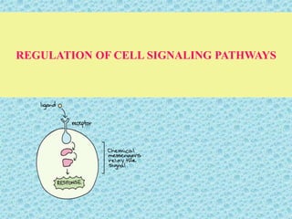
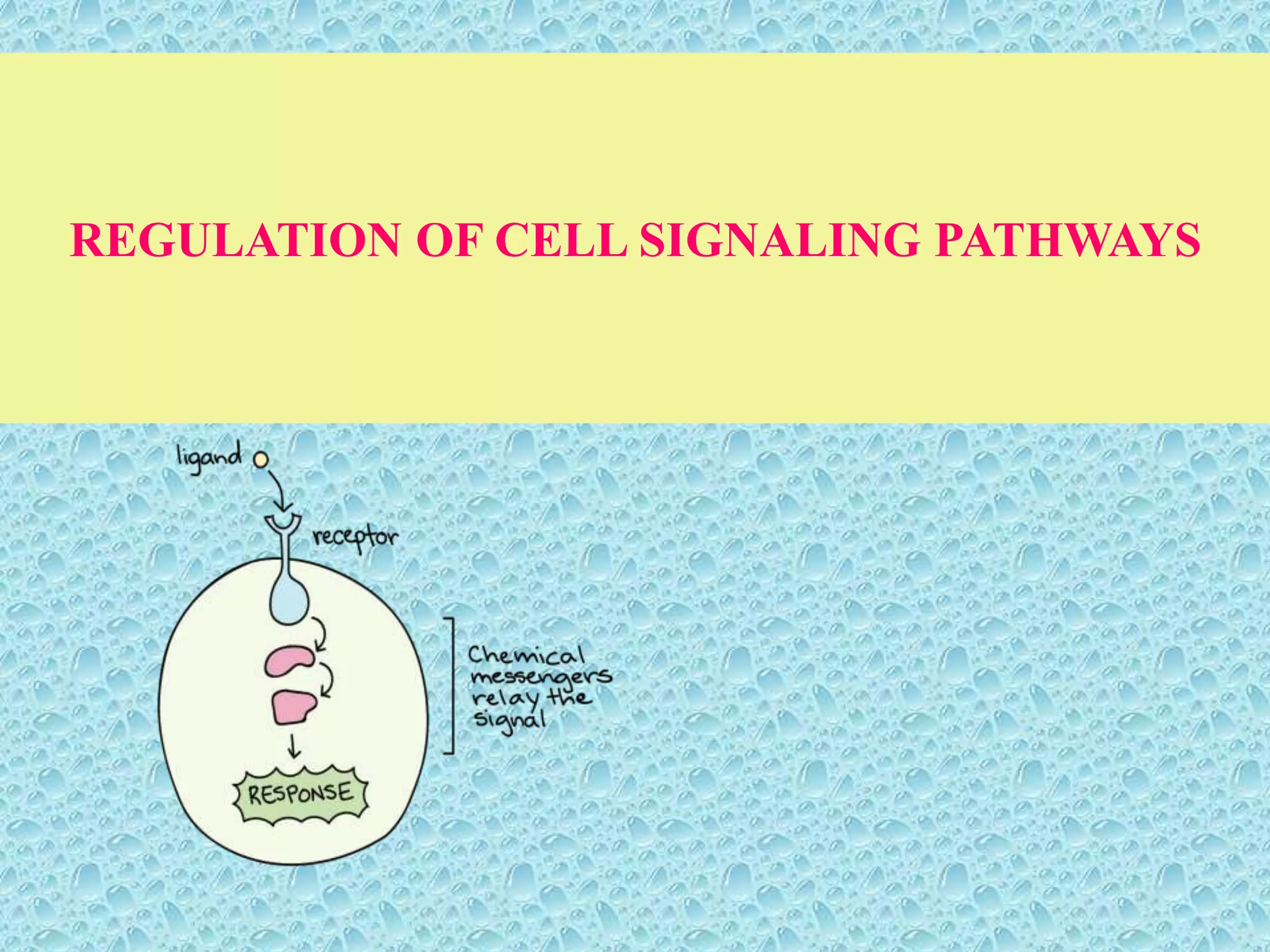
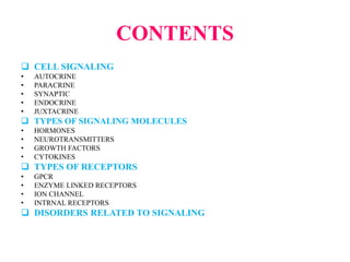
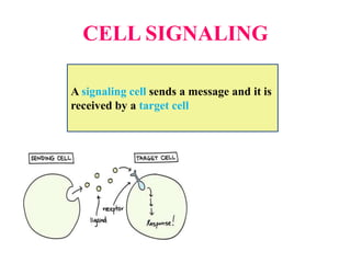
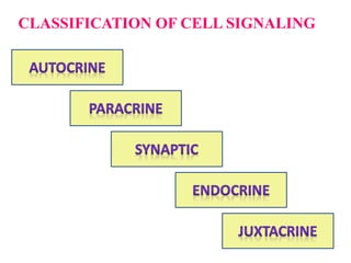
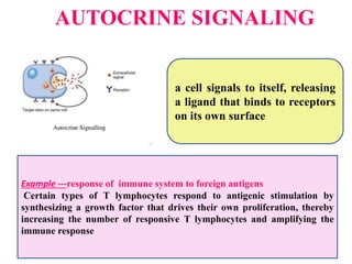
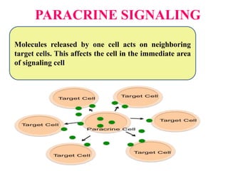
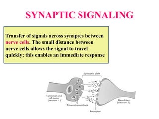
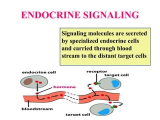
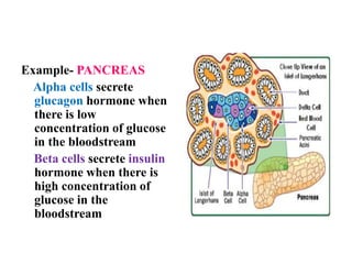
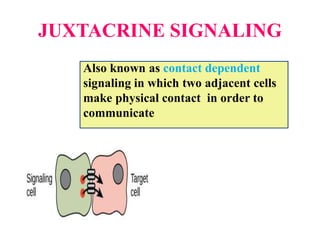
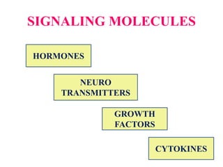
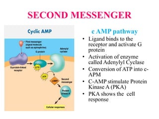
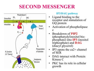
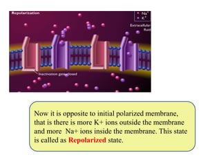
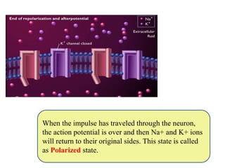
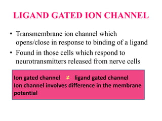
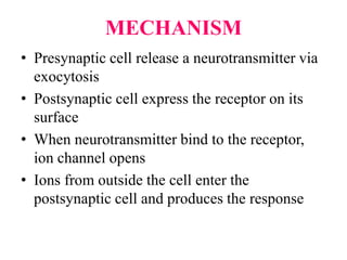
The document discusses cell signaling pathways and regulation. It describes different types of cell signaling including autocrine, paracrine, synaptic, endocrine, and juxtacrine signaling. It also discusses various signaling molecules like hormones, neurotransmitters, growth factors, and cytokines. The different types of cell surface receptors and intracellular receptors are described, including G protein-coupled receptors, enzyme-linked receptors, and ion channel receptors. The mechanisms of various cell signaling pathways such as G protein signaling and MAP kinase pathways are summarized. Disorders related to improper cell signaling are also mentioned.
Assignment on Need of cell signaling, Steps in cell signaling, Intercellular signaling pathways, Types of intercellular signaling pathways, Intracellular signaling pathways, Receptors, Intercellular and intracellular signaling pathways. Classification of receptor family and molecular structure ligand gated ion channels; Gprotein coupled receptors, tyrosine kinase receptors and nuclear receptors.
Cell signaling / Signal Transduction / Transmembrane signaling.
It is the process by which cells communicate with their environment and respond to external stimuli.
When a signaling molecule(ligand) binds to its receptor, it alters the shape or activity of the receptor, triggering a change inside of the cell such as alteration in the activity of a gene / cell division. Thus the original Intercellular Signal is converted into an Intracellular Signal that triggers as a response.
Introduction
Definition
History
Basic element in signal transduction
Basic Pathway of signal transduction
Types of signal transduction
Second messenger
Pathway of signal transduction
Conclusion
References
In biology, cell signaling is part of any communication process that governs basic activities of cells and coordinates multiple-cell actions. The ability of cells to perceive and correctly respond to their microenvironment is the basis of development, tissue repair, and immunity, as well as normal tissue homeostasis.
Ion channels have many features of typical membrane proteins. They are synthesized and inserted into the membrane of the endoplasmic reticulum, glycosylated in the Golgi, and transported and inserted into target membranes by membrane fusion. They are regulated by trafficking, phosphorylation, ubiquitination, reversible interactions with other signaling proteins and second messengers, proteolytic cleavage, and other modifications. Like other signaling proteins, ion channels are flexible molecules that undergo conformational changes between open (active) and closed (inactive) states. They evolve and increase in number through phylogeny and can be placed in gene families and super families according to their sequence similarities.
Cell Signalling Pathway (intra and extra cellular signalling)Aneela Rafiq
the way of communication cell to cell or cell to their environment. they produce some stimuli to correspond to surroundings for survival. Cell signalling helps to defend, survive, production of chemicals and lots of other things. signalling can external and internal. in this presentation, paths are elaborated externally and internally.
Hope it will help to understand cell signal in better way.
Molecular interaction, Regulation and Signalling receptors and vesiclesAnantha Kumar
1. Overview of Extracellular signalling
2. Signalling molecules operate over various distance in animals
3.Endocrine Signalling
4.Paracrine Signalling
5.Autocrine Signalling
6. Signalling by Plasma membrane attached proteins
7.Receptors
8 Properties of receptors
9.Cell surface receptors belong to four major classes
10.Signal transduction Mechanism
11. Second messenger
12. Contraction of skeletal Muscle cells mechanism
Cell signaling is the process where cell communicate with each other with the help of signaling molecules and receptors. Cell signaling is done by different types of signaling processes such as autocrine, paracrine, synaptic, endocrine, contact dependent signaling
The signal transduction pathway uses a network of interactions within cells, among cells, and throughout plant.
The external signals that affect plant growth and development include many aspects of the plant’s physical, chemical, and biological environments. Some external signals come from other plants.
Many signals interact cooperatively and synergistically with each other to produce the final response. Signal combinations that induce such complex plant responses include red and blue light, gravity and light, growth regulators and mineral nutrients .
For example the overall regulation of seed germination involves control by both external factors and internal signals.
The presentation illustrates the basic modes of cell signalling pathways for undergraduate students. It mentions variety of examples of cell signalling with different receptors, ligands and target molecules
This Presentation provides an outline knowledge about Cellular Communication, Steps involved, Its Types, Signal Transduction, Secondary Messenger , Receptors with some Interesting Facts and Current Trends. An assignment for the subject, Cellular and Molecular Pharmacology, 1st year M.Pharm, 1st semester.
GENERAL IDEA OF SIGNAL TRANSDUCTION
DEFINATION
WHAT DOES THE TERM SIGNAL TRANSDUCTION MEANS
HISTORY
BASIC ELEMENTS IN SIGNAL TRANSDUCTION
TYPES OF SIGNAL TRANSDUCTION
SIGNALLING MOLECULE
RECEPTOR MOLECULE
MODES OF CELL CELL SIGNALING
SECOND MESSENGER
SIGNAL TRANSDUCTION PATHWAY
SOME SIGNALING PATHWAYS
SIGNIFICANCE
CONCLUSION
REFERENCE
DERIVATION OF MODIFIED BERNOULLI EQUATION WITH VISCOUS EFFECTS AND TERMINAL V...Wasswaderrick3
In this book, we use conservation of energy techniques on a fluid element to derive the Modified Bernoulli equation of flow with viscous or friction effects. We derive the general equation of flow/ velocity and then from this we derive the Pouiselle flow equation, the transition flow equation and the turbulent flow equation. In the situations where there are no viscous effects , the equation reduces to the Bernoulli equation. From experimental results, we are able to include other terms in the Bernoulli equation. We also look at cases where pressure gradients exist. We use the Modified Bernoulli equation to derive equations of flow rate for pipes of different cross sectional areas connected together. We also extend our techniques of energy conservation to a sphere falling in a viscous medium under the effect of gravity. We demonstrate Stokes equation of terminal velocity and turbulent flow equation. We look at a way of calculating the time taken for a body to fall in a viscous medium. We also look at the general equation of terminal velocity.
Observation of Io’s Resurfacing via Plume Deposition Using Ground-based Adapt...Sérgio Sacani
Since volcanic activity was first discovered on Io from Voyager images in 1979, changes
on Io’s surface have been monitored from both spacecraft and ground-based telescopes.
Here, we present the highest spatial resolution images of Io ever obtained from a groundbased telescope. These images, acquired by the SHARK-VIS instrument on the Large
Binocular Telescope, show evidence of a major resurfacing event on Io’s trailing hemisphere. When compared to the most recent spacecraft images, the SHARK-VIS images
show that a plume deposit from a powerful eruption at Pillan Patera has covered part
of the long-lived Pele plume deposit. Although this type of resurfacing event may be common on Io, few have been detected due to the rarity of spacecraft visits and the previously low spatial resolution available from Earth-based telescopes. The SHARK-VIS instrument ushers in a new era of high resolution imaging of Io’s surface using adaptive
optics at visible wavelengths.
Earliest Galaxies in the JADES Origins Field: Luminosity Function and Cosmic ...Sérgio Sacani
We characterize the earliest galaxy population in the JADES Origins Field (JOF), the deepest
imaging field observed with JWST. We make use of the ancillary Hubble optical images (5 filters
spanning 0.4−0.9µm) and novel JWST images with 14 filters spanning 0.8−5µm, including 7 mediumband filters, and reaching total exposure times of up to 46 hours per filter. We combine all our data
at > 2.3µm to construct an ultradeep image, reaching as deep as ≈ 31.4 AB mag in the stack and
30.3-31.0 AB mag (5σ, r = 0.1” circular aperture) in individual filters. We measure photometric
redshifts and use robust selection criteria to identify a sample of eight galaxy candidates at redshifts
z = 11.5 − 15. These objects show compact half-light radii of R1/2 ∼ 50 − 200pc, stellar masses of
M⋆ ∼ 107−108M⊙, and star-formation rates of SFR ∼ 0.1−1 M⊙ yr−1
. Our search finds no candidates
at 15 < z < 20, placing upper limits at these redshifts. We develop a forward modeling approach to
infer the properties of the evolving luminosity function without binning in redshift or luminosity that
marginalizes over the photometric redshift uncertainty of our candidate galaxies and incorporates the
impact of non-detections. We find a z = 12 luminosity function in good agreement with prior results,
and that the luminosity function normalization and UV luminosity density decline by a factor of ∼ 2.5
from z = 12 to z = 14. We discuss the possible implications of our results in the context of theoretical
models for evolution of the dark matter halo mass function.
Comparing Evolved Extractive Text Summary Scores of Bidirectional Encoder Rep...University of Maribor
Slides from:
11th International Conference on Electrical, Electronics and Computer Engineering (IcETRAN), Niš, 3-6 June 2024
Track: Artificial Intelligence
https://www.etran.rs/2024/en/home-english/
Seminar of U.V. Spectroscopy by SAMIR PANDASAMIR PANDA
Spectroscopy is a branch of science dealing the study of interaction of electromagnetic radiation with matter.
Ultraviolet-visible spectroscopy refers to absorption spectroscopy or reflect spectroscopy in the UV-VIS spectral region.
Ultraviolet-visible spectroscopy is an analytical method that can measure the amount of light received by the analyte.
THE IMPORTANCE OF MARTIAN ATMOSPHERE SAMPLE RETURN.Sérgio Sacani
The return of a sample of near-surface atmosphere from Mars would facilitate answers to several first-order science questions surrounding the formation and evolution of the planet. One of the important aspects of terrestrial planet formation in general is the role that primary atmospheres played in influencing the chemistry and structure of the planets and their antecedents. Studies of the martian atmosphere can be used to investigate the role of a primary atmosphere in its history. Atmosphere samples would also inform our understanding of the near-surface chemistry of the planet, and ultimately the prospects for life. High-precision isotopic analyses of constituent gases are needed to address these questions, requiring that the analyses are made on returned samples rather than in situ.
This presentation explores a brief idea about the structural and functional attributes of nucleotides, the structure and function of genetic materials along with the impact of UV rays and pH upon them.
(May 29th, 2024) Advancements in Intravital Microscopy- Insights for Preclini...Scintica Instrumentation
Intravital microscopy (IVM) is a powerful tool utilized to study cellular behavior over time and space in vivo. Much of our understanding of cell biology has been accomplished using various in vitro and ex vivo methods; however, these studies do not necessarily reflect the natural dynamics of biological processes. Unlike traditional cell culture or fixed tissue imaging, IVM allows for the ultra-fast high-resolution imaging of cellular processes over time and space and were studied in its natural environment. Real-time visualization of biological processes in the context of an intact organism helps maintain physiological relevance and provide insights into the progression of disease, response to treatments or developmental processes.
In this webinar we give an overview of advanced applications of the IVM system in preclinical research. IVIM technology is a provider of all-in-one intravital microscopy systems and solutions optimized for in vivo imaging of live animal models at sub-micron resolution. The system’s unique features and user-friendly software enables researchers to probe fast dynamic biological processes such as immune cell tracking, cell-cell interaction as well as vascularization and tumor metastasis with exceptional detail. This webinar will also give an overview of IVM being utilized in drug development, offering a view into the intricate interaction between drugs/nanoparticles and tissues in vivo and allows for the evaluation of therapeutic intervention in a variety of tissues and organs. This interdisciplinary collaboration continues to drive the advancements of novel therapeutic strategies.
The ability to recreate computational results with minimal effort and actionable metrics provides a solid foundation for scientific research and software development. When people can replicate an analysis at the touch of a button using open-source software, open data, and methods to assess and compare proposals, it significantly eases verification of results, engagement with a diverse range of contributors, and progress. However, we have yet to fully achieve this; there are still many sociotechnical frictions.
Inspired by David Donoho's vision, this talk aims to revisit the three crucial pillars of frictionless reproducibility (data sharing, code sharing, and competitive challenges) with the perspective of deep software variability.
Our observation is that multiple layers — hardware, operating systems, third-party libraries, software versions, input data, compile-time options, and parameters — are subject to variability that exacerbates frictions but is also essential for achieving robust, generalizable results and fostering innovation. I will first review the literature, providing evidence of how the complex variability interactions across these layers affect qualitative and quantitative software properties, thereby complicating the reproduction and replication of scientific studies in various fields.
I will then present some software engineering and AI techniques that can support the strategic exploration of variability spaces. These include the use of abstractions and models (e.g., feature models), sampling strategies (e.g., uniform, random), cost-effective measurements (e.g., incremental build of software configurations), and dimensionality reduction methods (e.g., transfer learning, feature selection, software debloating).
I will finally argue that deep variability is both the problem and solution of frictionless reproducibility, calling the software science community to develop new methods and tools to manage variability and foster reproducibility in software systems.
Exposé invité Journées Nationales du GDR GPL 2024
4. CELL SIGNALING AND SIGNAL
TRANSDUCTION
CELL SIGNALING
• the communication
among different groups
of cells and tissues
• How one group of cell
inform other group of
cell- what to do
SIGNAL
TRANSDUCTION
• it refers to how the
presence of an
extracellular signal can
produce a change in the
intracellular state of the
cell without the initial
signal crossing the
membrane
5. Cell signaling is a part
of complex system of
communication that
governs basic cellular
activities and
coordinates cell actions.
The ability of cells to
perceive and correctly
respond to their micro-
environment is the basis
of development, tissue
repair and immunity as
well as normal tissue
homeostasis.
Errors in the cellular
information processing
are responsible for
diseases such as cancer,
autoimmunity and
diabetes.
By understanding cell
signaling, diseases may
be treated more
effectively
7. Cells do not behave as selfish entities but rather
tend to form «microsocieties» whose proper
functioning requires a precise coordination of
emission and reception of signals. So cells
communicate because-
Cell to cell signal encourage other cells to divide
and act as a dynamic part of tissue.
Cells need to know whether to live, die or divide
Neurotransmission- nerve cell activate adjacent
nerve cell to transmit the signal
Regulation of metabolism
Immune response
Sex determination and gonad development
9. AUTOCRINE SIGNALING
a cell signals to itself, releasing
a ligand that binds to receptors
on its own surface
Example ---response of immune system to foreign antigens
Certain types of T lymphocytes respond to antigenic stimulation by
synthesizing a growth factor that drives their own proliferation, thereby
increasing the number of responsive T lymphocytes and amplifying the
immune response
11. SYNAPTIC SIGNALING
Transfer of signals across synapses between
nerve cells. The small distance between
nerve cells allows the signal to travel
quickly; this enables an immediate response
13. Example- PANCREAS
Alpha cells secrete
glucagon hormone when
there is low
concentration of glucose
in the bloodstream
Beta cells secrete insulin
hormone when there is
high concentration of
glucose in the
bloodstream
14. JUXTACRINE SIGNALING
Also known as contact dependent
signaling in which two adjacent cells
make physical contact in order to
communicate
16. HORMONES
• These are the signaling molecules produced by
endocrine glands
• Transported by blood stream to the target cells
• These belong to different classes of chemical
structures- amino acids, peptides, proteins or
steroids
• Examples
Adrenaline (amino acid) – increase pulse rate and
blood pressure
Insulin (peptide) – carbohydrate catabolism
Progesterone (steroid) – preparation of endometrial
layer
17. NEUROTRANSMITTERS
• Also known as chemical messengers.
• Neurotransmitters are released from synaptic
vesicles in synapses into the synaptic cleft,
where they are received by receptors on the
target cells
• These transmit the signal from one neuron to
target neuron. These are synthesized from
amino acids.
18. Acetylcholine- an excitatory neurotransmitter at
the neuromuscular junction in skeletal muscle,
causing the muscle to contract.
Dopamine-this includes regulation of motor
behavior, pleasures related to motivation and also
emotional arousal
Gamma-aminobutyric acid (GABA)- during
anxiety, fear and pain GABA stimulate the
feelings of calm and relaxation
Serotonin- It functions to regulate appetite, sleep,
memory and learning and also temperature, mood
and behavior. Some depressed patients are seen
to have lower concentrations of serotonin in
their brain tissue
19. GROWTH FACTORS
• These are any group of proteins that stimulate the
growth of specific tissues
• These bind to the receptors on cell surface
• These play important role in promoting cellular
differentiation and cell division
• Example
Epidermal growth factor-stimulate the growth
of epithelial cells
Platelet derived growth factors- stimulate the
growth of muscle cells and connective tissues
Nerve growth factors- stimulate the growth of
neuronal cells
20. CYTOKINES
• These are signaling proteins that are extensively used
in immune function
• These are produced by immune cells (lymphocytes)
• Cytokines released from one cell affect the actions of
other cells by binding to receptors on their surface
• Examples
Interferons- these are proteins that inhibit virus
replication inside the cells
Interleukins – these regulate the immune and
inflammatory responses. Their functions include
growth, maturation and activation of immune cells
22. G- protein coupled receptors
• A large protein family of the receptors
• Detect molecules outside the cell
• Activate internal signal transduction
pathway and ultimately cellular
responses
• Ligands that bind and activate the
receptors are odors, pheromones,
hormones and neurotransmitters
23. STRUCTURE OF GPCR
• N terminal is present outside the cell
• 7 α helix is present on the plasma membrane
• C terminal is present inside the cell
24. STRUCTURE OF G- PROTEIN
• Contains 3 polypeptide subunits α, β and γ
• Guanine nucleotide binding site is present on α
subunit
• G- Proteins are held at plasma membrane by lipid
chains that covalently attached to α and γ subunits
26. MECHANISM
• Ligand binds to receptor
• Conformational changes in the cytoplasmic loops
• Affinity of the receptor to G-Protein
• Ligand bound receptor forms G- Protein Complex
• Conformational change in α subunit replace GDP
with GTP
27. G α subunit dissociates from the
complex and activate the target
Target releases second messenger )
GTPase hydrolyses the GTP back into GDP which
deactivates the target enzyme
28. SECOND MESSENGER
c AMP pathway
• Ligand binds to the
receptor and activate G
protein
• Activation of enzyme
called Adenylyl Cyclase
• Conversion of ATP into c-
APM
• C-AMP stimulate Protein
Kinase A (PKA)
• PKA shows the cell
response
29. SECOND MESSENGER
IP3/DAG pathway
• Ligand binding to the
receptor and stimulation of
GQ protein
• Activation of phospholipase
c
• Breakdown of PIP2
(phosphatydylinositol bis-
phosphate) into IP3 (inositol
triphosphate) and DAG
(diacyl glycerol)
• IP3 opens the ca2+ channel
of SER
• DAG interact with Protein
Kinase C
• PKC has its role in cellular
growth
30. Physiological roles of G protein
receptors
• Taste : GPCR in taste cells mediate the release
of gustducin in response to bitter and sweet
tasting substance
• Smell : receptors of olfactory epithelium bind
the odorants
• Behavior regulation : receptors in brain bind
several different neurotransmitters including
serotonin, dopamine, GABA and glutamate
31. ENZYME LINKED RECEPTORS
• Protein tyrosine kinases are the enzymes that
phosphorylate specific tyrosine residue
• The kinases are involved in cell regulation,
cell growth and cell division
Different growth factors act as ligand for signal
transduction.
• Vascular-endothelial growth factor (VEGF)
This growth factor promotes new blood vessel
growth, but is also important for maintenance
of endothelial cells in the delicate filtration
membrane of the kidney.
• Neurotrophins promote the differentiation of
neurons.
• Epidermal growth factor (EGF) stimulate
the growth of epithelial cells
32. MECHANISM
• The first step is that ligand binding
causes receptor dimerization: that is, binding
of the ligand brings together two receptors.
• The receptors, which are tyrosine kinases, get
phosphorylated with the help of ATP.
• The phosphotyrosine on the phosphorylated
receptor is a binding site for an adaptor
protein.
33.
34. RAS MAP KINASE PATHWAY
• The most common intracellular pathway triggered
by Receptor Tyrosine Kinase is known as
Mitogen Activated Protein Kinase Pathway
(MAPK)
• MAPK pathway is related to cell division and cell
proliferation
• It gives proteins and enzymes which are required
for cell growth
• Signaling molecules to initiate the pathway- EGF
and PDGF
• Receptors are embedded in the plasma membrane
36. All the signaling pathways have
“on” and “off” switch
If MAPK pathway
remains “ON”
continuously then what
will happen…..?
37. Due to RAS mutation RAS
permanently binds with GTP then
cell lose the control on cell division.
Cell will continuously grow and
divide which finally leads to
CANCER
38. ION CHANNEL RECEPTORS
• excitable cells (neurons, muscle cells,
and touch receptor cells)
• all of them use ion channel receptors to
convert chemical or mechanical
messages into electrical signals
• an excitable cell maintains a different
concentration of ions in its cytoplasm
than exists in its extracellular
environment
• concentration differences create a small
electrical potential across the plasma
membrane
• “specialized channels” in the plasma
membrane open and allow rapid ion
movement into or out of the cell
39.
40. ION CHANNELS
UNGATED CHANNEL Provide holes, through which ions can
diffuse across the membrane, they
remain open all the time. Example
ungated K+ and Cl- ion channel
VOLTAGE GATED CHANNEL Open or close in response to change in
the membrane potential. Example Na+
and K+ ion channels
MECHANICALLY GATED
CHANNEL
Include touch sensors in the skin and
vibration sensors in the inner ear
Some hollow organs like bladder and
intestine also
TEMPERATURE GATED
CHANNEL
Found in the sensory neuron in the skin,
channels open with an increase or
decrease in the temperature. This leads
to the sensations of warm and cold
LIGAND GATED CHANNEL These open in response to the binding of a
neurotransmitter
Example skeletal muscle cells
41. VOLTAGE GATED ION CHANNEL
• A class of trans-membrane proteins that form ion
channels that are activated by changes in the
electrical membrane potential near the channel
• The membrane potential alters the conformation
of the channel proteins, regulating their opening
and closing
• Cell membranes are generally impermeable
to ions
• Ions diffuse through the membrane through trans-
membrane protein channels
42. Resting membrane
potential in which there
is +ve charge outside
the cell and –ve charge
inside the cell. This is
called as Polarized
state. Activation gates
of Na+ and K+ ion
channels are closed
When a stimulus reaches
the resting neuron, Na+
channels open and allow
Na+ ions from outside to
the inside of the cell
43. When more Na+ ions
move into the cell, inside
become more +ve and
this state is called as
depolarization of the
cell
Now the K+ channels
open and K+ ions move
from inside to the
outside of the cell to
restore the electrical
balance
44. Now it is opposite to initial polarized membrane,
that is there is more K+ ions outside the membrane
and more Na+ ions inside the membrane. This state
is called as Repolarized state.
45. When the impulse has traveled through the neuron,
the action potential is over and then Na+ and K+ ions
will return to their original sides. This state is called
as Polarized state.
46. LIGAND GATED ION CHANNEL
• Transmembrane ion channel which
opens/close in response to binding of a ligand
• Found in those cells which respond to
neurotransmitters released from nerve cells
Ion gated channel ≠ ligand gated channel
Ion channel involves difference in the membrane
potential
47. MECHANISM
• Presynaptic cell release a neurotransmitter via
exocytosis
• Postsynaptic cell express the receptor on its
surface
• When neurotransmitter bind to the receptor,
ion channel opens
• Ions from outside the cell enter the
postsynaptic cell and produces the response
48.
49. INTERNAL RECEPTORS
• Internal receptors, also known as
intracellular or cytoplasmic receptors
• found in the cytoplasm of the cell
• respond to hydrophobic ligand molecules
that are able to travel across the plasma
membrane.
51. MECHANISM
• Being lipids, steroid hormones enter the cell by
simple diffusion across the plasma membrane
• Once inside the cell, ligand binds to the
internal receptor
• The ligand receptors complex move into the
nucleus
• Then bind to the specific regulatory regions of
the chromosomal DNA and promotes initiation
of transcription
52. • Transcription is the process of copying the
information in a cell DNA into a special form
of RNA called as messenger RNA
• m-RNA then moves into the cytoplasm and
associates with ribosomes to link specific
amino acids in correct order and produce
protein
55. BIOLOGICAL FUNCTIONS OF
INTERNAL RECEPTORS
• Regulations of growth and embryonic
development
• Maintenance of phenotype
• Regulation of metabolic processes such as
cholesterol and bile acid metabolism
56. When Cell Communication Goes Wrong
The cells in our bodies are constantly sending out and receiving
signals.
But what if a cell fails to send out a signal at the proper time?
What if a signal doesn't reach its target?
What if a target cell does not respond to a signal, or a cell
responds even though it has not received a signal?
These are just a few ways in which cell
communication can go wrong, resulting in disease.
In fact, most diseases involve at least one
breakdown in cell communication.
57. DISORDERS
• Night Blindness
mutations in G-protein.
It affects the response of
rod cells to light
• Pseudohypoparathyroi
dism -the genetic loss
of G(s) protein, results
in non-responsiveness
to parathyroid hormone
58. • Testotoxicosis- mutation in the
receptor for luteinizing hormone can
over-stimulate G(s) proteins, resulting
in the excessive production of
testosterone
• Adenomas G proteins lose their
ability to hydrolyse GTP through
mutation, resulting in the excessive
secretion of growth hormone and the
increased proliferation of cells
59. ALZHEIMER’S DISEASE
It is chronic neuro-degenerative disease that
usually starts slowly and worsens over time
Symptoms-
Short term memory loss
Mood swings
Loss of motivation
Behavioral issues
60. Causes of Alzheimer
Plaques
Due to gene mutation
clumps of protein
called β amyloid
damage and destroy
the brain cell and
interfere with cell to
cell communication
61. Causes of Alzheimer
Tangles
brain cells depend on
internal support and
transport system to carry the
nutrients throughout their
long extensions.
This requires a normal
functioning proteins called
In Alzheimer, protein
threads twist into tangles
leads to failure of transport
and death of brain cells
tau
62. MULTIPLE SCLEROSIS
• A disease in which the protective wrappings
around nerve cells in the brain and spinal cord
are destroyed.
• Affected nerve cell can no longer transmit
signals from one area of the brain to other area
Muscle weakness
Blurred vision
Depression
63.
64. DIABETES
• Type I and type II diabetes
have very similar
symptoms, but they have
different causes
• People who have type I
diabetes are unable to
produce the insulin signal,
• Those with type II diabetes
do produce insulin.
However, the cells of type II
diabetics have lost the
ability to respond to insulin.
• The end result is the same:
blood sugar levels become
dangerously high
65. BARTTER SYNDROME
• It is an Autosomal
recessive disease
• It is caused by
mutations in the genes
coding for K+ and Ca++
channel subunits
• It causes reduced
potassium and excessive
calcium in the urine
Symptoms
• polyuria
• polydipsia
• dehydration
• vomiting
• growth retardation
66. CONCLUSION
In order to response to changes in their
immediate environment, cells must be able to
receive and process signals that originate outside
their borders.
Individual cells often receive many signals
simultaneously and then they integrate the
information they receive into a unified action
plan
Cells also send out the signals to the other cells
both near and far
67. Once the receptor protein receives a signal, it
undergoes a conformational change, which in
turn launches a series of biochemical
reactions within the cell.
The genes encoding for the proteins in cells
can mutate during the cell growth which can
cause disturbance in cell signaling pathway.
The disturbance can lead to several diseases
like Multiple Sclerosis, Alzheimer disease,
Cancer and many more.
68. REFERENCES
• Catterall A (2000) From ionic current to molecular
mechanism: the structure and function of voltage
gated sodium channels. Neuron 26: 13-25
• Dingledine R, Borges K and Bowei D (1999) The
glutamate receptor ion channel. Pharmacol Rev 51: 7-
61
• Jeremy B, Tymoczko J and Stryer L (ed) (2007).
Biochemistry. pp 389-90. Sara Tenney, New York
• Lemmon A and Schlessinger J (2010) Cell signaling
by receptor tyrosine kinases. Cell 141: 1117-134
69. • Niemeyer B and Mery L (2001) Ion channels in
health and diseases. J Embo Rep 2: 568-73
• Ralph A and Edward A eds (2010) Handbook of cell
signaling. Vol 67, pp 63-69. Academic press,
Netherland.
• Tonks K and Neel G (1996) From form to function:
signaling by protein tyrosine phosphates. Cell 87:
365-68
• Yan K, Gao L, Zhang Y and Zhou X (2016) The
cyclic AMP signaling pathway: Exploring targets for
successful drug discovery. J Mol Med Rep 13: 3715-
723