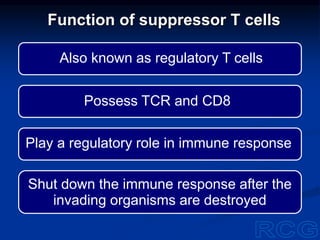
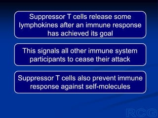
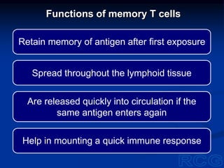
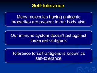
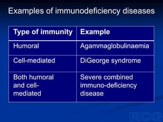
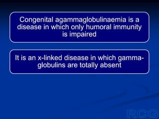
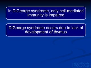
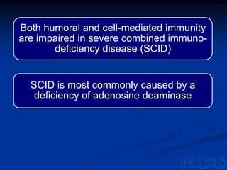
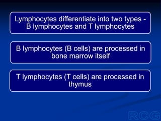
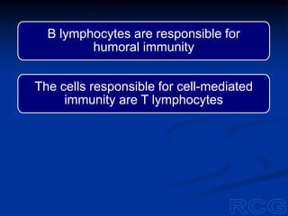
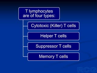
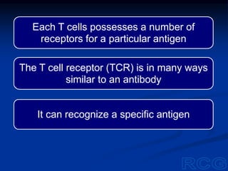
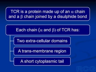
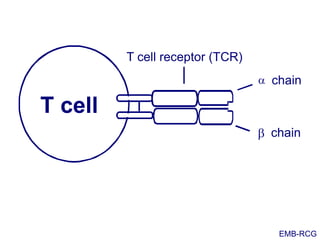
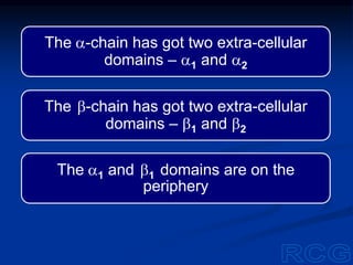
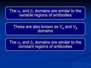
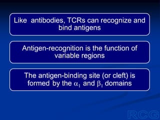
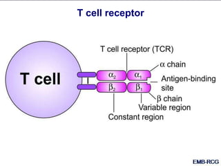
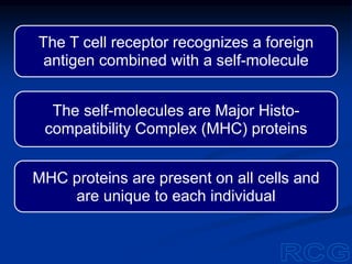
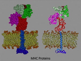
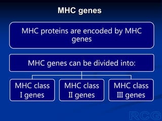
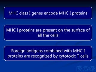
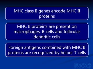
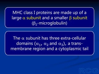
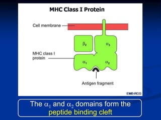
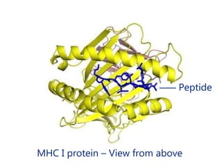
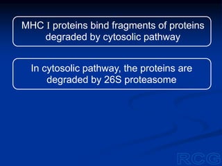
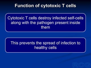
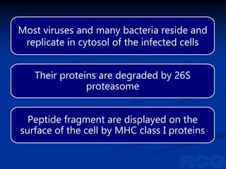
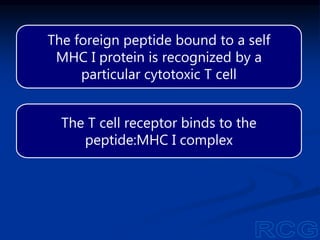
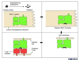
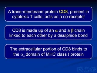
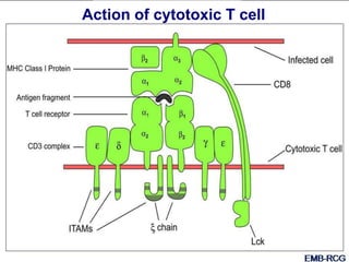
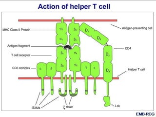
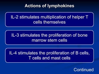
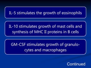
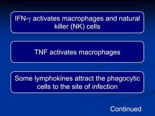
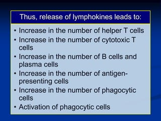
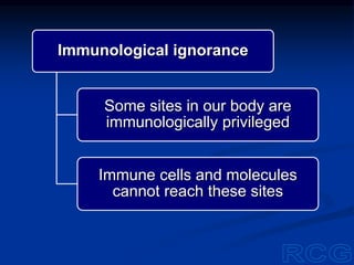
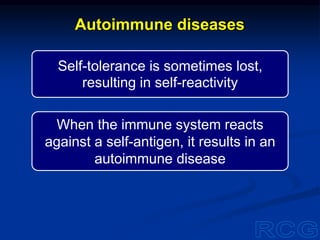
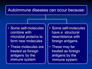
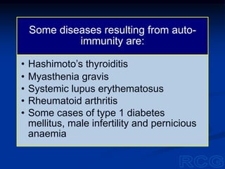
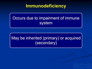
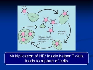
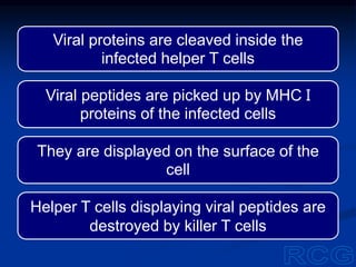
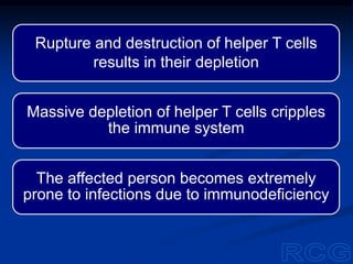
Cell-mediated Immunity
R. C. Gupta
M.D. (Biochemistry)
Jaipur, India
Our defenses against infections include
innate immunity and acquired immunity
Innate immunity acts non-specifically
against all pathogens
Acquired immunity is specific; one cell or
molecule acts against one pathogen
Acquired immunity comprises humoral
immunity and cell-mediated immunity
Humoral immunity acts against pathogens
that haven’t entered any self-cell
Cell-mediated immunity acts against
pathogens that have entered some self-cell
Lymphocytes play a key role in humoral
as well as cell-mediated immunity
Lymphocytes are formed from stem cells
in bone marrow
T lymphocytes (T cells) are processed in
thymus
B lymphocytes (B cells) are processed in
bone marrow itself
Lymphocytes differentiate into two types -
B lymphocytes and T lymphocytes
The cells responsible for cell-mediated
immunity are T lymphocytes
B lymphocytes are responsible for
humoral immunity
T lymphocytes
are of four types:
Cytotoxic (Killer) T cells
Helper T cells
Suppressor T cells
Memory T cells
Each T cells possesses a number of
receptors for a particular antigen
The T cell receptor (TCR) is in many ways
similar to an antibody
It can recognize a specific antigen
Each chain (a and b) of TCR has:
Two extra-cellular domains
A trans-membrane region
A short cytoplasmic tail
TCR is a protein made up of an a chain
and a b chain joined by a disulphide bond
chain
b chain
T cell
T cell receptor (TCR)
a
EMB-RCG
The a-chain has got two extra-cellular
domains – a1 and a2
The b-chain has got two extra-cellular
domains – b1 and b2
The a1 and b1 domains are on the
periphery
The a1 and b1 domains are similar to the
variable regions of antibodies
These are also known as Va and Vb
domains
The a2 and b2 domains are similar to the
constant regions of antibodies
Like antibodies, TCRs can recognize and
bind antigens
Antigen-recognition is the function of
variable regions
The antigen-binding site (or cleft) is
formed by the a1 and b1 domains
T cell receptor
T cell receptors have a diversity similar to
antibody diversity
TCR diversity also arises from gene re-
arrangement
TCR diversity
One of the 70 V segments
Thousands of different combinations are
possible
The gene for the variable region of a
chain (Va) is constructed from:
One of the 61 J segments
One of the 52 V segments
One of the 2 D segments
One of the 13 J segments
Thousands of different combinations
are possible
The gene for the variable region of b
chain (Vb) is constructed from:
When an a chain combines with a b chain,
millions of combinations are possible
A given T cell has one combination, and is
specific for one particular antigen
The T cell receptor cannot bind a free
antigen
The antigen must be attached to a cell, and
it should be a self-cell
If a T cell is transferred from one person to
another, it will not recognize the antigen
MHC proteins and MHC genes
The T cell receptor recognizes a foreign
antigen combined with a self-molecule
The self-molecules are Major Histo-
compatibility Complex (MHC) proteins
MHC proteins are present on all cells and
are unique to each individual
MHC Proteins
MHC genes
MHC proteins are encoded by MHC
genes
MHC genes can be divided into:
MHC class
I genes
MHC class
II genes
MHC class
III genes
MHC class I genes encode MHC I proteins
MHC I proteins are present on the surface of
all the cells
Foreign antigens combined with MHC I
proteins are recognized by cytotoxic T cells
MHC class II genes encode MHC II
proteins
MHC II proteins are present on
macrophages, B cells and follicular
dendritic cells
Foreign antigens combined with MHC II
proteins are recognized by helper T cells
MHC class III genes
encode:
Complement
proteins
Some other
plasma proteins
MHC proteins are trans-membrane proteins
They are synthesized on the endoplasmic
reticulum
They get inserted in the cell membrane
They have a peptide (antigen) binding cleft
on their extracellular surface
MHC proteins
While on their way to the cell membrane,
MHC proteins pick up peptide fragments
formed by degradation of:
The fragments are bound firmly and
displayed on the surface of the cell
Endogenous
proteins
Exogenous
proteins
Endogenous peptides bound to MHC
proteins are ignored by the immune
system
If a T cell finds a foreign peptide bound
to an MHC protein, cell-mediated immunity
comes into operation
MHC class I proteins are made up of a
large a subunit and a smaller b subunit
(b2-microglobulin)
The a subunit has three extra-cellular
domains (a1, a2 and a3), a trans-
membrane region and a cytoplasmic tail
The a1 and a2 domains form the
peptide binding cleft
MHC I protein – View from above
‒‒‒ Peptide
MHC I proteins bind fragments of proteins
degraded by cytosolic pathway
In cytosolic pathway, the proteins are
degraded by 26S proteasome
MHC class II proteins are made up of
almost equal sized a subunit and
b subunit
Each subunit has two extracellular
domains, a trans-membrane region and
a cytoplasmic tail
The a1 and b1 domains form the peptide
binding cleft
‒ Peptide
MHC II protein – View from above
MHC class II proteins bind fragments of
proteins degraded by lysosomal pathway
These proteins first enter intracellular
vesicles called endosomes
The endosomes fuse with lysosomes to
form endolysosomes
The proteins present in endolysosomes
are degraded by lysosomal enzymes
The lysosomal proteolytic enzymes are
cathepsins
The proteins present in
endosomes include:
Proteins of extracellular pathogens
Proteins of pathogens that reside
in endosomes
Proteins taken up by the cells by
endocytosis
MHC Class II
Protein
Antigen
fragment
a2 b2
b1
Cell membrane
Newly synthesized MHC II proteins bind
peptide fragments, and go to the cell
membrane
MHC II proteins get inserted in the cell
membrane, displaying the bound peptide
on the surface of the cell
Cytotoxic T cells destroy infected self-cells
along with the pathogen present inside
them
This prevents the spread of infection to
healthy cells
Function of cytotoxic T cells
Most viruses and many bacteria reside and
replicate in cytosol of the infected cells
Their proteins are degraded by 26S
proteasome
Peptide fragment are displayed on the
surface of the cell by MHC class I proteins
The foreign peptide bound to a self
MHC I protein is recognized by a
particular cytotoxic T cell
The T cell receptor binds to the
peptide:MHC I complex
A trans-membrane protein CD8, present in
cytotoxic T cells, acts as a co-receptor
CD8 is made up of an a and a b chain
linked to each other by a disulphide bond
The extracellular portion of CD8 binds to
the a3 domain of MHC class I protein
The cytoplasmic tail of CD8 is associated
with Lck, a cytosolic tyrosine kinase
The cytoplasmic tail of T cell receptor is
associated with a trans-membrane protein,
CD3 complex
CD3 complex consists of a g chain, a d
chain, two e chains and two x chains
Cytoplasmic portions of these chains have
immunoreceptor tyrosine-based activation
motifs (ITAMs)
Phosphorylated ITAMs act as a docking site
for ZAP-70 (zeta associated protein of 70 kD)
On binding of T cell receptor and CD8 to
MHC I protein and antigen fragment, Lck
becomes active
Active Lck phosphorylates the ITAMs of CD3
complex
ZAP-70 phosphorylates the tyrosine
residues of some target proteins in the
cytotoxic T cell
This results in release of stored granules
from the cytotoxic T cell targeted at the
infected cell
The granules contain perforin, granzyme
and granulysin
Action of cytotoxic T cell
Several perforin molecules get inserted in
the cell membrane of the infected cell
These are polymerized to form trans-
membrane pores
Granzyme and granulysin can enter the cell
through the pores
Cell contents can leak out of the pores
Granzyme is a serine protease
It hydrolyse the proteins of the infected cell
and the pathogen
Granulysin activates apoptosis of the
infected cell
Thus, combined action of perforin,
granzyme and granulysin results in:
Destruction of the infected cell
Destruction of the pathogen
present in the infected cell
Function of helper T cells
Helper T cells share some common
features with cytotoxic T cells
But they also differ in some respects
Helper T cells and cytotoxic T cells
share the following common features:
• Their TCRs are made up of a and b
chains
• Cytoplasmic tail of TCRs is associated
with CD3 complex having ITAMs
• They have co-receptors associated
with Lck
Helper T cells and cytotoxic T cells
differ in the following respects:
• The co-receptor in helper T cells is
CD4 instead of CD8
• Their TCR recognizes a foreign
peptide bound to MHC class II
protein
CD4:
Is made up of a single
polypeptide chain
Has four domains – D1,
D2, D3 and D4
D3
D4
D1
D2
Four domains in
CD4 protein
Antigen-presenting cells
MCH II proteins are present on
macrophages, follicular dendritic cells
and B lymphocytes
These cells are known as antigen-
presenting cells (APCs)
Antigen-presenting cells
Dendritic cell
Macrophage
B lymphocyte
TCR of a helper T cell binds to an antigen
fragment displayed by MHC II protein of APC
The D1 domain of CD4 binds to b1 domain of
MHC II protein
After this binding, intracellular Fyn is activated
by CD45
Fyn phosphorylates ITAMs of CD3 complex
Phosphorylated ITAMs are the docking site
for ZAP-70
ZAP-70 binds to phosphorylated ITAMs and
is phosphorylated by Lck
ZAP-70 phosphorylates the tyrosine
residues of some target proteins
This results in the release of cytokines
(lymphokines) from the helper T cell
Action of helper T cell
Lymphokines include:
• Interleukins (ILs) e.g. IL-2, IL-3, Il-4,
IL-5, IL-6 etc
• Granulocyte colony stimulating
factor (G-CSF)
• Granulocyte-macrophage colony
stimulating factor (GM-CSF)
• Tumour necrosis factor (TNF)
• Interferon-g (IFNg)
Actions of lymphokines
IL-2 stimulates multiplication of helper T
cells themselves
IL-3 stimulates the proliferation of bone
marrow stem cells
IL-4 stimulates the proliferation of B cells,
T cells and mast cells
Continued
IL-5 stimulates the growth of eosinophils
IL-10 stimulates growth of mast cells and
synthesis of MHC II proteins in B cells
GM-CSF stimulates growth of granulo-
cytes and macrophages
Continued
IFN-g activates macrophages and natural
killer (NK) cells
TNF activates macrophages
Some lymphokines attract the phagocytic
cells to the site of infection
Continued
Thus, release of lymphokines leads to:
• Increase in the number of helper T cells
• Increase in the number of cytotoxic T
cells
• Increase in the number of B cells and
plasma cells
• Increase in the number of antigen-
presenting cells
• Increase in the number of phagocytic
cells
• Activation of phagocytic cells
Thus, both innate and adaptive immunity
are geared up to fight against the pathogen
A co-ordinated and effective immune
response is mounted against the pathogen
Helper T cells respond to calls for help
The call is given by antigen-presenting
cells
The call is in the form of an antigen
fragment displayed on MHC II protein
Helper T cells have no direct role in
destroying the antigen
They activate different arms of the defense
system and co-ordinate their actions
Helper T cells are described as the
Commanding Officer of the defense force
If helper T cells are depleted or debilitated,
the entire immune system is crippled
Also known as regulatory T cells
Shut down the immune response after the
invading organisms are destroyed
Play a regulatory role in immune response
Possess TCR and CD8
Function of suppressor T cells
Suppressor T cells release some
lymphokines after an immune response
has achieved its goal
Suppressor T cells also prevent immune
response against self-molecules
This signals all other immune system
participants to cease their attack
Functions of memory T cells
Retain memory of antigen after first exposure
Spread throughout the lymphoid tissue
Are released quickly into circulation if the
same antigen enters again
Help in mounting a quick immune response
Different arms of defense system do not act
in isolation
Innate immunity acts in co-ordination with
adaptive immunity
Humoral immunity and cell-mediated
immunity also act in co-ordination
Integrated immune response
Many molecules having antigenic
properties are present in our body also
Our immune system doesn’t act against
these self-antigens
Tolerance to self-antigens is known as
self-tolerance
Self-tolerance
Suppressor T cells have some role in self-
tolerance
They suppress immune reaction against
self-antigens
But there is a second and more important
mechanism
Newly-formed lymphocytes are processed
during foetal life
The purpose of this processing is two-fold:
To allow only the immunologically
competent lymphocytes to mature
To eliminate those lymphocytes
that can react with self-antigens
Developing T cells express the genes
for TCR, CD4 and CD8
Some of the TCRs are incapable of
recognizing any self-MHC protein
Such T cells would be functionally
useless, if allowed to mature
Processing of T cells
Developing T cells are first subjected to
positive selection
If the TCR and CD8 bind to some
MHC I protein, the cell is allowed to
mature into a cytotoxic T cell
Expression of CD4 gene is suppressed
Positive selection
If the TCR and CD4 bind to some
MHC II protein, the cell is allowed to
mature into a helper T cell
Expression of CD8 gene is suppressed
The positively selected cells are subjected
to negative selection
If the TCR binds some self-antigen:self-MHC
complex, the T cell is pushed into apoptosis
Thus T cells capable of acting against self-
antigens are eliminated
Negative selection
Receptor editing
Clonal deletion
Anergy
Immunological ignorance
Developing B cells recognizing self-
antigens can have the following fates:
Processing of B cells
Receptor editing
Developing B cells that can react with
multivalent self-antigens undergo further
light chain gene re-arrangement
Re-arrangement continues until the
antigen receptor is so modified that it
cannot bind the self-antigen
Multivalent antigen
Antigen
Clonal deletion
If self-reactivity is not lost by
receptor editing, the developing
B cell is pushed into apoptosis
Thus, the entire clone of self-reacting
B cells is deleted
Anergy
Developing B cells that encounter
self-antigens of low valency
become anergic
Their antigen receptors remain
within the cell and signal
transduction is impaired
Antigen of low valency
Antigen
Epitope Epitope
Immunological ignorance
Some sites in our body are
immunologically privileged
Immune cells and molecules
cannot reach these sites
Immunologically privileged sites
Brain Eye
Testes Foetus
Self-antigens present in these areas do
not come in contact with immune system
The immune system remains ignorant of
such self-antigens
This results in self-tolerance
Thus, potentially self-reacting
T cells and B cells are:
Deleted or
Modified or
Incapacitated
Self-tolerance is sometimes lost,
resulting in self-reactivity
When the immune system reacts
against a self-antigen, it results in an
autoimmune disease
Autoimmune diseases
• Some self-molecules
have a structural
resemblance with
foreign antigens
• These may be
treated as foreign
antigens by the
immune system
Autoimmune diseases can occur because:
• Some self-molecules
combine with
microbial proteins to
form new molecules
• These molecules are
treated as foreign
antigens by the
immune system
Some diseases resulting from auto-
immunity are:
• Hashimoto’s thyroiditis
• Myasthenia gravis
• Systemic lupus erythematosus
• Rheumatoid arthritis
• Some cases of type 1 diabetes
mellitus, male infertility and pernicious
anaemia
Occurs due to impairment of immune
system
May be inherited (primary) or acquired
(secondary)
Immunodeficiency
Both
Cell-mediated immunity or
Humoral immunity or
Inherited immunodeficiency
may affect:
Examples of immunodeficiency diseases
Type of immunity Example
Humoral Agammaglobulinaemia
Cell-mediated DiGeorge syndrome
Both humoral
and cell-
mediated
Severe combined
immuno-deficiency
disease
Congenital agammaglobulinaemia is a
disease in which only humoral immunity
is impaired
It is an x-linked disease in which gamma-
globulins are totally absent
In DiGeorge syndrome, only cell-mediated
immunity is impaired
DiGeorge syndrome occurs due to lack of
development of thymus
Both humoral and cell-mediated immunity
are impaired in severe combined immuno-
deficiency disease (SCID)
SCID is most commonly caused by a
deficiency of adenosine deaminase
Acquired immunodeficiency
may result from:
Severe malnutrition
Irradiation
Immunosuppressive drugs
Viral infections
Acquired immunodeficiency
A very sinister form of acquired immuno-
deficiency is caused by human immuno-
deficiency virus (HIV)
The disease caused by HIV is known as
acquired immuno deficiency syndrome
(AIDS)
HIV and AIDS
HIV is a retrovirus
Retroviruses have an RNA genome
They also possess reverse transcriptase
After infection by a retrovirus:
Its genomic RNA enters the
infected cell
Reverse transcriptase also
enters the infected cell
Reverse transcriptase prepares a DNA
copy of the viral genome
This copy is incorporated in the DNA of
the infected cell
Viral genome becomes a part of the host
cell genome
The structure is similar to that of other
retroviruses
Its core is made up of two copies of its
RNA genome and some enzymes
The genomic RNA and the enzymes are
surrounded by a conical capsid
Structure of HIV
The capsid is made up of core proteins
(p 24, p 7 and p 6)
The capsid is surrounded by a spherical
envelope
The envelope is made up of lipid bilayer
and some proteins
The envelope is formed when the virus
buds out of an infected cell
The virus takes a part of the host-cell
membrane with it
The envelope contains two viral glyco-
proteins, gp 120 and gp 41
The glycoprotein gp 41 is embedded in
the lipid bilayer
The other glycoprotein, gp 120 is attached
to gp 41 by non-covalent interactions
gp120 gp41
p6
Integrase
Protease
Reverse
transcriptase
p24
p17
p7
Genomic
RNA
Lipid bilayer
The HIV genome is made up of 9,749
nucleotides
Three genes, env, gag and pol, are
common to all retroviruses
There are six accessory genes unique
to HIV
Genome of HIV
The accessory genes in HIV are: (i) tat, (ii)
rev, (iii) nef, (iv) vif, (v) vpu and (vi) vpr
Fifteen proteins are synthesized from
these nine genes
HIV genes
The name of env gene is derived from
envelope
It encodes a poly-protein, gp160
After translation, gp160 is cleaved by
protease
The products of cleavage are gp120 and
gp 41
gag gene is named after group-specific
antigen
It encodes the core proteins of the virus
It is translated into a poly-protein
The poly-protein encoded by gag gene is
cleaved into – p 24, p 17, p 7 and p 6
p17 forms the inner lining of lipid bilayer
The other proteins are present in the
capsid
The name of pol gene is derived from
polymerase
It encodes the viral enzymes
It is translated into a poly-protein
The polyprotein encoded by pol
gene is cleaved into:
Reverse transcriptase
Integrase
Protease
Reverse transcriptase possesses
ribonuclease H activity also
HIV infection and immunodeficiency
HIV is present in infected persons in:
Some blood cells
Genital secretions
An infected person can transmit the virus
to other persons
HIV can be transmitted from an infected
person to an uninfected person through:
Sexual contact (vaginal or anal)
Transfusion of infected blood
Sharing of contaminated needles
Mother to foetus transmission in
pregnancy
The molecule exposed on the surface of
HIV is gp 120
This happens to have a structure comple-
mentary to that of CD4
Due to this complementarity, gp 120 binds
avidly to CD4
CD4 molecules are present on the surface
of helper T cells
HIV and helper T cell
gp 120 of HIV binds avidly to CD4 of helper T cells
Lipid bilayer surrounding HIV fuses with
the cell membrane of helper T cell
The virus sheds its envelope and its core
enters the helper T cell
Viral reverse transcriptase synthesizes a
complementary DNA strand
A cDNA-RNA hybrid is formed
The ribonuclease H activity of the enzyme
hydrolyses the genomic RNA
The single DNA strand then acts as a
template
A second complementary DNA strand is
synthesized
Thus, a double-stranded DNA is formed
This is a DNA copy of the viral genome
This is incorporated in the host cell DNA
by the viral integrase
Thus, the viral genome becomes a
permanent part of the host DNA
The viral genome incorporated in host cell
genome is called proviral DNA
The proviral DNA is transcribed to mRNA
by the host cell
The mRNA is translated to form viral poly-
proteins
These are cleaved by protease
The mRNA and the viral enzymes are
surrounded by the core proteins
The core is surrounded by p 17 and
envelope proteins to form a new virus
The virus buds out of the cell
Thus, a new virus is released by the
infected cell
The new virus infects another uninfected
helper T cell
This cycle is repeated
New virus particles are formed and
released
More and more helper T cells are infected
Multiplication of HIV inside helper T cells
leads to rupture of cells
Viral proteins are cleaved inside the
infected helper T cells
Viral peptides are picked up by MHC I
proteins of the infected cells
They are displayed on the surface of the
cell
Helper T cells displaying viral peptides are
destroyed by killer T cells
Rupture and destruction of helper T cells
results in their depletion
Massive depletion of helper T cells cripples
the immune system
The affected person becomes extremely
prone to infections due to immunodeficiency
Cell mediated immunity

Cell mediated immunity

  • 1.
    Cell-mediated Immunity R. C.Gupta M.D. (Biochemistry) Jaipur, India
  • 2.
    Our defenses againstinfections include innate immunity and acquired immunity Innate immunity acts non-specifically against all pathogens Acquired immunity is specific; one cell or molecule acts against one pathogen
  • 3.
    Acquired immunity compriseshumoral immunity and cell-mediated immunity Humoral immunity acts against pathogens that haven’t entered any self-cell Cell-mediated immunity acts against pathogens that have entered some self-cell
  • 4.
    Lymphocytes play akey role in humoral as well as cell-mediated immunity Lymphocytes are formed from stem cells in bone marrow
  • 5.
    T lymphocytes (Tcells) are processed in thymus B lymphocytes (B cells) are processed in bone marrow itself Lymphocytes differentiate into two types - B lymphocytes and T lymphocytes
  • 6.
    The cells responsiblefor cell-mediated immunity are T lymphocytes B lymphocytes are responsible for humoral immunity
  • 7.
    T lymphocytes are offour types: Cytotoxic (Killer) T cells Helper T cells Suppressor T cells Memory T cells
  • 8.
    Each T cellspossesses a number of receptors for a particular antigen The T cell receptor (TCR) is in many ways similar to an antibody It can recognize a specific antigen
  • 9.
    Each chain (aand b) of TCR has: Two extra-cellular domains A trans-membrane region A short cytoplasmic tail TCR is a protein made up of an a chain and a b chain joined by a disulphide bond
  • 10.
    chain b chain T cell Tcell receptor (TCR) a EMB-RCG
  • 11.
    The a-chain hasgot two extra-cellular domains – a1 and a2 The b-chain has got two extra-cellular domains – b1 and b2 The a1 and b1 domains are on the periphery
  • 12.
    The a1 andb1 domains are similar to the variable regions of antibodies These are also known as Va and Vb domains The a2 and b2 domains are similar to the constant regions of antibodies
  • 13.
    Like antibodies, TCRscan recognize and bind antigens Antigen-recognition is the function of variable regions The antigen-binding site (or cleft) is formed by the a1 and b1 domains
  • 14.
  • 15.
    T cell receptorshave a diversity similar to antibody diversity TCR diversity also arises from gene re- arrangement TCR diversity
  • 16.
    One of the70 V segments Thousands of different combinations are possible The gene for the variable region of a chain (Va) is constructed from: One of the 61 J segments
  • 17.
    One of the52 V segments One of the 2 D segments One of the 13 J segments Thousands of different combinations are possible The gene for the variable region of b chain (Vb) is constructed from:
  • 18.
    When an achain combines with a b chain, millions of combinations are possible A given T cell has one combination, and is specific for one particular antigen
  • 19.
    The T cellreceptor cannot bind a free antigen The antigen must be attached to a cell, and it should be a self-cell If a T cell is transferred from one person to another, it will not recognize the antigen MHC proteins and MHC genes
  • 20.
    The T cellreceptor recognizes a foreign antigen combined with a self-molecule The self-molecules are Major Histo- compatibility Complex (MHC) proteins MHC proteins are present on all cells and are unique to each individual
  • 21.
  • 22.
    MHC genes MHC proteinsare encoded by MHC genes MHC genes can be divided into: MHC class I genes MHC class II genes MHC class III genes
  • 23.
    MHC class Igenes encode MHC I proteins MHC I proteins are present on the surface of all the cells Foreign antigens combined with MHC I proteins are recognized by cytotoxic T cells
  • 24.
    MHC class IIgenes encode MHC II proteins MHC II proteins are present on macrophages, B cells and follicular dendritic cells Foreign antigens combined with MHC II proteins are recognized by helper T cells
  • 25.
    MHC class IIIgenes encode: Complement proteins Some other plasma proteins
  • 26.
    MHC proteins aretrans-membrane proteins They are synthesized on the endoplasmic reticulum They get inserted in the cell membrane They have a peptide (antigen) binding cleft on their extracellular surface MHC proteins
  • 27.
    While on theirway to the cell membrane, MHC proteins pick up peptide fragments formed by degradation of: The fragments are bound firmly and displayed on the surface of the cell Endogenous proteins Exogenous proteins
  • 28.
    Endogenous peptides boundto MHC proteins are ignored by the immune system If a T cell finds a foreign peptide bound to an MHC protein, cell-mediated immunity comes into operation
  • 29.
    MHC class Iproteins are made up of a large a subunit and a smaller b subunit (b2-microglobulin) The a subunit has three extra-cellular domains (a1, a2 and a3), a trans- membrane region and a cytoplasmic tail
  • 30.
    The a1 anda2 domains form the peptide binding cleft
  • 31.
    MHC I protein– View from above ‒‒‒ Peptide
  • 32.
    MHC I proteinsbind fragments of proteins degraded by cytosolic pathway In cytosolic pathway, the proteins are degraded by 26S proteasome
  • 33.
    MHC class IIproteins are made up of almost equal sized a subunit and b subunit Each subunit has two extracellular domains, a trans-membrane region and a cytoplasmic tail The a1 and b1 domains form the peptide binding cleft
  • 35.
    ‒ Peptide MHC IIprotein – View from above
  • 36.
    MHC class IIproteins bind fragments of proteins degraded by lysosomal pathway These proteins first enter intracellular vesicles called endosomes The endosomes fuse with lysosomes to form endolysosomes
  • 37.
    The proteins presentin endolysosomes are degraded by lysosomal enzymes The lysosomal proteolytic enzymes are cathepsins
  • 38.
    The proteins presentin endosomes include: Proteins of extracellular pathogens Proteins of pathogens that reside in endosomes Proteins taken up by the cells by endocytosis
  • 39.
    MHC Class II Protein Antigen fragment a2b2 b1 Cell membrane Newly synthesized MHC II proteins bind peptide fragments, and go to the cell membrane MHC II proteins get inserted in the cell membrane, displaying the bound peptide on the surface of the cell
  • 40.
    Cytotoxic T cellsdestroy infected self-cells along with the pathogen present inside them This prevents the spread of infection to healthy cells Function of cytotoxic T cells
  • 41.
    Most viruses andmany bacteria reside and replicate in cytosol of the infected cells Their proteins are degraded by 26S proteasome Peptide fragment are displayed on the surface of the cell by MHC class I proteins
  • 42.
    The foreign peptidebound to a self MHC I protein is recognized by a particular cytotoxic T cell The T cell receptor binds to the peptide:MHC I complex
  • 44.
    A trans-membrane proteinCD8, present in cytotoxic T cells, acts as a co-receptor CD8 is made up of an a and a b chain linked to each other by a disulphide bond The extracellular portion of CD8 binds to the a3 domain of MHC class I protein
  • 45.
    The cytoplasmic tailof CD8 is associated with Lck, a cytosolic tyrosine kinase The cytoplasmic tail of T cell receptor is associated with a trans-membrane protein, CD3 complex
  • 46.
    CD3 complex consistsof a g chain, a d chain, two e chains and two x chains Cytoplasmic portions of these chains have immunoreceptor tyrosine-based activation motifs (ITAMs)
  • 47.
    Phosphorylated ITAMs actas a docking site for ZAP-70 (zeta associated protein of 70 kD) On binding of T cell receptor and CD8 to MHC I protein and antigen fragment, Lck becomes active Active Lck phosphorylates the ITAMs of CD3 complex
  • 48.
    ZAP-70 phosphorylates thetyrosine residues of some target proteins in the cytotoxic T cell This results in release of stored granules from the cytotoxic T cell targeted at the infected cell The granules contain perforin, granzyme and granulysin
  • 49.
  • 50.
    Several perforin moleculesget inserted in the cell membrane of the infected cell These are polymerized to form trans- membrane pores Granzyme and granulysin can enter the cell through the pores
  • 51.
    Cell contents canleak out of the pores Granzyme is a serine protease It hydrolyse the proteins of the infected cell and the pathogen Granulysin activates apoptosis of the infected cell
  • 52.
    Thus, combined actionof perforin, granzyme and granulysin results in: Destruction of the infected cell Destruction of the pathogen present in the infected cell
  • 53.
    Function of helperT cells Helper T cells share some common features with cytotoxic T cells But they also differ in some respects
  • 54.
    Helper T cellsand cytotoxic T cells share the following common features: • Their TCRs are made up of a and b chains • Cytoplasmic tail of TCRs is associated with CD3 complex having ITAMs • They have co-receptors associated with Lck
  • 55.
    Helper T cellsand cytotoxic T cells differ in the following respects: • The co-receptor in helper T cells is CD4 instead of CD8 • Their TCR recognizes a foreign peptide bound to MHC class II protein
  • 56.
    CD4: Is made upof a single polypeptide chain Has four domains – D1, D2, D3 and D4
  • 57.
  • 58.
    Antigen-presenting cells MCH IIproteins are present on macrophages, follicular dendritic cells and B lymphocytes These cells are known as antigen- presenting cells (APCs)
  • 59.
  • 60.
    TCR of ahelper T cell binds to an antigen fragment displayed by MHC II protein of APC The D1 domain of CD4 binds to b1 domain of MHC II protein After this binding, intracellular Fyn is activated by CD45
  • 61.
    Fyn phosphorylates ITAMsof CD3 complex Phosphorylated ITAMs are the docking site for ZAP-70 ZAP-70 binds to phosphorylated ITAMs and is phosphorylated by Lck
  • 62.
    ZAP-70 phosphorylates thetyrosine residues of some target proteins This results in the release of cytokines (lymphokines) from the helper T cell
  • 63.
  • 64.
    Lymphokines include: • Interleukins(ILs) e.g. IL-2, IL-3, Il-4, IL-5, IL-6 etc • Granulocyte colony stimulating factor (G-CSF) • Granulocyte-macrophage colony stimulating factor (GM-CSF) • Tumour necrosis factor (TNF) • Interferon-g (IFNg)
  • 65.
    Actions of lymphokines IL-2stimulates multiplication of helper T cells themselves IL-3 stimulates the proliferation of bone marrow stem cells IL-4 stimulates the proliferation of B cells, T cells and mast cells Continued
  • 66.
    IL-5 stimulates thegrowth of eosinophils IL-10 stimulates growth of mast cells and synthesis of MHC II proteins in B cells GM-CSF stimulates growth of granulo- cytes and macrophages Continued
  • 67.
    IFN-g activates macrophagesand natural killer (NK) cells TNF activates macrophages Some lymphokines attract the phagocytic cells to the site of infection Continued
  • 68.
    Thus, release oflymphokines leads to: • Increase in the number of helper T cells • Increase in the number of cytotoxic T cells • Increase in the number of B cells and plasma cells • Increase in the number of antigen- presenting cells • Increase in the number of phagocytic cells • Activation of phagocytic cells
  • 69.
    Thus, both innateand adaptive immunity are geared up to fight against the pathogen A co-ordinated and effective immune response is mounted against the pathogen
  • 70.
    Helper T cellsrespond to calls for help The call is given by antigen-presenting cells The call is in the form of an antigen fragment displayed on MHC II protein
  • 71.
    Helper T cellshave no direct role in destroying the antigen They activate different arms of the defense system and co-ordinate their actions
  • 72.
    Helper T cellsare described as the Commanding Officer of the defense force If helper T cells are depleted or debilitated, the entire immune system is crippled
  • 73.
    Also known asregulatory T cells Shut down the immune response after the invading organisms are destroyed Play a regulatory role in immune response Possess TCR and CD8 Function of suppressor T cells
  • 74.
    Suppressor T cellsrelease some lymphokines after an immune response has achieved its goal Suppressor T cells also prevent immune response against self-molecules This signals all other immune system participants to cease their attack
  • 75.
    Functions of memoryT cells Retain memory of antigen after first exposure Spread throughout the lymphoid tissue Are released quickly into circulation if the same antigen enters again Help in mounting a quick immune response
  • 76.
    Different arms ofdefense system do not act in isolation Innate immunity acts in co-ordination with adaptive immunity Humoral immunity and cell-mediated immunity also act in co-ordination Integrated immune response
  • 77.
    Many molecules havingantigenic properties are present in our body also Our immune system doesn’t act against these self-antigens Tolerance to self-antigens is known as self-tolerance Self-tolerance
  • 78.
    Suppressor T cellshave some role in self- tolerance They suppress immune reaction against self-antigens But there is a second and more important mechanism
  • 79.
    Newly-formed lymphocytes areprocessed during foetal life The purpose of this processing is two-fold: To allow only the immunologically competent lymphocytes to mature To eliminate those lymphocytes that can react with self-antigens
  • 80.
    Developing T cellsexpress the genes for TCR, CD4 and CD8 Some of the TCRs are incapable of recognizing any self-MHC protein Such T cells would be functionally useless, if allowed to mature Processing of T cells
  • 81.
    Developing T cellsare first subjected to positive selection If the TCR and CD8 bind to some MHC I protein, the cell is allowed to mature into a cytotoxic T cell Expression of CD4 gene is suppressed Positive selection
  • 82.
    If the TCRand CD4 bind to some MHC II protein, the cell is allowed to mature into a helper T cell Expression of CD8 gene is suppressed
  • 83.
    The positively selectedcells are subjected to negative selection If the TCR binds some self-antigen:self-MHC complex, the T cell is pushed into apoptosis Thus T cells capable of acting against self- antigens are eliminated Negative selection
  • 84.
    Receptor editing Clonal deletion Anergy Immunologicalignorance Developing B cells recognizing self- antigens can have the following fates: Processing of B cells
  • 85.
    Receptor editing Developing Bcells that can react with multivalent self-antigens undergo further light chain gene re-arrangement Re-arrangement continues until the antigen receptor is so modified that it cannot bind the self-antigen
  • 86.
  • 87.
    Clonal deletion If self-reactivityis not lost by receptor editing, the developing B cell is pushed into apoptosis Thus, the entire clone of self-reacting B cells is deleted
  • 88.
    Anergy Developing B cellsthat encounter self-antigens of low valency become anergic Their antigen receptors remain within the cell and signal transduction is impaired
  • 89.
    Antigen of lowvalency Antigen Epitope Epitope
  • 90.
    Immunological ignorance Some sitesin our body are immunologically privileged Immune cells and molecules cannot reach these sites
  • 91.
  • 92.
    Self-antigens present inthese areas do not come in contact with immune system The immune system remains ignorant of such self-antigens
  • 93.
    This results inself-tolerance Thus, potentially self-reacting T cells and B cells are: Deleted or Modified or Incapacitated
  • 94.
    Self-tolerance is sometimeslost, resulting in self-reactivity When the immune system reacts against a self-antigen, it results in an autoimmune disease Autoimmune diseases
  • 95.
    • Some self-molecules havea structural resemblance with foreign antigens • These may be treated as foreign antigens by the immune system Autoimmune diseases can occur because: • Some self-molecules combine with microbial proteins to form new molecules • These molecules are treated as foreign antigens by the immune system
  • 96.
    Some diseases resultingfrom auto- immunity are: • Hashimoto’s thyroiditis • Myasthenia gravis • Systemic lupus erythematosus • Rheumatoid arthritis • Some cases of type 1 diabetes mellitus, male infertility and pernicious anaemia
  • 97.
    Occurs due toimpairment of immune system May be inherited (primary) or acquired (secondary) Immunodeficiency
  • 98.
    Both Cell-mediated immunity or Humoralimmunity or Inherited immunodeficiency may affect:
  • 99.
    Examples of immunodeficiencydiseases Type of immunity Example Humoral Agammaglobulinaemia Cell-mediated DiGeorge syndrome Both humoral and cell- mediated Severe combined immuno-deficiency disease
  • 100.
    Congenital agammaglobulinaemia isa disease in which only humoral immunity is impaired It is an x-linked disease in which gamma- globulins are totally absent
  • 101.
    In DiGeorge syndrome,only cell-mediated immunity is impaired DiGeorge syndrome occurs due to lack of development of thymus
  • 102.
    Both humoral andcell-mediated immunity are impaired in severe combined immuno- deficiency disease (SCID) SCID is most commonly caused by a deficiency of adenosine deaminase
  • 103.
    Acquired immunodeficiency may resultfrom: Severe malnutrition Irradiation Immunosuppressive drugs Viral infections Acquired immunodeficiency
  • 104.
    A very sinisterform of acquired immuno- deficiency is caused by human immuno- deficiency virus (HIV) The disease caused by HIV is known as acquired immuno deficiency syndrome (AIDS) HIV and AIDS
  • 105.
    HIV is aretrovirus Retroviruses have an RNA genome They also possess reverse transcriptase
  • 106.
    After infection bya retrovirus: Its genomic RNA enters the infected cell Reverse transcriptase also enters the infected cell
  • 107.
    Reverse transcriptase preparesa DNA copy of the viral genome This copy is incorporated in the DNA of the infected cell Viral genome becomes a part of the host cell genome
  • 108.
    The structure issimilar to that of other retroviruses Its core is made up of two copies of its RNA genome and some enzymes The genomic RNA and the enzymes are surrounded by a conical capsid Structure of HIV
  • 109.
    The capsid ismade up of core proteins (p 24, p 7 and p 6) The capsid is surrounded by a spherical envelope The envelope is made up of lipid bilayer and some proteins
  • 110.
    The envelope isformed when the virus buds out of an infected cell The virus takes a part of the host-cell membrane with it
  • 111.
    The envelope containstwo viral glyco- proteins, gp 120 and gp 41 The glycoprotein gp 41 is embedded in the lipid bilayer The other glycoprotein, gp 120 is attached to gp 41 by non-covalent interactions
  • 112.
  • 113.
    The HIV genomeis made up of 9,749 nucleotides Three genes, env, gag and pol, are common to all retroviruses There are six accessory genes unique to HIV Genome of HIV
  • 114.
    The accessory genesin HIV are: (i) tat, (ii) rev, (iii) nef, (iv) vif, (v) vpu and (vi) vpr Fifteen proteins are synthesized from these nine genes
  • 115.
  • 116.
    The name ofenv gene is derived from envelope It encodes a poly-protein, gp160 After translation, gp160 is cleaved by protease The products of cleavage are gp120 and gp 41
  • 117.
    gag gene isnamed after group-specific antigen It encodes the core proteins of the virus It is translated into a poly-protein
  • 118.
    The poly-protein encodedby gag gene is cleaved into – p 24, p 17, p 7 and p 6 p17 forms the inner lining of lipid bilayer The other proteins are present in the capsid
  • 119.
    The name ofpol gene is derived from polymerase It encodes the viral enzymes It is translated into a poly-protein
  • 120.
    The polyprotein encodedby pol gene is cleaved into: Reverse transcriptase Integrase Protease Reverse transcriptase possesses ribonuclease H activity also
  • 121.
    HIV infection andimmunodeficiency HIV is present in infected persons in: Some blood cells Genital secretions An infected person can transmit the virus to other persons
  • 122.
    HIV can betransmitted from an infected person to an uninfected person through: Sexual contact (vaginal or anal) Transfusion of infected blood Sharing of contaminated needles Mother to foetus transmission in pregnancy
  • 123.
    The molecule exposedon the surface of HIV is gp 120 This happens to have a structure comple- mentary to that of CD4 Due to this complementarity, gp 120 binds avidly to CD4 CD4 molecules are present on the surface of helper T cells
  • 124.
    HIV and helperT cell gp 120 of HIV binds avidly to CD4 of helper T cells
  • 125.
    Lipid bilayer surroundingHIV fuses with the cell membrane of helper T cell The virus sheds its envelope and its core enters the helper T cell Viral reverse transcriptase synthesizes a complementary DNA strand A cDNA-RNA hybrid is formed
  • 126.
    The ribonuclease Hactivity of the enzyme hydrolyses the genomic RNA The single DNA strand then acts as a template A second complementary DNA strand is synthesized
  • 127.
    Thus, a double-strandedDNA is formed This is a DNA copy of the viral genome This is incorporated in the host cell DNA by the viral integrase Thus, the viral genome becomes a permanent part of the host DNA
  • 128.
    The viral genomeincorporated in host cell genome is called proviral DNA The proviral DNA is transcribed to mRNA by the host cell The mRNA is translated to form viral poly- proteins These are cleaved by protease
  • 129.
    The mRNA andthe viral enzymes are surrounded by the core proteins The core is surrounded by p 17 and envelope proteins to form a new virus The virus buds out of the cell Thus, a new virus is released by the infected cell
  • 131.
    The new virusinfects another uninfected helper T cell This cycle is repeated New virus particles are formed and released More and more helper T cells are infected
  • 132.
    Multiplication of HIVinside helper T cells leads to rupture of cells
  • 133.
    Viral proteins arecleaved inside the infected helper T cells Viral peptides are picked up by MHC I proteins of the infected cells They are displayed on the surface of the cell Helper T cells displaying viral peptides are destroyed by killer T cells
  • 134.
    Rupture and destructionof helper T cells results in their depletion Massive depletion of helper T cells cripples the immune system The affected person becomes extremely prone to infections due to immunodeficiency