COMPLEMENT SYSTEM
WHAT IS COMPLEMENT SYSTEM
• Complement system is a part of the
immune system called the innate
immune system that is not adaptable
and does not change over the course of
an individual's lifetime.
• However, it can be recruited and brought
into action by the adaptive immune
system.
HISTORY
Research on complement began
in the 1890s, when Jules Bordet
showed that sheep antiserum
to the bacterium Vibrio
cholerae caused lysis of the
bacteria and that heating the
antiserum destroyed its
bacteriolytic activity.
Surprisingly, the ability to lyse
the bacteria was restored to
the heated serum by adding
fresh serum that contained no
antibodies.
JULES BORDET
HISTORY
Paul Ehrlich in Berlin
independently carried out
similar experiments and coined
the term complement, defining
it as “the activity of blood
serum that completes the
action of antibody.” In ensuing
years, researchers discovered
that the action of complement
was the result of interactions of
a large and complex group of
proteins.
PAUL EHRLICH
OVERVIEW
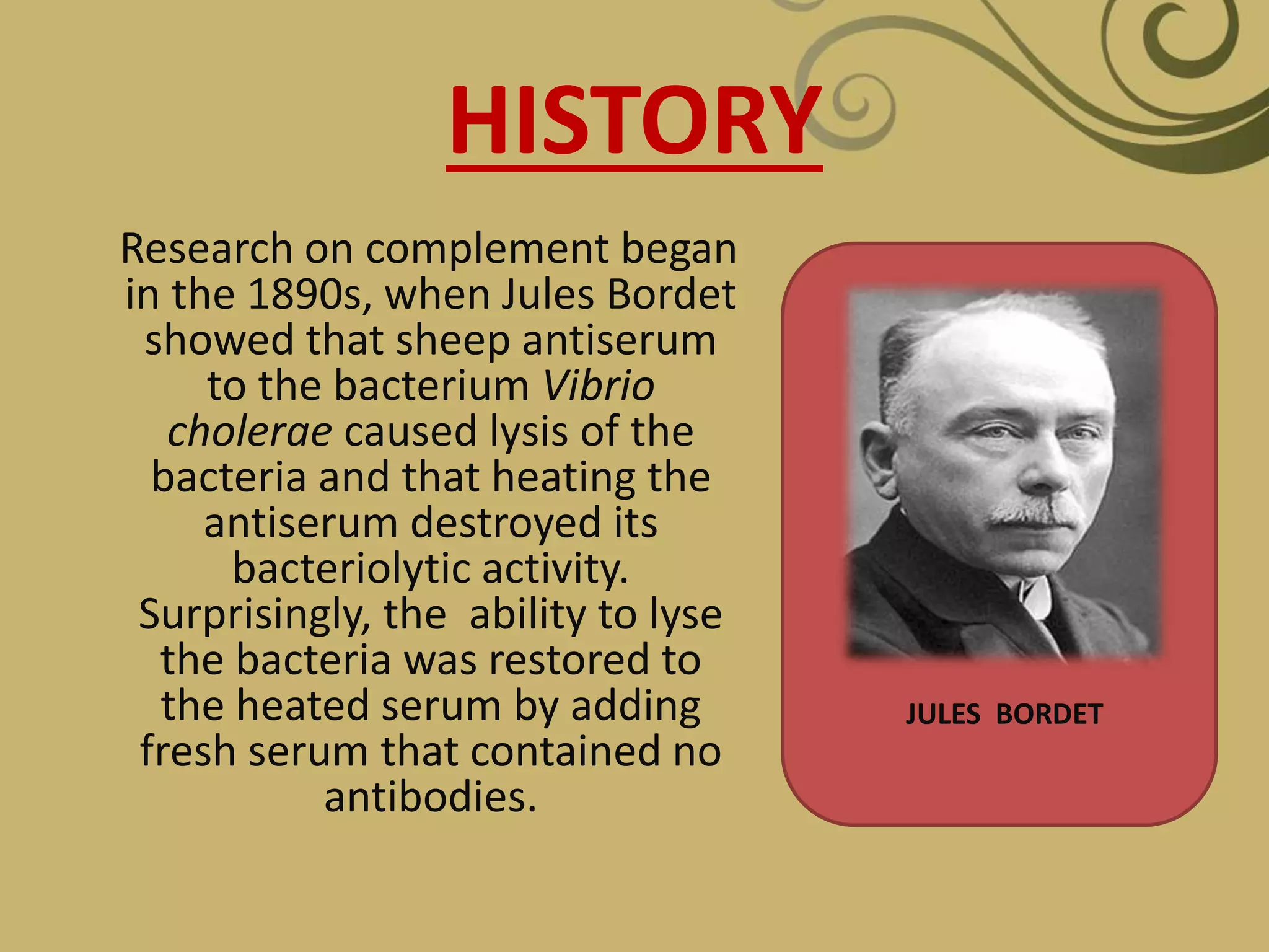
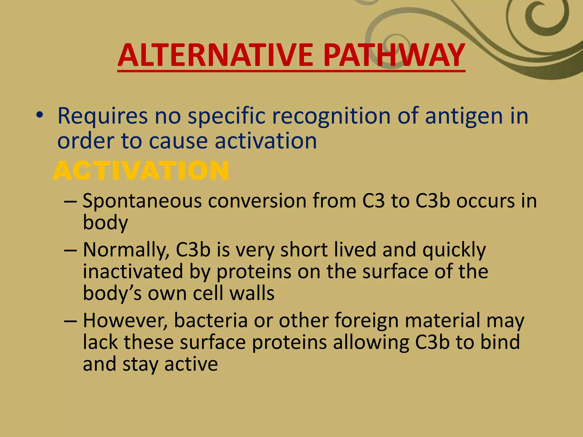
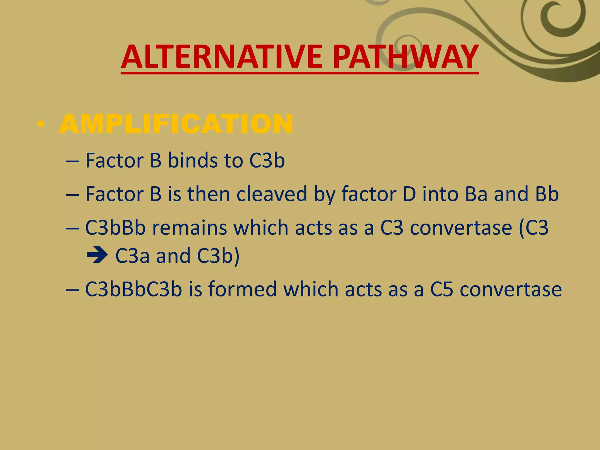
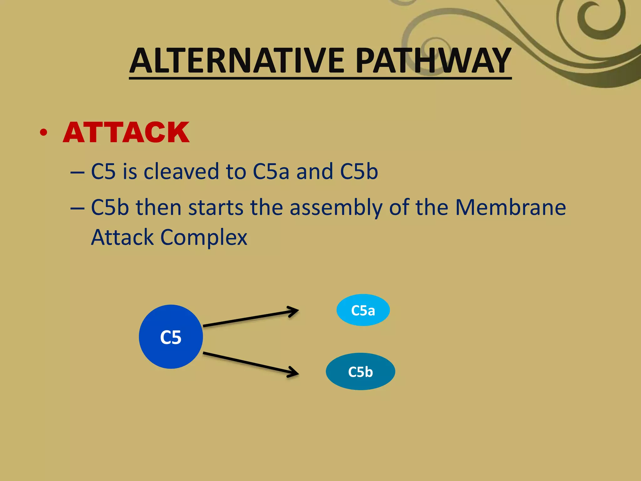
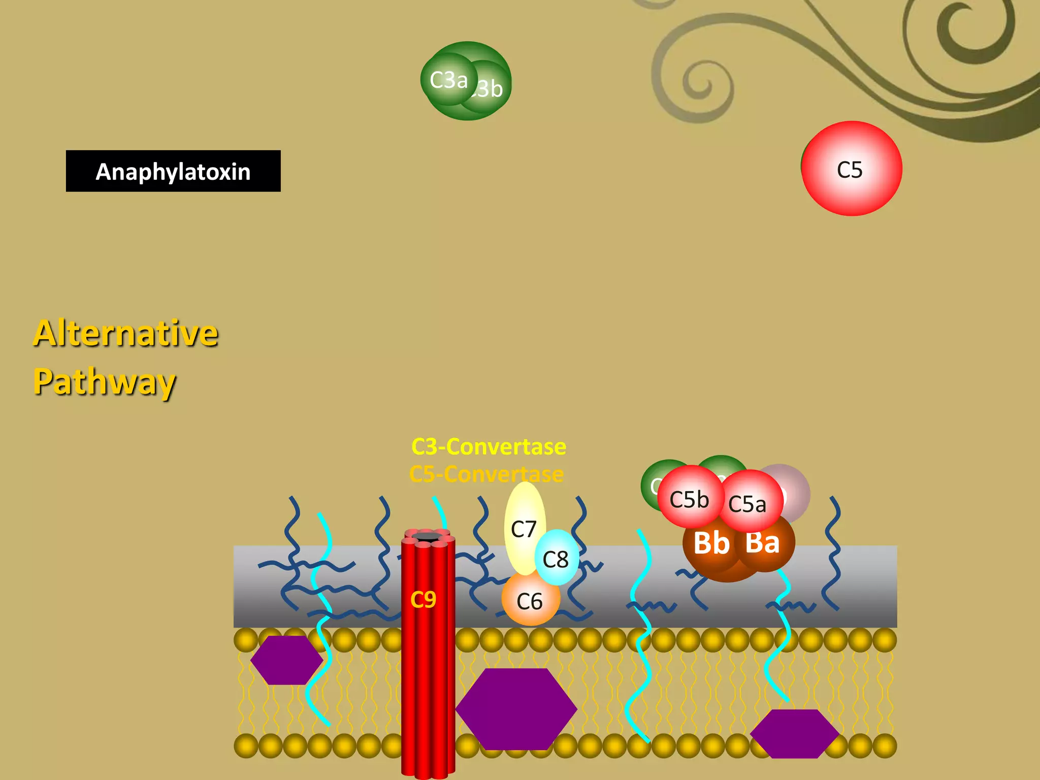
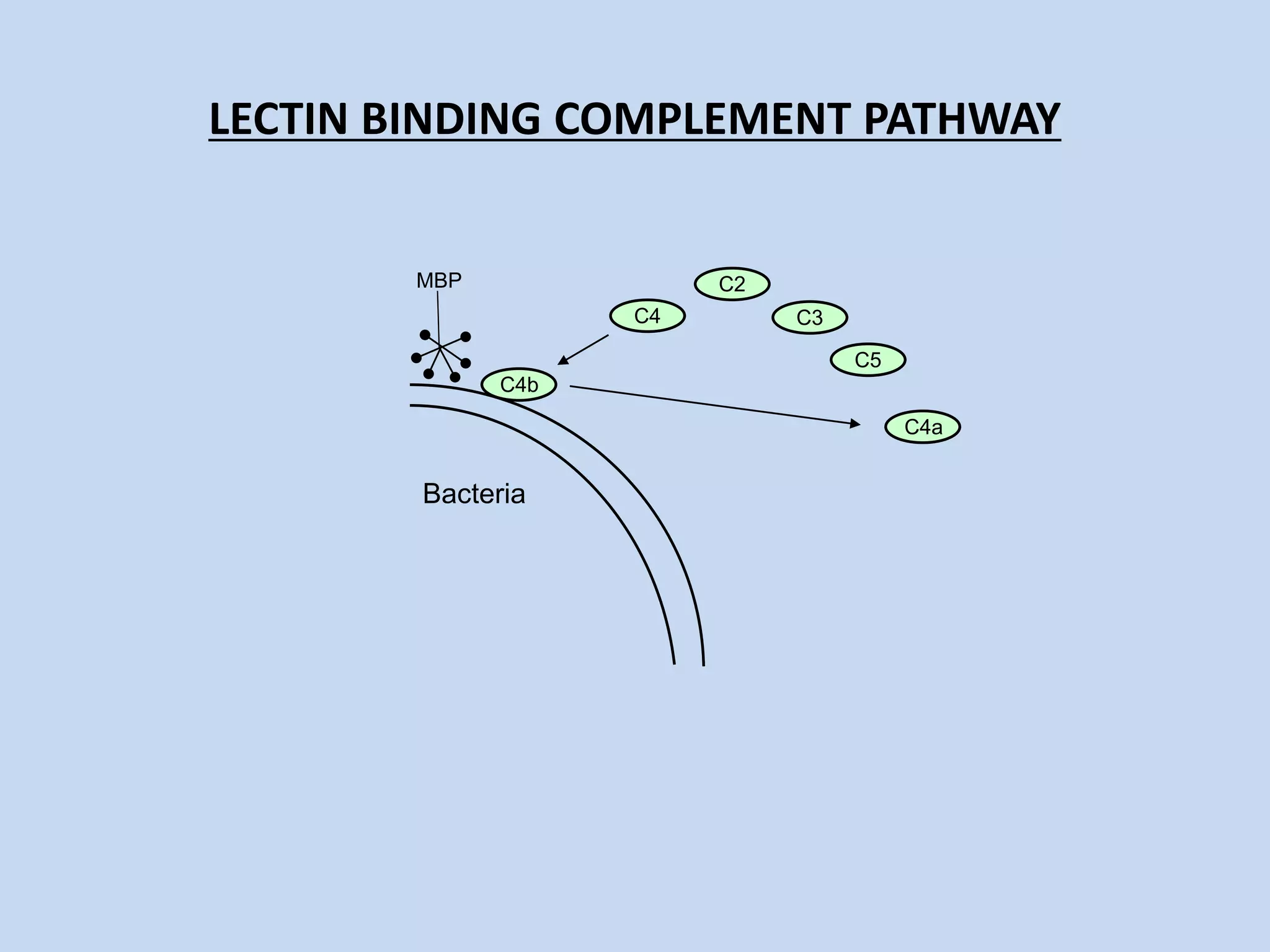
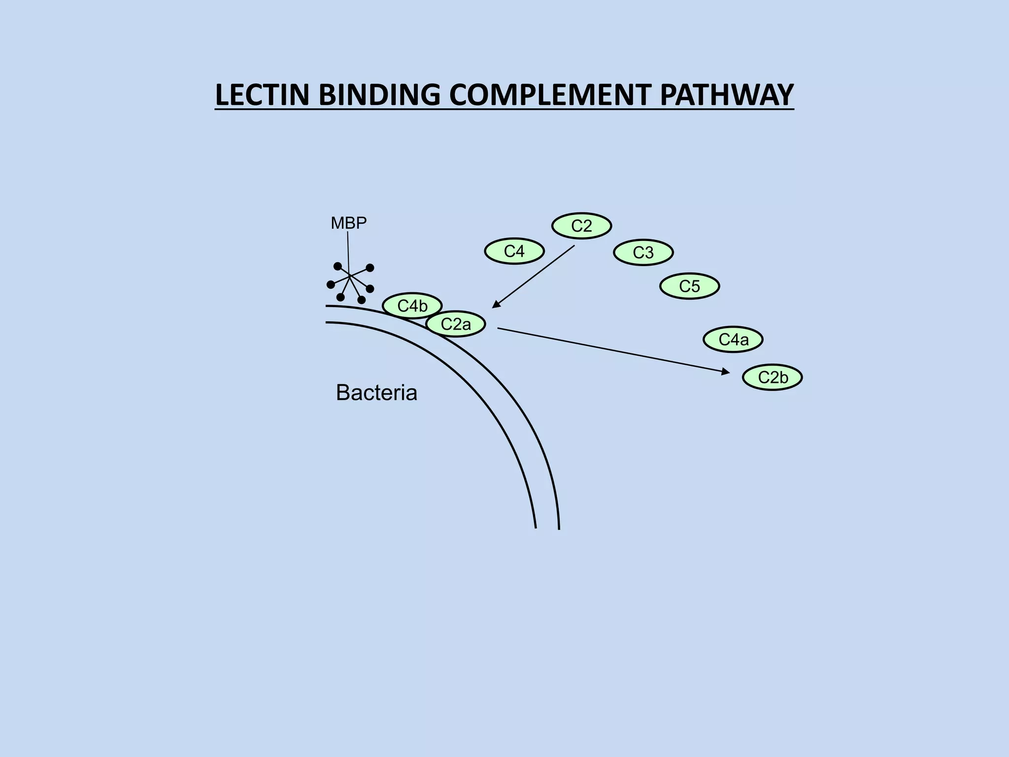
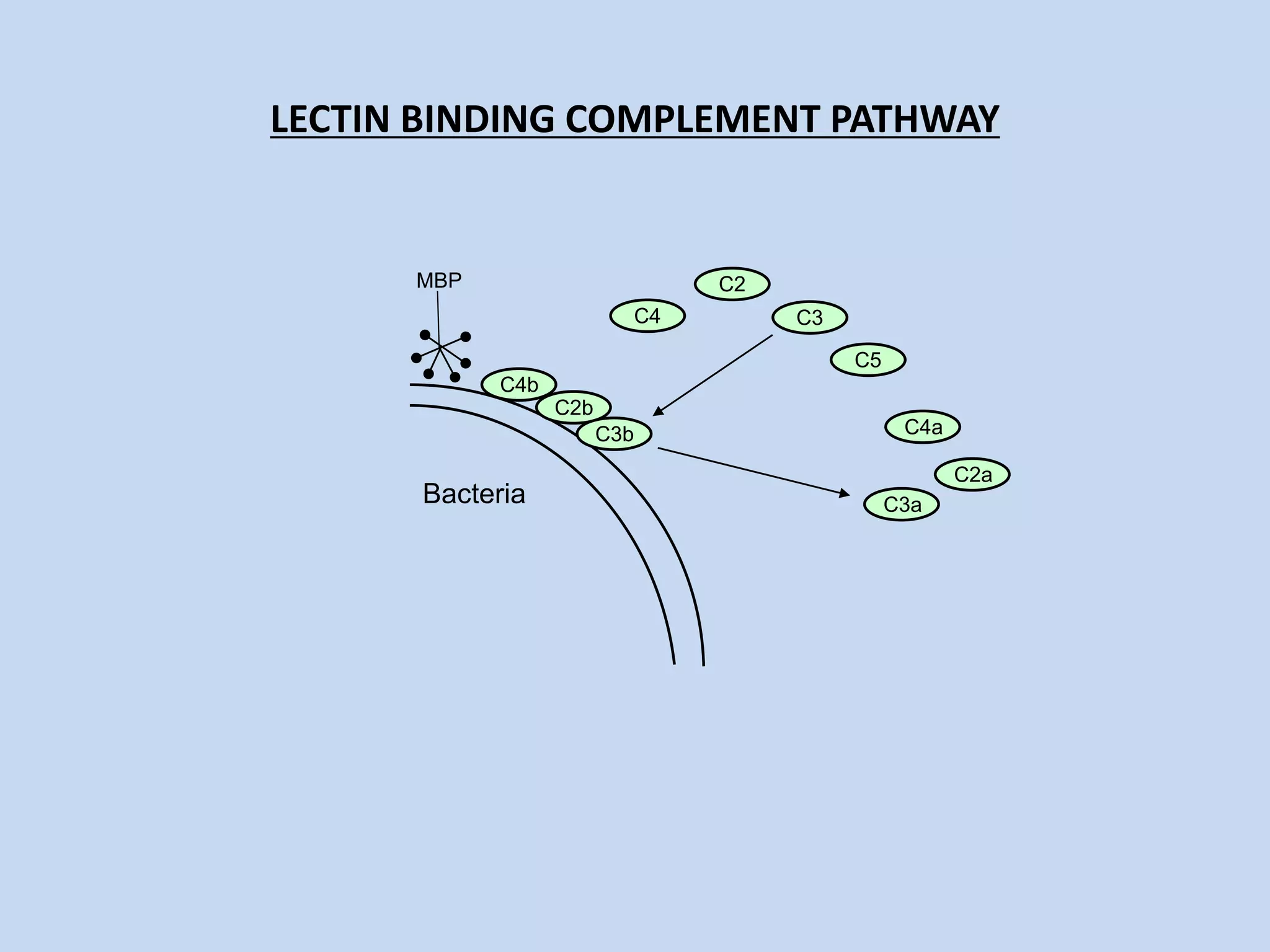
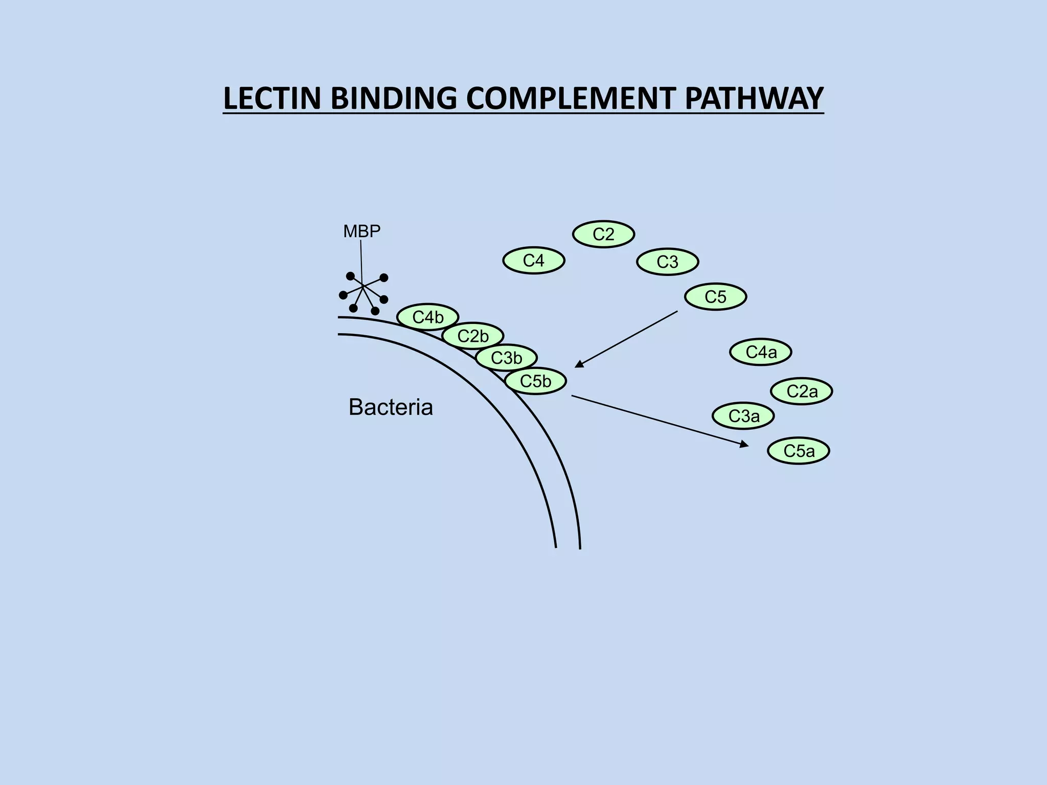
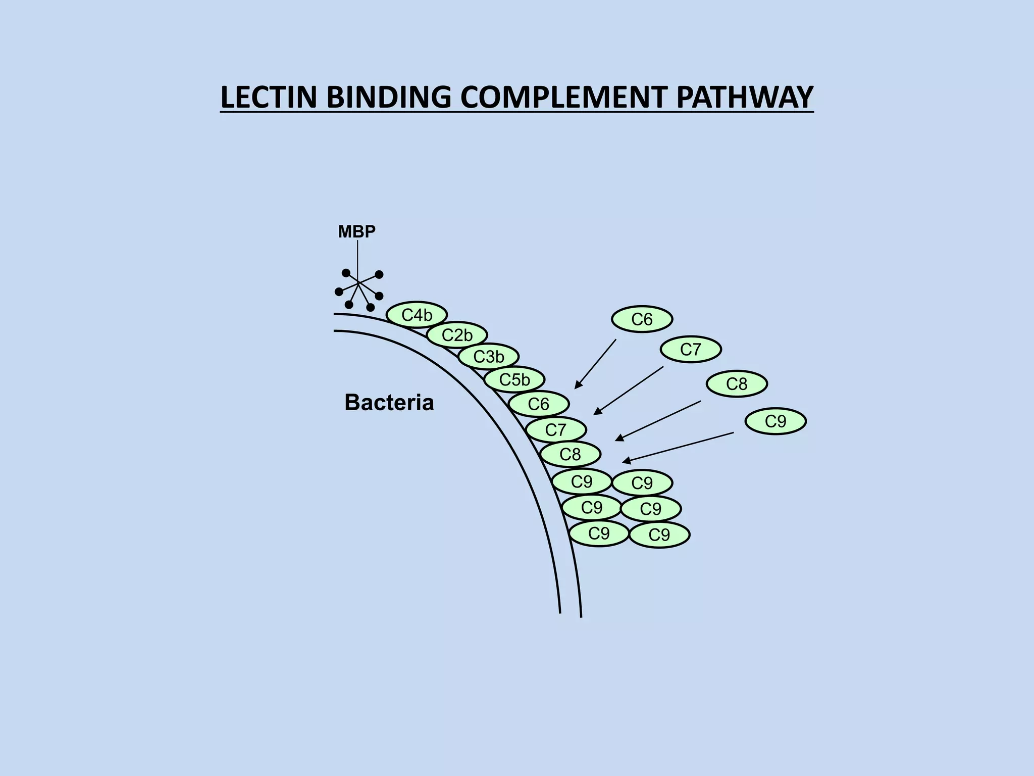
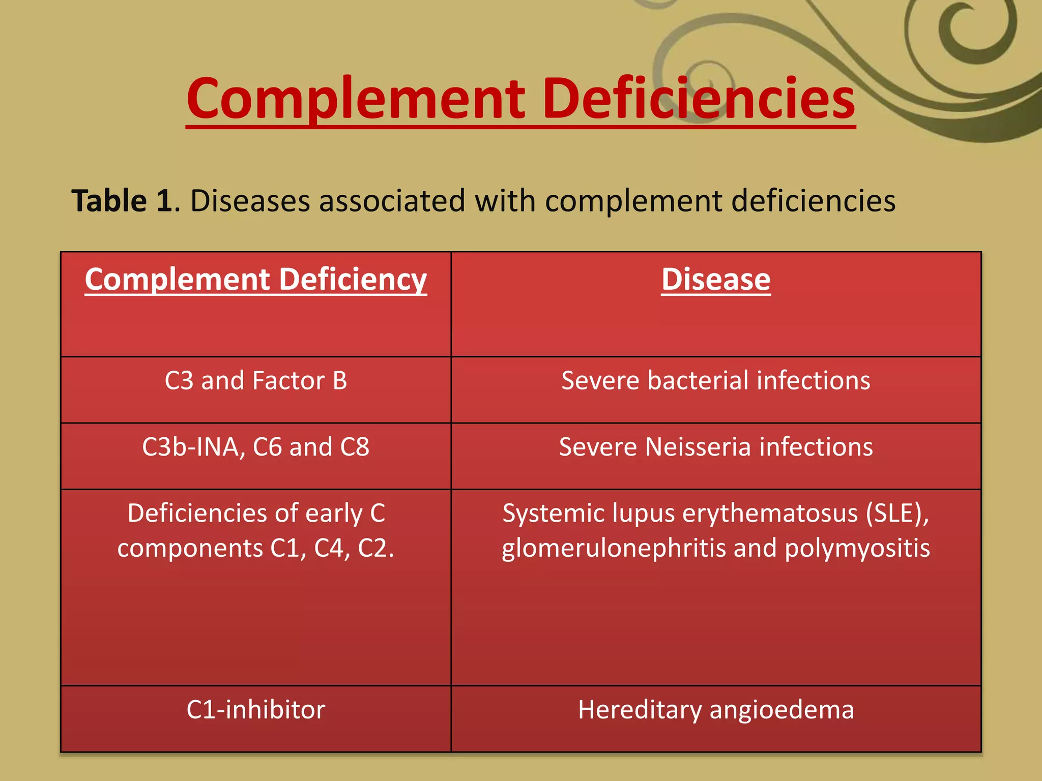
• The complement system is part of the innate
immune system.
• It is named “complement system” because it
was first identified as a heat-labile component
of serum that “complemented” antibodies in
the killing of bacteria
• It is now known that it consists of over 30
proteins and contributes 3 g/L to overall
serum protein quantities
Function of Complement
• After initial activation, the various complement
components interact, in a highly regulated cascade, to
carry out a number of basic function.
1. Lysis of cells, bacteria and viruses.
2. Opsonization, which promots phagocytosis of perticulate
antigens
3. Binding to specific complement receptors on cells of the
immune system, triggering specific cell function,
inflammation, and secretion of immunoregulatory
molecules.
4. Immune clearance, which removes immune complexes
from the circulation and diposit them in spleen and liver.
Components of Complement
The soluble proteins and glycoproteins that constitute complement
system are synthesize mainly by liver hepatocytes, although significant
amount are produced by blood monocytes, tissue macrophages and
epithelial cells of GI tract.
• Components are designated by munerals(C1-C9), by latter symbols
(factor D),
• Peptide fragments formed by activation of a component are denoted
by small letters. In most cases, the smaller fragment resulting from
cleavage of a component is designated “a” and the larger fragment
designated “b” (e.g., C3a, C3b; note that C2 is an exception: C2a is the
larger cleavage fragment).
• The larger fragments bind to the target near the site of activation, and
the smaller fragments diffuse from the site and can initiate localized
inflammatory responses by binding to specific receptors. The
complement fragments interact with one another to form functional
complexes. Those complexes that have enzymatic activity are
designated by a bar over the number or symbol (e.g., C4b2a, C3bBb).
Complement Activation
CLASSICAL PATHWAY
• Begins with antibody binding to a cell surface and ends with
the lysis of the cell
• The proteins in this pathway are named C1-C9 (the order they
were discovered and not the order of the reaction)
• When complement is activated it is split into two parts
– a – smaller of the two
– B – larger part and usually the active part (except with factor 2)
• Three steps in this pathway-
– ACTIVATION
– AMPLIFICATION
– ATTACK
CLASSICAL PATHWAY
• ACTIVATION
– C1q portion of C1 attaches to the Fc portion of an
antibody
– Only IgG and IgM can activate complement
– Once activated C1s is eventually cleaved which
activates C4 and C2
– C4b & C2a come together to form the C4b2a
which is the C3 convertase
– C3 convertase activates C3 to C3a and C3b
CLASSICAL PATHWAY
• ACTIVATION
–C3a binds to receptors on basophils and
mast cells triggering them to release
there vasoactive compounds (enhances
vasodilation and vasopermeability)
–C3a is called an anaphylatoxin
–C3b serves as an opsonin which
facilitates immune complex clearance
CLASSICAL PATHWAY
• AMPLIFICATION
– Each C1s creates many C4b and C2a fragments
– Each C4bC2a creates many C3b (activated C3)
– Each C3b goes on to create many Membrane Attack
Complexes
– Example
• 1 C1S makes 100 C4bC2a
• 100 C4bC2b makes 10,000 C3b
• 10,000 C3b makes 1,000,000 MAC
CLASSICAL PATHWAY
• ATTACK
– Most C3b serves an opsonin function
– Some C3b binds to C4bC2a to form the C5
convertase C4bC2aC3b
– C5 convertase cleaves C5 leading to the formation
of the Membrane attack Complex (C5-C6-C7-C8-C9)
– The MAC “punches holes” in cell walls resulting in
lysis
C1q
C2C4
2a
2b4b
4a
C3-convertase
C3
C3a
C3b
C5-Convertase
C3a binds to receptors on
basophils and mast cells
triggering them to release there
vasoactive compounds (enhances
vasodilation and
vasopermeability) -
ANAPHYLATOXIN
C5
C5aC5b
C5a is a:
1. Potent anaphylatoxin
2. Chemoattractant for
neutrophils
C6
C7
C8
C9
Classical
Pathway
ALTERNATIVE PATHWAY
• Requires no specific recognition of antigen in
order to cause activation
ACTIVATION
– Spontaneous conversion from C3 to C3b occurs in
body
– Normally, C3b is very short lived and quickly
inactivated by proteins on the surface of the
body’s own cell walls
– However, bacteria or other foreign material may
lack these surface proteins allowing C3b to bind
and stay active
ALTERNATIVE PATHWAY
• AMPLIFICATION
– Factor B binds to C3b
– Factor B is then cleaved by factor D into Ba and Bb
– C3bBb remains which acts as a C3 convertase (C3
 C3a and C3b)
– C3bBbC3b is formed which acts as a C5 convertase
ALTERNATIVE PATHWAY
• ATTACK
– C5 is cleaved to C5a and C5b
– C5b then starts the assembly of the Membrane
Attack Complex
C5
C5a
C5b
C3C3bC3a
Anaphylatoxin
B
D
Bb Ba
C3
C3a C3bC5-Convertase
C3-Convertase
C5
C5aC5b
Alternative
Pathway
C6
C7
C8
C9
LECTIN PATHWAY
• Lectins are proteins that recognize and bind to specific
carbohydrate targets.
• The lectin pathway, like the alternative pathway, does not
depend on antibody for its activation.
• The lectin pathway is activated by the binding of
mannose-binding lectin (MBL) to mannose residues on
glycoproteins or carbohydrates on the surface of
microorganisms including certain Salmonella, Listeria,
and Neisseria strains, MBL is an acute phase protein
produced in inflammatory responses.
LECTIN PATHWAY
• Its function in the complement pathway is similar to that of
C1q, which it resembles in structure.
• After MBL binds to the surface of a cell or pathogen, MBL-
associated serine proteases,MASP-1 and MASP-2, bind to
MBL.
• The active complex formed by this association causes
cleavage and activation of C4 and C2.
• The MASP-1 and -2 proteins have structural similarity to C1r
and C1s and mimic their activities.
• This means of activating the C2–C4 components to form a C5
convertase without need for specific antibody binding
represents an important innate defense mechanism
comparable to the alternative pathway, but utilizing the
elements of the classical pathway except for the C1 proteins.
LECTIN BINDING COMPLEMENT PATHWAY
Bacteria
C4
C2
C3
C5
MBP
C4b
C4a
LECTIN BINDING COMPLEMENT PATHWAY
Bacteria
C4
C2
C3
C5
MBP
C4b
C4a
C2a
C2b
LECTIN BINDING COMPLEMENT PATHWAY
Bacteria
C4
C2
C3
C5
MBP
C4b
C4a
C2b
C2a
C3b
C3a
LECTIN BINDING COMPLEMENT PATHWAY
Bacteria
C4
C2
C3
C5
MBP
C4b
C4a
C2b
C2a
C3b
C3a
C5b
C5a
LECTIN BINDING COMPLEMENT PATHWAY
Bacteria
C6
C7
C8
C9
MBP
C4b
C2b
C3b
C5b
C6
C7
C8
C9
C9
C9
C9
C9
C9
BIOLOGIC FUNCTION
• anaphylatoxins: C3a and C5a: mast cell degranulation
– smooth muscle contraction
– mast cell degranulation mediator release (histamine,
leukotrienes)
– vascular changes: dilation, increased permeability (edema)
– C5a also leukocyte adhesion and chemotaxis (recruitment)
• opsonization: C3b, C3bi, C3d: (binding to complement receptors
and enhanced phagocytosis by neutrophils and macrophages)
• clearance of circulating immune complexes
• membrane attack complex: C5b-C9 (cell lysis)
Complement Deficiencies
Role of Complement in Disease
The complement system plays a critical role in
inflammation and defence against some bacterial
infections. Complement may also be activated during
reactions against incompatible blood transfusions, and
during the damaging immune responses that
accompany autoimmune disease. Deficiencies of
individual complement components or inhibitors of the
system can lead to a variety of diseases (Table 1), which
gives some indication of their role in protection against
disease.
Complement Deficiencies
Complement Deficiency Disease
C3 and Factor B Severe bacterial infections
C3b-INA, C6 and C8 Severe Neisseria infections
Deficiencies of early C
components C1, C4, C2.
Systemic lupus erythematosus (SLE),
glomerulonephritis and polymyositis
C1-inhibitor Hereditary angioedema
Table 1. Diseases associated with complement deficiencies
Referance
KUBI IMMUNOLOGY 6th EDITION
IMMUOLOGY, NANDINI SHETTY, 2nd EDITION
https://www.google.co.in/#q=complement+system+ppt
I would like to express thankful gratitude to
Dr.Amit Chakravarty, Principal of IGE as well
as Dr.Sudipa Chakravarty,Vice Principal of
IGE.I would also like to thanks Jayasmita
Mahapatra, help me to making this
presentation and I also helped me to doing
this. THANKS AGAIN TO ALL WHO HELPED
ME.
THANK
YOU

Complement system

  • 1.
  • 2.
    WHAT IS COMPLEMENTSYSTEM • Complement system is a part of the immune system called the innate immune system that is not adaptable and does not change over the course of an individual's lifetime. • However, it can be recruited and brought into action by the adaptive immune system.
  • 3.
    HISTORY Research on complementbegan in the 1890s, when Jules Bordet showed that sheep antiserum to the bacterium Vibrio cholerae caused lysis of the bacteria and that heating the antiserum destroyed its bacteriolytic activity. Surprisingly, the ability to lyse the bacteria was restored to the heated serum by adding fresh serum that contained no antibodies. JULES BORDET
  • 4.
    HISTORY Paul Ehrlich inBerlin independently carried out similar experiments and coined the term complement, defining it as “the activity of blood serum that completes the action of antibody.” In ensuing years, researchers discovered that the action of complement was the result of interactions of a large and complex group of proteins. PAUL EHRLICH
  • 5.
    OVERVIEW • The complementsystem is part of the innate immune system. • It is named “complement system” because it was first identified as a heat-labile component of serum that “complemented” antibodies in the killing of bacteria • It is now known that it consists of over 30 proteins and contributes 3 g/L to overall serum protein quantities
  • 6.
    Function of Complement •After initial activation, the various complement components interact, in a highly regulated cascade, to carry out a number of basic function. 1. Lysis of cells, bacteria and viruses. 2. Opsonization, which promots phagocytosis of perticulate antigens 3. Binding to specific complement receptors on cells of the immune system, triggering specific cell function, inflammation, and secretion of immunoregulatory molecules. 4. Immune clearance, which removes immune complexes from the circulation and diposit them in spleen and liver.
  • 7.
    Components of Complement Thesoluble proteins and glycoproteins that constitute complement system are synthesize mainly by liver hepatocytes, although significant amount are produced by blood monocytes, tissue macrophages and epithelial cells of GI tract. • Components are designated by munerals(C1-C9), by latter symbols (factor D), • Peptide fragments formed by activation of a component are denoted by small letters. In most cases, the smaller fragment resulting from cleavage of a component is designated “a” and the larger fragment designated “b” (e.g., C3a, C3b; note that C2 is an exception: C2a is the larger cleavage fragment). • The larger fragments bind to the target near the site of activation, and the smaller fragments diffuse from the site and can initiate localized inflammatory responses by binding to specific receptors. The complement fragments interact with one another to form functional complexes. Those complexes that have enzymatic activity are designated by a bar over the number or symbol (e.g., C4b2a, C3bBb).
  • 8.
    Complement Activation CLASSICAL PATHWAY •Begins with antibody binding to a cell surface and ends with the lysis of the cell • The proteins in this pathway are named C1-C9 (the order they were discovered and not the order of the reaction) • When complement is activated it is split into two parts – a – smaller of the two – B – larger part and usually the active part (except with factor 2) • Three steps in this pathway- – ACTIVATION – AMPLIFICATION – ATTACK
  • 9.
    CLASSICAL PATHWAY • ACTIVATION –C1q portion of C1 attaches to the Fc portion of an antibody – Only IgG and IgM can activate complement – Once activated C1s is eventually cleaved which activates C4 and C2 – C4b & C2a come together to form the C4b2a which is the C3 convertase – C3 convertase activates C3 to C3a and C3b
  • 10.
    CLASSICAL PATHWAY • ACTIVATION –C3abinds to receptors on basophils and mast cells triggering them to release there vasoactive compounds (enhances vasodilation and vasopermeability) –C3a is called an anaphylatoxin –C3b serves as an opsonin which facilitates immune complex clearance
  • 11.
    CLASSICAL PATHWAY • AMPLIFICATION –Each C1s creates many C4b and C2a fragments – Each C4bC2a creates many C3b (activated C3) – Each C3b goes on to create many Membrane Attack Complexes – Example • 1 C1S makes 100 C4bC2a • 100 C4bC2b makes 10,000 C3b • 10,000 C3b makes 1,000,000 MAC
  • 12.
    CLASSICAL PATHWAY • ATTACK –Most C3b serves an opsonin function – Some C3b binds to C4bC2a to form the C5 convertase C4bC2aC3b – C5 convertase cleaves C5 leading to the formation of the Membrane attack Complex (C5-C6-C7-C8-C9) – The MAC “punches holes” in cell walls resulting in lysis
  • 13.
    C1q C2C4 2a 2b4b 4a C3-convertase C3 C3a C3b C5-Convertase C3a binds toreceptors on basophils and mast cells triggering them to release there vasoactive compounds (enhances vasodilation and vasopermeability) - ANAPHYLATOXIN C5 C5aC5b C5a is a: 1. Potent anaphylatoxin 2. Chemoattractant for neutrophils C6 C7 C8 C9 Classical Pathway
  • 14.
    ALTERNATIVE PATHWAY • Requiresno specific recognition of antigen in order to cause activation ACTIVATION – Spontaneous conversion from C3 to C3b occurs in body – Normally, C3b is very short lived and quickly inactivated by proteins on the surface of the body’s own cell walls – However, bacteria or other foreign material may lack these surface proteins allowing C3b to bind and stay active
  • 15.
    ALTERNATIVE PATHWAY • AMPLIFICATION –Factor B binds to C3b – Factor B is then cleaved by factor D into Ba and Bb – C3bBb remains which acts as a C3 convertase (C3  C3a and C3b) – C3bBbC3b is formed which acts as a C5 convertase
  • 16.
    ALTERNATIVE PATHWAY • ATTACK –C5 is cleaved to C5a and C5b – C5b then starts the assembly of the Membrane Attack Complex C5 C5a C5b
  • 17.
  • 18.
    LECTIN PATHWAY • Lectinsare proteins that recognize and bind to specific carbohydrate targets. • The lectin pathway, like the alternative pathway, does not depend on antibody for its activation. • The lectin pathway is activated by the binding of mannose-binding lectin (MBL) to mannose residues on glycoproteins or carbohydrates on the surface of microorganisms including certain Salmonella, Listeria, and Neisseria strains, MBL is an acute phase protein produced in inflammatory responses.
  • 19.
    LECTIN PATHWAY • Itsfunction in the complement pathway is similar to that of C1q, which it resembles in structure. • After MBL binds to the surface of a cell or pathogen, MBL- associated serine proteases,MASP-1 and MASP-2, bind to MBL. • The active complex formed by this association causes cleavage and activation of C4 and C2. • The MASP-1 and -2 proteins have structural similarity to C1r and C1s and mimic their activities. • This means of activating the C2–C4 components to form a C5 convertase without need for specific antibody binding represents an important innate defense mechanism comparable to the alternative pathway, but utilizing the elements of the classical pathway except for the C1 proteins.
  • 20.
    LECTIN BINDING COMPLEMENTPATHWAY Bacteria C4 C2 C3 C5 MBP C4b C4a
  • 21.
    LECTIN BINDING COMPLEMENTPATHWAY Bacteria C4 C2 C3 C5 MBP C4b C4a C2a C2b
  • 22.
    LECTIN BINDING COMPLEMENTPATHWAY Bacteria C4 C2 C3 C5 MBP C4b C4a C2b C2a C3b C3a
  • 23.
    LECTIN BINDING COMPLEMENTPATHWAY Bacteria C4 C2 C3 C5 MBP C4b C4a C2b C2a C3b C3a C5b C5a
  • 24.
    LECTIN BINDING COMPLEMENTPATHWAY Bacteria C6 C7 C8 C9 MBP C4b C2b C3b C5b C6 C7 C8 C9 C9 C9 C9 C9 C9
  • 25.
    BIOLOGIC FUNCTION • anaphylatoxins:C3a and C5a: mast cell degranulation – smooth muscle contraction – mast cell degranulation mediator release (histamine, leukotrienes) – vascular changes: dilation, increased permeability (edema) – C5a also leukocyte adhesion and chemotaxis (recruitment) • opsonization: C3b, C3bi, C3d: (binding to complement receptors and enhanced phagocytosis by neutrophils and macrophages) • clearance of circulating immune complexes • membrane attack complex: C5b-C9 (cell lysis)
  • 26.
    Complement Deficiencies Role ofComplement in Disease The complement system plays a critical role in inflammation and defence against some bacterial infections. Complement may also be activated during reactions against incompatible blood transfusions, and during the damaging immune responses that accompany autoimmune disease. Deficiencies of individual complement components or inhibitors of the system can lead to a variety of diseases (Table 1), which gives some indication of their role in protection against disease.
  • 27.
    Complement Deficiencies Complement DeficiencyDisease C3 and Factor B Severe bacterial infections C3b-INA, C6 and C8 Severe Neisseria infections Deficiencies of early C components C1, C4, C2. Systemic lupus erythematosus (SLE), glomerulonephritis and polymyositis C1-inhibitor Hereditary angioedema Table 1. Diseases associated with complement deficiencies
  • 28.
    Referance KUBI IMMUNOLOGY 6thEDITION IMMUOLOGY, NANDINI SHETTY, 2nd EDITION https://www.google.co.in/#q=complement+system+ppt
  • 29.
    I would liketo express thankful gratitude to Dr.Amit Chakravarty, Principal of IGE as well as Dr.Sudipa Chakravarty,Vice Principal of IGE.I would also like to thanks Jayasmita Mahapatra, help me to making this presentation and I also helped me to doing this. THANKS AGAIN TO ALL WHO HELPED ME.
  • 30.