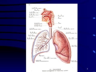
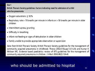
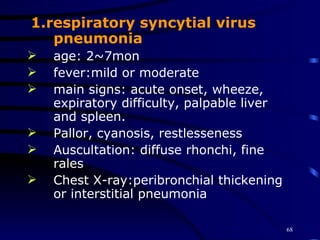
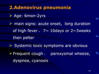
The Second Affiliated Hospital of Sun  Yat-sen University Tan Wei-ping Infections of the Respiratory Tract
Contents Upper Respiratory Infection Acute infectious Laryngitis Acute Bronchitis Bronchiolitis Pneumonia
Anatomy upper respiratory tract(URT) Cricoid Cartilage lower   respiratory tract(LRT) URT  : Nose 、 pharynx  、  Uvula  、  Larynx  、  auditory tube 、 paranasal sinuses LRT  : Trachea 、 bronchia  、 bronchiole 、 the respiratory bronchiole 、 the alveolar duct and the alveolus
 
 
Anatomy   characteristics upper respiratory tract Nose: cilia↓, capillary↑  Sinuses: mucous Auditory tube tonsil Larynx
lower   respiratory tract : Weak Cartilage Supporting , ciliary function impairment Right bronchus :straight , large Collagen and elastin fibers , capillary ,  interstitial tissue Thorax : chest wall,  respiratory muscle ; diaphragm , mediastinum
Physiological characteristics The compensatory ability ↓  Respiratory  rate↑ Pattern of respiration Gas-exchanging membrane  :diffuse rate CO 2 > O 2 Resistance of airway: child  > adult
Arterial blood gas examination (1)  pH↓acidosis ; (2) PaO 2 ↓ SaO 2  ↓ hypoxemia ; (3)PaCO 2 ↑  carbon dioxide retention   mal-ventilation
The Immunological Characteristics : Impaired mucociliary clearance function SIgA↓ 、 IgA↓ 、 IgG↓ subtype of IgG ↓ Alveolar macrophages ↓ , lysozyme ↓  lactoferrin ↓  、 interferon ↓  complement ↓
Upper Respiratory Infection (URI, or Common Cold) 1.   Etiology  90%   viruses, the majority of colds. rhinoviruses ,coronaviruses. parainfluenza virus, adenovirus, enterovirus, respiratory syncytial virus. 2.   season   fall and winter 3.   Frequency  three   to eight colds a year.
Inoculation by virus Invasion of epithelium of Release of  nasopharynx,sinuses and URT  cellular damage Inflammatory  of nasopharynx mediators in  Cholinergic nasal secretions  stimulation Increased  Increased  vascular  mucus Permeability  production  bronchial  constriction Nasal  Rhinorrhea  postnasal Stuffiness  drip  sore cough  throat  Pathophysiology  of  the  common  cold
The clinical manifestation   Common cold   congestion  a runny nose  sneezing  Cough,sore throat  sometimes vomiting and diarrhea  Fever,malaise,abdominal pain
Specific type of URI Herpangina: coxsackie-viruses group A.  summer /fall.High fever, sore throat , 1-4mm  vesicles /ulcers  on anterior tonsillar pillars, softpalate,uvula,tonsils pharyngeal wall.  Pharyngoconjunctival fever : type 3,7 adenovirus,  spring /summer. High fever, sore throat , pharyngitis, conjunctivitis, cervical lymphadenopathy.
Complications sinusitis  otitis media  cervical lymphadenopathy Mesentery lymphadenopathy retropharngeal abscess pneumonia  rheumatic fever  acute glomerulonephritis
Differential  diagnosis Flu Appendicitis Early phase of acute infectious disease
Sometimes  sore throat  Sore throat   Fatigue, may persist  Mild fatigue  severe aches and pains  Slight aches and pains   Cough, may progress   Mild, hacking cough  Sometimes sneezing  Sneezing   Sometimes stuffy nose   Stuffy, runny nose  Commonly  headache  Sometimes  headache  High fever  Low or no fever  Flu Cold
Treatment increased fluid intake avoidance of secondhand smoke Saline nose drops  bulb syringe remove the mucus cool mist humidifier antipyretics, such as acetaminophen,ibuprofen, decrease the discomfort of colds. Do not give aspirin (associated with Reye syndrome)
Prevention Keep your child away from a person with a cold. Encourage your child to wash his/her hands frequently and not to touch his/her mouth, eyes, or nose until their hands are washed. Make sure toys and play areas are properly cleaned, especially if multiple children are playing together.
Acute infectious Laryngitis Etiology Virus or bacteria Typical anatomy
Clinical manifestations Barking cough, hoarseness, inspiratory stridor ,  nasal flaring,  suprasternal , infrasternal, intercostal retraction Fever, dyspnea, cyanosis restlessness, tachycardia .  Worsen at night.  Congestion of pharynx,   vocal cord edema
Grade of laryngic obstruction Ⅰ :  inspiratory stridor, respiratory  difficulty only after activity Ⅱ : symptoms occurs at rest.  tachycardia,  rhonchi Ⅲ : Ⅱ + hypoxia,  diminished breath sounds Ⅳ :  exhaustion, lethargy , pallor , breath sounds diminish or  disappear. dull of heart sounds, arrhythmia
Diagnosis & differential diagnosis laryngeal diphtheria laryngeal spasm bronchial foreign bodies
treatment Maintaining of airway  : steam inhalation,  clearing of secretion. antibiotics  corticosteroid oxygen supply sedatives (phenergan) Ⅲ ↑  tracheotomy
Acute Bronchitis( tracheobronchitis ) Etiology Virus or bacteria Allergy, climate, air pollution, chronic infection of URT, particularly sinusitis. Rickets, malnutrition
Clinical manifestations unproductive cough 3~4days -> productive cough, purulent sputum -> 5~10days Chest pain, shortness of breath Vomiting  Physical finding: low-grade fever, roughening of breath sounds, rhonchi, coarse moist rales.
Asthmatic bronchitis : ﹤ 3yrs old, eczema, allergy symptoms  resemble asthma recurrent episodes reduced after 3~4yrs old.
Treatment increased fluid intake frequent shifts of position antibiotics cough suppressants  expectorants , antihistamines acetaminophen
Bronchiolitis Etiology and epidemiology 50 %  respiratory syncytial virus(RSV) parainfluenza 3 virus, mycoplasma, adenoviruses north——winter and early spring Guangdong——spring ,summer and early autumn
2.Pathophysiology Lesion: small air passages (diameter75 ~ 300um) Edema, accumulation of mucus and cellular debris, spasm of smooth mussle-> ↑ resistance of small airway ->expiratory difficulty ->overinflation  or atelectasis  ->hypoxemia,  hypercapnia, acidosis
Clinical manifestation Infant ﹤2yrs old, peak at 3~ 6 mo of age, male, non breast-feed URI 3 ~ 4days paroxysmal wheezy cough dyspnea develop rapidly mild or moderate fever
Physical examination R 60~80/min HR  160 ~ 200/min Nasal flare, intercostal and subcostal retractions Pallor, cyanosis Hyperexpanded chest , expiratory phase ↑ wheeze , widespread fine crackles Liver and spleen palpable below the costal margin Critical phase  48 ~ 72h afer onset of dyspnea; recover during 5 ~ 15days
Laboratory examination WBC and differential cells count normal Virus detected by antigen detection, PCR, or culture. X-ray: hyperinflation of the lungs, emphysema, scattered areas of consolidation
Differential diagnosis Asthma Cystic fibrosis Heart failure Foreign body in the trachea pertussis
 
两侧肺纹理粗重。两肺中内带多数小斑片状及小结节病灶影。无病灶之肺部透过度增高。
Treament Supportive treatment cool, humidified oxygen supply, maintain SaO2  94%~96%; increased fluid intake : oral intake, intravenous solutions head and chest slightly elevated
Ribavirin Antibiotics corticosteroids
Pneumonia
What’s Pneumonia An abnormal inflammatory condition of the lung infections (bacterial, viral or fungal)   chemical injury (gastric acid/ aspiration of food/ hydrocarbon and lipoid pneumonia/ radiation induced pneumonia)
Definition Defined by clinical features or with the addition of radiologic findings Tachypnea : indicator of pneumonia(WHO) < 2m,  R ≥60/min 2~12m,  R ≥50/min > 12m,  R ≥40/min sensitivity of 74% and a specificity of 67% compared with radiology
Epidemiology A leading killer of  children ≤5yr 1.9 million death worldwide/year
Etiology Viruses:40% ( < 2yr) Bacteria:27~44% Streptococcus pneumoniae Staphylococcus aureus, Moraxhella catarrhalis, group A Streptococci, and Haemophilus  MP, CP:9~14% Mixed:23%
Contributing Etiology:  risk foctors   Malnutrition Rickets Iron-deficiency Immuno-deficency Congenital heart disease Low born bodyweight
Clinical manifestation fever cough  Dyspnea (Tachypnea,cyanosis) Localized crackles(fine moist rales) Abnormal chest x-ray
Classification Pathologic  ( anatomic changes)   lobar  pneumonia :  involves a single lobe   Bronchial pneumonia :  patches around the tubes   Interstitial  pneumonia :  areas between the alveoli   Pathogenic  ( microorganisms ) bacteria viruses fungi parasites
Classification course of disease   Acute subacute chronic   State of disease   common severe
Classification Clinical  manifestation Typical atipical The combined clinical classification community-acquired pneumonia(CAP)  hospital-acquired pneumonia (HAP)
Bronchopneumonia 1.Etiology   viruses, bacteria, MP, CP.  2~3yrs old 2.pathology   edema,infiltration


3 pathophysiology hypoxemia,  hypercapnia, toxemia respiratory insufficiency acidosis myocarditis, heart failure, shock, DIC toxic encephalopathy toxic intestinal paralysis
4.Clinical manifestation ( 1) mild (commone)pneumonia : fever cough  dyspnea ( tachypnea, cyanosis) crackles Chest x-ray
两肺纹理增粗。两肺中下野中内带见斑片状阴影,密度较均匀。部分病灶融合成范围稍大的片状影
(2)  Severe pneumonia : other systems involved myocarditis  heart failure toxic encephalopathy toxic intestinal paralysis
Heart failure R  > 60/min HR  > 180/min Sudden onset of restlesseness,Pallor, cyanosis, delayed capillary refill( > 3~5s) Dull heart sound, gallop rhythm, Jugular vein congestion  Liver enlarged rapidly Oliguria or anuria, edema
5.Complications Empyema Pyopneumothorax pneumatocele
6.Laboratory findings: WBC, NBT,CRP Pathogen(antigen and/or antibody) chest x-ray
7.diagnosis Fever, cough, tachypnea, dyspnea, localized fine moist rales, chest x-ray 8.differential diagnosis acute bronchitis  tuberculosis  foreign bodies of trachea
 
男 2 岁半。 4 个月前吃蚕豆后发生呛咳,经常发烧,左侧呼吸音低。胸片:左侧阻塞性肺气肿。心 影稍右移,经支气管检查于左支气管取出蚕豆碎块。
 
9. treament (1)  general therapy (2) antipathogen therapy antibiotics antivirus
(3)symptomatic therapy oxygen supply airway management abdominal distention fever (4) Corticosteroid  (5) complications /underling disease   (6)Immunotherapy
who should be admitted to hispital
Characteristics of pneumonia caused by different pathogen
1.respiratory syncytial virus pneumonia age: 2~7mon fever:mild or moderate main signs: acute onset, wheeze, expiratory difficulty, palpable liver and spleen. Pallor, cyanosis, restlesseness Auscultation: diffuse rhonchi, fine rales Chest X-ray:peribronchial thickening or interstitial pneumonia
2.Adenovirus pneumonia Age: 6mon-2yrs main signs: acute onset,  long duration of high fever , 7~ 10days or 2~3weeks  then pelter Systemic toxic symptoms are obvious Frequent cough ,  paroxysmal wheeze, dyspnea, cyanosis
late appearance  of rales(3~7days)  myocarditis, heart failure, and encephalopathy X-ray changes early
右肺中上野散在小灶状浸润阴影。左肺野中外带见大片状融合状阴影。其余肺野含气量增高。
3.Staphylococcal aureus pneumonia < 1 year are most commonly affected Acute onset, severe systemic symtoms High fever, respiratory distress, GI . Physical examinations: early appearance of rales  Effusion, empyema, pyopneumothorax , abscess of other organs ,  Sepsis   WBC↑, polymorphonuclear cells ↑ chest x-ray:infiltration, multiple abscesses
 
 
4. Gram-negative bacillary pneumonia, GNBP Haemophilus influenza , pneumonia bacilli severe, hard to treament, poor prognosis Systemic toxic symptoms ,  shock Rales, Infiltration, consolidation, hemorrhagic necrosis  X-ray:  lobar, or segmental, effusion, abscess DIC
5. Mycoplasma pneumonia Usually over 5yrs,  also infant sore throat, headache, myalgia Mild or moderate fever, 1~3weeks Unproductive cough, wheeze in infant Multiple system damage Auscultation:  scattered rhonchi or rales. X-ray: “walking pneumonia”, effusions
6. Chlamydial pneumonia  (c.Trachomatis) age:  2~12weeks Chronic onset, nasal stuffiness, cough, tachypnea, rales, few wheezes no fever Eye sticky
THANKS

C:\Documents And Settings\Administrator\桌面\13 Uri

  • 1.
    The Second AffiliatedHospital of Sun Yat-sen University Tan Wei-ping Infections of the Respiratory Tract
  • 2.
    Contents Upper RespiratoryInfection Acute infectious Laryngitis Acute Bronchitis Bronchiolitis Pneumonia
  • 3.
    Anatomy upper respiratorytract(URT) Cricoid Cartilage lower respiratory tract(LRT) URT : Nose 、 pharynx 、 Uvula 、 Larynx 、 auditory tube 、 paranasal sinuses LRT : Trachea 、 bronchia 、 bronchiole 、 the respiratory bronchiole 、 the alveolar duct and the alveolus
  • 4.
  • 5.
  • 6.
    Anatomy characteristics upper respiratory tract Nose: cilia↓, capillary↑ Sinuses: mucous Auditory tube tonsil Larynx
  • 7.
    lower respiratory tract : Weak Cartilage Supporting , ciliary function impairment Right bronchus :straight , large Collagen and elastin fibers , capillary , interstitial tissue Thorax : chest wall, respiratory muscle ; diaphragm , mediastinum
  • 8.
    Physiological characteristics Thecompensatory ability ↓ Respiratory rate↑ Pattern of respiration Gas-exchanging membrane :diffuse rate CO 2 > O 2 Resistance of airway: child > adult
  • 9.
    Arterial blood gasexamination (1) pH↓acidosis ; (2) PaO 2 ↓ SaO 2 ↓ hypoxemia ; (3)PaCO 2 ↑ carbon dioxide retention mal-ventilation
  • 10.
    The Immunological Characteristics: Impaired mucociliary clearance function SIgA↓ 、 IgA↓ 、 IgG↓ subtype of IgG ↓ Alveolar macrophages ↓ , lysozyme ↓ lactoferrin ↓ 、 interferon ↓ complement ↓
  • 11.
    Upper Respiratory Infection(URI, or Common Cold) 1. Etiology 90% viruses, the majority of colds. rhinoviruses ,coronaviruses. parainfluenza virus, adenovirus, enterovirus, respiratory syncytial virus. 2. season fall and winter 3. Frequency three to eight colds a year.
  • 12.
    Inoculation by virusInvasion of epithelium of Release of nasopharynx,sinuses and URT cellular damage Inflammatory of nasopharynx mediators in Cholinergic nasal secretions stimulation Increased Increased vascular mucus Permeability production bronchial constriction Nasal Rhinorrhea postnasal Stuffiness drip sore cough throat Pathophysiology of the common cold
  • 13.
    The clinical manifestation Common cold congestion a runny nose sneezing Cough,sore throat sometimes vomiting and diarrhea Fever,malaise,abdominal pain
  • 14.
    Specific type ofURI Herpangina: coxsackie-viruses group A. summer /fall.High fever, sore throat , 1-4mm vesicles /ulcers on anterior tonsillar pillars, softpalate,uvula,tonsils pharyngeal wall. Pharyngoconjunctival fever : type 3,7 adenovirus, spring /summer. High fever, sore throat , pharyngitis, conjunctivitis, cervical lymphadenopathy.
  • 15.
    Complications sinusitis otitis media cervical lymphadenopathy Mesentery lymphadenopathy retropharngeal abscess pneumonia rheumatic fever acute glomerulonephritis
  • 16.
    Differential diagnosisFlu Appendicitis Early phase of acute infectious disease
  • 17.
    Sometimes sorethroat Sore throat Fatigue, may persist Mild fatigue severe aches and pains Slight aches and pains Cough, may progress Mild, hacking cough Sometimes sneezing Sneezing Sometimes stuffy nose Stuffy, runny nose Commonly headache Sometimes headache High fever Low or no fever Flu Cold
  • 18.
    Treatment increased fluidintake avoidance of secondhand smoke Saline nose drops bulb syringe remove the mucus cool mist humidifier antipyretics, such as acetaminophen,ibuprofen, decrease the discomfort of colds. Do not give aspirin (associated with Reye syndrome)
  • 19.
    Prevention Keep yourchild away from a person with a cold. Encourage your child to wash his/her hands frequently and not to touch his/her mouth, eyes, or nose until their hands are washed. Make sure toys and play areas are properly cleaned, especially if multiple children are playing together.
  • 20.
    Acute infectious LaryngitisEtiology Virus or bacteria Typical anatomy
  • 21.
    Clinical manifestations Barkingcough, hoarseness, inspiratory stridor , nasal flaring, suprasternal , infrasternal, intercostal retraction Fever, dyspnea, cyanosis restlessness, tachycardia . Worsen at night. Congestion of pharynx, vocal cord edema
  • 22.
    Grade of laryngicobstruction Ⅰ : inspiratory stridor, respiratory difficulty only after activity Ⅱ : symptoms occurs at rest. tachycardia, rhonchi Ⅲ : Ⅱ + hypoxia, diminished breath sounds Ⅳ : exhaustion, lethargy , pallor , breath sounds diminish or disappear. dull of heart sounds, arrhythmia
  • 23.
    Diagnosis & differentialdiagnosis laryngeal diphtheria laryngeal spasm bronchial foreign bodies
  • 24.
    treatment Maintaining ofairway : steam inhalation, clearing of secretion. antibiotics corticosteroid oxygen supply sedatives (phenergan) Ⅲ ↑ tracheotomy
  • 25.
    Acute Bronchitis( tracheobronchitis) Etiology Virus or bacteria Allergy, climate, air pollution, chronic infection of URT, particularly sinusitis. Rickets, malnutrition
  • 26.
    Clinical manifestations unproductivecough 3~4days -> productive cough, purulent sputum -> 5~10days Chest pain, shortness of breath Vomiting Physical finding: low-grade fever, roughening of breath sounds, rhonchi, coarse moist rales.
  • 27.
    Asthmatic bronchitis :﹤ 3yrs old, eczema, allergy symptoms resemble asthma recurrent episodes reduced after 3~4yrs old.
  • 28.
    Treatment increased fluidintake frequent shifts of position antibiotics cough suppressants expectorants , antihistamines acetaminophen
  • 29.
    Bronchiolitis Etiology andepidemiology 50 % respiratory syncytial virus(RSV) parainfluenza 3 virus, mycoplasma, adenoviruses north——winter and early spring Guangdong——spring ,summer and early autumn
  • 30.
    2.Pathophysiology Lesion: smallair passages (diameter75 ~ 300um) Edema, accumulation of mucus and cellular debris, spasm of smooth mussle-> ↑ resistance of small airway ->expiratory difficulty ->overinflation or atelectasis ->hypoxemia, hypercapnia, acidosis
  • 31.
    Clinical manifestation Infant﹤2yrs old, peak at 3~ 6 mo of age, male, non breast-feed URI 3 ~ 4days paroxysmal wheezy cough dyspnea develop rapidly mild or moderate fever
  • 32.
    Physical examination R60~80/min HR 160 ~ 200/min Nasal flare, intercostal and subcostal retractions Pallor, cyanosis Hyperexpanded chest , expiratory phase ↑ wheeze , widespread fine crackles Liver and spleen palpable below the costal margin Critical phase 48 ~ 72h afer onset of dyspnea; recover during 5 ~ 15days
  • 33.
    Laboratory examination WBCand differential cells count normal Virus detected by antigen detection, PCR, or culture. X-ray: hyperinflation of the lungs, emphysema, scattered areas of consolidation
  • 34.
    Differential diagnosis AsthmaCystic fibrosis Heart failure Foreign body in the trachea pertussis
  • 35.
  • 36.
  • 37.
    Treament Supportive treatmentcool, humidified oxygen supply, maintain SaO2 94%~96%; increased fluid intake : oral intake, intravenous solutions head and chest slightly elevated
  • 38.
  • 39.
  • 40.
    What’s Pneumonia Anabnormal inflammatory condition of the lung infections (bacterial, viral or fungal) chemical injury (gastric acid/ aspiration of food/ hydrocarbon and lipoid pneumonia/ radiation induced pneumonia)
  • 41.
    Definition Defined byclinical features or with the addition of radiologic findings Tachypnea : indicator of pneumonia(WHO) < 2m, R ≥60/min 2~12m, R ≥50/min > 12m, R ≥40/min sensitivity of 74% and a specificity of 67% compared with radiology
  • 42.
    Epidemiology A leadingkiller of children ≤5yr 1.9 million death worldwide/year
  • 43.
    Etiology Viruses:40% (< 2yr) Bacteria:27~44% Streptococcus pneumoniae Staphylococcus aureus, Moraxhella catarrhalis, group A Streptococci, and Haemophilus MP, CP:9~14% Mixed:23%
  • 44.
    Contributing Etiology: risk foctors Malnutrition Rickets Iron-deficiency Immuno-deficency Congenital heart disease Low born bodyweight
  • 45.
    Clinical manifestation fevercough Dyspnea (Tachypnea,cyanosis) Localized crackles(fine moist rales) Abnormal chest x-ray
  • 46.
    Classification Pathologic ( anatomic changes) lobar pneumonia : involves a single lobe Bronchial pneumonia : patches around the tubes Interstitial pneumonia : areas between the alveoli Pathogenic ( microorganisms ) bacteria viruses fungi parasites
  • 47.
    Classification course ofdisease Acute subacute chronic State of disease common severe
  • 48.
    Classification Clinical manifestation Typical atipical The combined clinical classification community-acquired pneumonia(CAP) hospital-acquired pneumonia (HAP)
  • 49.
    Bronchopneumonia 1.Etiology viruses, bacteria, MP, CP. 2~3yrs old 2.pathology edema,infiltration
  • 50.
  • 51.
  • 52.
  • 53.
    3 pathophysiology hypoxemia, hypercapnia, toxemia respiratory insufficiency acidosis myocarditis, heart failure, shock, DIC toxic encephalopathy toxic intestinal paralysis
  • 54.
    4.Clinical manifestation (1) mild (commone)pneumonia : fever cough dyspnea ( tachypnea, cyanosis) crackles Chest x-ray
  • 55.
  • 56.
    (2) Severepneumonia : other systems involved myocarditis heart failure toxic encephalopathy toxic intestinal paralysis
  • 57.
    Heart failure R > 60/min HR > 180/min Sudden onset of restlesseness,Pallor, cyanosis, delayed capillary refill( > 3~5s) Dull heart sound, gallop rhythm, Jugular vein congestion Liver enlarged rapidly Oliguria or anuria, edema
  • 58.
  • 59.
    6.Laboratory findings: WBC,NBT,CRP Pathogen(antigen and/or antibody) chest x-ray
  • 60.
    7.diagnosis Fever, cough,tachypnea, dyspnea, localized fine moist rales, chest x-ray 8.differential diagnosis acute bronchitis tuberculosis foreign bodies of trachea
  • 61.
  • 62.
    男 2 岁半。4 个月前吃蚕豆后发生呛咳,经常发烧,左侧呼吸音低。胸片:左侧阻塞性肺气肿。心 影稍右移,经支气管检查于左支气管取出蚕豆碎块。
  • 63.
  • 64.
    9. treament (1) general therapy (2) antipathogen therapy antibiotics antivirus
  • 65.
    (3)symptomatic therapy oxygensupply airway management abdominal distention fever (4) Corticosteroid (5) complications /underling disease (6)Immunotherapy
  • 66.
    who should beadmitted to hispital
  • 67.
    Characteristics of pneumoniacaused by different pathogen
  • 68.
    1.respiratory syncytial viruspneumonia age: 2~7mon fever:mild or moderate main signs: acute onset, wheeze, expiratory difficulty, palpable liver and spleen. Pallor, cyanosis, restlesseness Auscultation: diffuse rhonchi, fine rales Chest X-ray:peribronchial thickening or interstitial pneumonia
  • 69.
    2.Adenovirus pneumonia Age:6mon-2yrs main signs: acute onset, long duration of high fever , 7~ 10days or 2~3weeks then pelter Systemic toxic symptoms are obvious Frequent cough , paroxysmal wheeze, dyspnea, cyanosis
  • 70.
    late appearance of rales(3~7days) myocarditis, heart failure, and encephalopathy X-ray changes early
  • 71.
  • 72.
    3.Staphylococcal aureus pneumonia< 1 year are most commonly affected Acute onset, severe systemic symtoms High fever, respiratory distress, GI . Physical examinations: early appearance of rales Effusion, empyema, pyopneumothorax , abscess of other organs , Sepsis WBC↑, polymorphonuclear cells ↑ chest x-ray:infiltration, multiple abscesses
  • 73.
  • 74.
  • 75.
    4. Gram-negative bacillarypneumonia, GNBP Haemophilus influenza , pneumonia bacilli severe, hard to treament, poor prognosis Systemic toxic symptoms , shock Rales, Infiltration, consolidation, hemorrhagic necrosis X-ray: lobar, or segmental, effusion, abscess DIC
  • 76.
    5. Mycoplasma pneumoniaUsually over 5yrs, also infant sore throat, headache, myalgia Mild or moderate fever, 1~3weeks Unproductive cough, wheeze in infant Multiple system damage Auscultation: scattered rhonchi or rales. X-ray: “walking pneumonia”, effusions
  • 77.
    6. Chlamydial pneumonia (c.Trachomatis) age: 2~12weeks Chronic onset, nasal stuffiness, cough, tachypnea, rales, few wheezes no fever Eye sticky
  • 78.