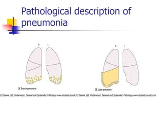
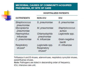
Diseases of Respiratory System
Li yonghuai
From the department of Respiratory medicine
of the first affiliated hospital of Anhui Medical
University
Pulmonary medicine
we have learned the basic medical courses
Now we are learning the clinic medical courses,
then we are on the way to become a true
doctor
Now let’s begin
 The course have 8 weeks class, every
time has 3 periods
 We will learn some respiratory diseases
 Pulmonary medicine is also linked to the
field of critical care medicine, so we
must work hard to study it.
Background
 Pulmonary diseases are major contributors to
morbidity and mortality in the general
population.
 Although advances in the diagnosis and
treatment of many common pulmonary
disorders have improved the lives of patients,
these complex illnesses continue to affect a
large segment of the global population.
Pulmonary diseases can be
divided into the following:
 Respiratory infectious diseases
 Airways diseases (reactive and no-reactive )
 Interstitial lung diseases
 Pulmonary vascular disease
 Primary pulmonary tumors
 Pleural diseases
Respiratory infectious diseases
 Acute upper respiratory tract infection
(common cold and flu)
 Acute tracheobronchitis
 Pneumonia
 Tuberculosis
 Bronchiectasis
 Lung abscess
Airways diseases
 Asthma
 COPD (chronic obstructive pulmonary
disease)
 OSAHS (obstructive sleep apnea-
hypopnea syndrome )
Interstitial lung diseases
 unknown cause
 known cause
Pulmonary vascular disease
 Pulmonary Embolism
 Primary Pulmonary Hypertension
Primary pulmonary tumors
Non-small cell carcinoma
Squamous cell carcinoma
Adenocarcinoma
Large cell carcinoma
Small cell carcinoma
Pleural diseases
 Pleural effusion
 pneumothorax
How to diagnose
 symptoms
 physical examination
 CHEST IMAGING (chest radiograph or
computer tomography)
 pulmonary function testing
 bronchoscopic examination
 laboratory techniques
symptoms
 Cough
 Sputum
 Hemoptysis
 Apnea
 Chest pain
physical examination
 Inspect
 palpate
 percuss
 auscultate
chest radiograph or computer
tomography
pulmonary function testing
bronchoscopic examination.
laboratory techniques
 Gram’s Stain and Culture of
Sputum(a sputum sample must have
>25 neutrophils and <10 squamous
epithelial cells per low-power field)
 Blood,Pleural effusion and so on
Routine Test
How to treat
 Abandon smoking
 Oxygen therapy
 Expansion of bronchus
How to learn
 What is the disease (definition, symptoms,
physical examination, assistant examinations)
 What cause the disease (etiology,
pathogenesis, pathology)
 How to diagnose the disease( diagnostic
reasons, differential diagnosis )
 How to treat the disease (therapeutic
principle, chemotherapy, prevention)
 The scientific basis of many pulmonary
medicine is rapidly expanding.
 Novel diagnostic and therapeutic approaches
populate the published literature with great
frequency.
 Maintaining updated knowledge of these
evolving areas is essential for the optimal
care of patients with lung diseases
Reference book
 Clinic Diagnostics
 Pathophysiology
 Pharmacology
 Harrison’s Pulmonary and Critical Care
Medicine
 You can do
 You can do best
Let’s have a rest
Chapter 1
Respiratory Infections
 Acute respiratory tract infection
 Upper respiratory tract
 Lower respiratory tract
 Of the upper and lower airway boundary
is throat
The functions of respiratory
tract
 Ventilatory flow
 Defence to infection
Respiratory tract defences
 Cough
 Mucociliary clearance mechanisms
 Mucosal immune system
If the defense was broke up, respiratory
tract would be infected
Acute upper respiratory tract
infections
 What is acute upper respiratory tract
infections? (definition)
 It is a kind of acute inflammation of upper
respiratory tract (nasal cavity, pharynx, and
laryngeal)
 Most of it caused by
viral infection, less with
bacteria involvment
Definition of a disease contains
the following elements
 The aetiology of diseases or (and)
pathogenesis
 Remarkable clinic features (symptoms,
signs, assistant examinations)
types of acute upper respiratory tract infections
 The common cold
 Acute pharyngitis and laryngitis
 Herpangina
 Pharyngoconjunctival fever
 Acute pharyngitis and tonsillitis
The common cold
Definition
 The common cold is a mind, self-limited, viral
infection of upper respiratory tract
 A common cold is usually harmless, although it
may not feel that way.
 it's a runny nose, sore throat , cough, watery
eyes, sneezing and congestion — or maybe all of
the above.
 Due to any one of more than 200
viruses can cause a common cold,
symptoms tend to vary greatly.
 Most adults are likely to have a
common cold two to four times a year.
Children, especially preschoolers, may
have a common cold as many as six to
10 times annually.
 Most people recover from a common
cold in about a week or two.
 If symptoms don't improve, maybe
there are complications.
Causes
 Although more than 200 viruses can cause a common
cold, the rhinovirus is the most common culprit, and
it's highly contagious.
 A cold virus enters your body through your mouth or
nose.
 The virus can spread through droplets in the air
when someone who is sick coughs, sneezes or talks.
 But it also spreads by hand-to-hand contact with
someone who has a cold or by using shared objects,
such as utensils, towels, toys or telephones.
 Touch your eyes, nose or mouth after such contact or
exposure, and you're likely to "catch" a cold.
Symptoms
 Symptoms of a common cold usually appear about one to three
days after exposure to a cold virus.
 Signs and symptoms of a common cold may include:
 Runny or stuffy nose
 Itchy or sore throat
 Cough
 Congestion
 Slight body aches or a mild headache
 Sneezing
 Watery eyes
 Low-grade fever (sometime up to 39 ℃)
 Mild fatigue
Complications
 Acute ear infection (otitis media).
 Ear infection occurs when bacteria or viruses infiltrate
the space behind the eardrum. It's a frequent
complication of common colds in children.
 Typical signs and symptoms include earaches and, in
some cases, a green or
 yellow discharge from the nose or the return of a
fever following a common cold. Children who are too
young to verbalize their distress may simply cry or
sleep restlessly.
 Ear pulling is not a reliable sign.

 Wheezing.
 A cold can trigger wheezing in children with asthma.
 Sinusitis.
 In adults or children, a common cold that doesn't
resolve may lead to
 sinusitis — inflammation and infection of the sinuses.
 Other secondary infections. These include strep
throat (streptococcal pharyngitis), pneumonia,
bronchitis in adults and croup in children. These
infections need to be treated by a doctor.
Diagnosis
 History
 Clinic features
 Assistant examinations
Differential diagnosis
 Acute lower respiratory tract infections
(Acute tracheobronchitis)
 Sputum or not
 Influenza (flu)
See the next ppt
What is differential diagnosis
 There are similar symptoms, physical
examinations, and assistant examinations
between the disease that will be diagnosed and
that other diseases
 we will differentiate these diseases according to
symptom, physical examination, assistant
examination, then, we can find the differences
among those diseases, at last, we can detect the
right diagnosis
The Distinction
Influenza .VS. Common Cold
· a runny or stuffy nose
· sneezing
· sore throat
· watery eyes
· a feeling that your ears are blocked
The symptoms of a common cold include:
· irritation in the throat or lungs
· headache
· high fever
· extreme fatigue
· severe muscle aches
· vomiting
· diarrhea
The symptoms of influenza include:
Symptoms Cold Flu
Fever
Sometimes, usually mild Typical, higher
( 38.8-40°C , especially in young
children)persist 3-4 days
Headache Occasionally Common
General Aches Slight Usual; often and serious
General Fatigue Sometimes Usual; early; can last 2 to 3 weeks
Stuffy Nose Common Sometimes
Sneezing Usual Sometimes
Sore Throat Common Sometimes
Cough Mild to moderate; productive
cough
Common; even serious
Complications middle ear infection; generally
no serious complication.
bronchitis, ear infection, pneumonia; can
be life-threatening
Influenza .VS. Common Cold
↓
Treatment
Cold Flu
★There is no cure for the common
cold
★getting plenty of rest
★drinking a lot of liquids
★gargling with warm salt water
★using cough drops, throat sprays,
or cold medicines.
★take medications such as
paracetamol .pain relievers, or fever
reducers are available over the
counter
★ prescription antiviral drugs for flu
may be given in some cases
★avoid using alcohol and tobacco
★ young children should avoid
taking aspirin during an influenza
infection
Influenza .VS. Common Cold
↓
Prevention
Cold Flu
★successful immunization is highly
improbable
★wash hands regularly
★ avoid close contact with anyone with
a cold
★avoid touching the mouth and face
★sleep for 7-8 hours per night
★regular exercise
★Get Vaccinated
The single best way to prevent the flu
is to get a flu vaccination each fall.
About two weeks after vaccination,
antibodies develop that protect against
influenza virus infection.
Treatments and drugs
 There's no cure for the common cold.
 Antibiotics are of no use against cold viruses.
 But the Other secondary infections. These
include strep throat (streptococcal
pharyngitis), pneumonia, bronchitis in adults
and croup in children. These infections need
to be treated
Acute tracheobronchitis
Definition
 Is an inflammation of the tracheo-bronchial
tree
 Usually in association with bacterial or viral
infection, but physical or chemical irritants,
antigens aspiration can also play a role
 Cough and sputum are prominent
manifestation
Aetiology and pathogenesis
 Infection
 Can be caused by bacterial (such as
pneumococcus, hemophilies influenzae,
streptococcus, and staphylococci )
 or viral (such as adenovirus, influenza virus,
respiratory syncytial virus, and parainfluenza
virus) infection
 Is a commom complication of acute upper
respiratory tract infection
Physical or chemical factors
 Aspiration of cold air, dust, irritant gas
or smoke (such as: haze, PM2.5)
Anaphylaxis
 Varied allergens can produce the
inflammation
 Such as: pollen, fungal spore, organic
dust, tropina, and the migration of
parasites in lung
Clinical manifestations
 Cough and mucoid sputum (sometimes
is purulent sputum)
 Rhonchi and coarse crackles
 The symptoms will persist 2 to 3 weeks
Laboratory findings
 White blood cell count and differential
count are normol but will be increased
in the severe bacterial infection
 Sputum smear and culture may detect
the pathogenic organisms
 Chest radiograph may present lung
marking increase in most cases
Diagnosis
 The diagnosis can be established bases
on the history, clinical features, and
combined with the laboratory detections.
 Sputum smear and culture may get the
pathogens.
Differential diagnosis
 Acute upper respiratory tract infection
 Sputum or not sputum
 Nasopharynx symptoms or not
 Physical examination and chest X-ray
are normal or abnormal
 Bronchopneumonia (a type of pneumonia)
Chest x-ray shows the irregular patch
infiltration shadows go along with the lung
markings or just shows the lung
marking increase
Treatment
 Most cases require measures directed only
at relieving cough, for it is self-limited
disease
 For patients with fever or a predominant
tracheitis and purulent sputum, should give
antibiotic therapy and the sputum should be
Gram stained and cultured
Pneumonia
Infection of pulmonary
parenchyma with consolidation
 Definition
 Pathophysiology
 Pathology
 Community-Acquired Pneumonia
 Etiology
 Epidemiology
 Clinical Manifestations
 Diagnosis
 Prognosis
 Prevention .
Definition
 Infection of pulmonary parenchyma
with consolidation
Definition
 Gr. “disease of the lungs”
 Infection involving the distal airspaces
usually with inflammatory exudation
(“localised oedema”).
 Fluid filled spaces lead to consolidation
Classification of Pneumonia
 By clinical setting
(e.g. community acquired pneumonia---
CAP;hospital acquired pneumonia----
HAP)
 By organism
(e.g. pneumococcal ,mycoplasma, etc)
 By morphology
(lobar pneumonia, bronchopneumonia,
interstitial pneumonia )
Organisms
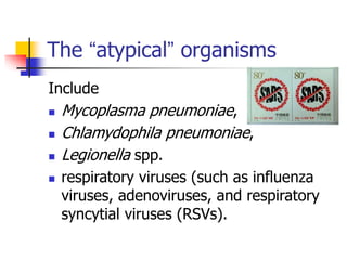
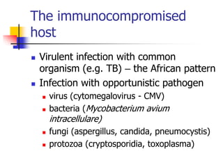
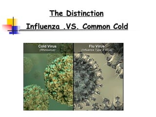
 Viruses – influenza, parainfluenza,
measles, varicella-zoster, respiratory
syncytial virus (RSV). Common, often
self limiting but can be complicated
 Bacteria
 Chlamydia[klə'mɪdɪə] ,
mycoplasma[,maɪko'plɑzmə]
 Fungi['fʌŋgi:]
Lobar Pneumonia
 Confluent consolidation involving a
complete lung lobe
 Most often due to Streptococcus
pneumoniae (pneumococcus)
 Can be seen with other organisms
(Klebsiella, Legionella)
Clinical Setting
 Usually community acquired
 Classically in otherwise healthy young
adults
Pathophysiology
 Pneumonia results from
the proliferation of microbial pathogens at the
alveolar level
VS
the host’s response to those pathogens.
the host’s respon
the proliferation
of microbial
pathogens
Microorganisms gain access to the lower
respiratory tract in several ways
 The most common way : aspiration from the
oropharynx [,ɔro'færɪŋks] .
Small-volume aspiration occurs frequently during sleep
(especially in the elderly) and in patients with decreased
levels of consciousness
 via hematogenous spread
 Contiguous extension from an infected
pleural or mediastinal space
Pathological description of
pneumonia
Pathology
 A classical acute inflammatory response
 Exudation of fibrin-rich fluid
 Neutrophil infiltration
 Macrophage infiltration
 Resolution
 Immune system plays a part antibodies
lead to opsonisation, phagocytosis of
bacteria
Macroscopic pathology
 Heavy lung
 Congestion
 Red hepatisation
 Grey hepatisation
 Resolution
The classical pathway
CAP
(Community-acquired
pneumona)
ETIOLOGY
 bacteria (Streptococcus pneumoniae is most
common)
 mycoplasma , chlamydia , legionella
 fungi
 Viruses
 protozoa
The “typical” bacterial pathogens
includes :
 S.pneumoniae,
 Haemophilus influenzae,
 S. aureus
 gram-negative bacilli (such as Klebsiella
pneumoniae and Pseudomonas aeruginosa)
The “atypical” organisms
Include
 Mycoplasma pneumoniae,
 Chlamydophila pneumoniae,
 Legionella spp.
 respiratory viruses (such as influenza
viruses, adenoviruses, and respiratory
syncytial viruses (RSVs).
 Anti-SARS
 In the ∼10–15% of CAP cases that are
polymicrobial
 the etiology often includes a combination
of typical and atypical pathogens.
 Unfortunately, it is usually impossible to
predict the pathogen in a case of CAP
with any degree of certainty;
 more than half of cases, a specific
etiology is never determined.
 So it is important to consider
epidemiologic and risk factors that
might suggest certain pathogens
EPIDEMIOLOGY
 In USA, about 80% of the 4 million CAP
cases that occur annually are treated on
an outpatient ;about 20% are treated in
the hospital
Risk factors for CAP
 Alcoholism
 Chronic disease
 Immunosuppression
 An age of 70 years or older
CLINICAL MANIFESTATIONS
 can vary from indolent to fulminant in
presentation
 and vary from mild to fatal in severity
symptom
 fever with a tachycardic response
 may have chills or sweats
 cough that is either nonproductive or
productive of mucoid, purulent, or
blood-tinged sputum.
 Severity of infection, the patient may
not be able to speak in full sentences or
may be very short of breath.
 If the pleura is involved, the patient
may be chest pain.
 Up to 20% of patients may have
gastrointestinal symptoms such as
nausea, vomiting, or diarrhea.
 Other symptoms may include fatigue,
headache, myalgias, and arthralgias.
physical examination
vary with
 the degree of pulmonary consolidation
 and the presence or absence of a
significant pleural effusion
 An increased respiratory rate and use of accessory
muscles of respiration are common.
 Palpation may reveal increased or decreased tactile
fremitus,
 the percussion note can vary from dull to flat,
reflecting underlying consolidated lung and pleural
fluid, respectively.
 Crackles, bronchial breath sounds, and possibly a
pleural friction rub may be heard on auscultation.
 The clinical presentation may not be so obvious in
the elderly
DIAGNOSIS
 When confronted with possible CAP
 the physician must ask two questions:
 Is this pneumonia?
 and, if so, what is the etiology?
 the former question is typically
answered by clinical and radiographic
methods
 the latter requires the aid of laboratory
techniques
 Whereas the former question is typically
answered by clinical and radiographic
methods,
 the latter requires the aid of laboratory
techniques
Clinical Diagnosis
 The newly emergence of cough, sputum, or
with chest pain ,hemoptysis
 Fever
 Related sings of pulmonary consolidation,
such as: decreased or increased fremitus,
the percussion note can vary from dull to
flat, Crackles ,etc
laboratory techniques
 WBC > 10×109 or <4×10⁹
 standard postero-anteriorand lateral
chest radiography: localized alveolar
infiltrates and consolidation
Complications
 Organisation (fibrous scarring)
 Abscess
 Bronchiectasis
 Empyema (pus in the pleural cavity)
Viral pneumonia
 Gives a pattern of acute
injury similar to adult
respiratory distress
syndrome (ARDS)
 Acute inflammatory
infiltration less obvious
 Viral inclusions
sometimes seen in
epithelial cells
The immunocompromised
host
 Virulent infection with common
organism (e.g. TB) – the African pattern
 Infection with opportunistic pathogen
 virus (cytomegalovirus - CMV)
 bacteria (Mycobacterium avium
intracellulare)
 fungi (aspergillus, candida, pneumocystis)
 protozoa (cryptosporidia, toxoplasma)
Diagnosis
 High index of suspicion
 Teamwork (physician, microbiologist,
pathologist)
 Broncho-alveolar lavage
 Biopsy (with lots of special stains!)
Immunosuppressed patient – fatal haemorrhage
into Aspergillus-containing cavity
HIV-positive patient CMV (cytomegalovirus) and
“pulmonary oedema” on transbronchial
biopsy….
Special stain also shows Pneumocystis
Pulmonary diseasDDDDDDDDDDDDDDDDDDes.ppt

Pulmonary diseasDDDDDDDDDDDDDDDDDDes.ppt

  • 1.
    Diseases of RespiratorySystem Li yonghuai From the department of Respiratory medicine of the first affiliated hospital of Anhui Medical University Pulmonary medicine
  • 2.
    we have learnedthe basic medical courses Now we are learning the clinic medical courses, then we are on the way to become a true doctor
  • 3.
    Now let’s begin The course have 8 weeks class, every time has 3 periods  We will learn some respiratory diseases  Pulmonary medicine is also linked to the field of critical care medicine, so we must work hard to study it.
  • 4.
    Background  Pulmonary diseasesare major contributors to morbidity and mortality in the general population.  Although advances in the diagnosis and treatment of many common pulmonary disorders have improved the lives of patients, these complex illnesses continue to affect a large segment of the global population.
  • 5.
    Pulmonary diseases canbe divided into the following:  Respiratory infectious diseases  Airways diseases (reactive and no-reactive )  Interstitial lung diseases  Pulmonary vascular disease  Primary pulmonary tumors  Pleural diseases
  • 6.
    Respiratory infectious diseases Acute upper respiratory tract infection (common cold and flu)  Acute tracheobronchitis  Pneumonia  Tuberculosis  Bronchiectasis  Lung abscess
  • 7.
    Airways diseases  Asthma COPD (chronic obstructive pulmonary disease)  OSAHS (obstructive sleep apnea- hypopnea syndrome )
  • 8.
    Interstitial lung diseases unknown cause  known cause
  • 9.
    Pulmonary vascular disease Pulmonary Embolism  Primary Pulmonary Hypertension
  • 10.
    Primary pulmonary tumors Non-smallcell carcinoma Squamous cell carcinoma Adenocarcinoma Large cell carcinoma Small cell carcinoma
  • 11.
    Pleural diseases  Pleuraleffusion  pneumothorax
  • 12.
    How to diagnose symptoms  physical examination  CHEST IMAGING (chest radiograph or computer tomography)  pulmonary function testing  bronchoscopic examination  laboratory techniques
  • 13.
    symptoms  Cough  Sputum Hemoptysis  Apnea  Chest pain
  • 14.
    physical examination  Inspect palpate  percuss  auscultate
  • 15.
    chest radiograph orcomputer tomography
  • 16.
  • 17.
  • 18.
    laboratory techniques  Gram’sStain and Culture of Sputum(a sputum sample must have >25 neutrophils and <10 squamous epithelial cells per low-power field)  Blood,Pleural effusion and so on Routine Test
  • 19.
    How to treat Abandon smoking  Oxygen therapy  Expansion of bronchus
  • 20.
    How to learn What is the disease (definition, symptoms, physical examination, assistant examinations)  What cause the disease (etiology, pathogenesis, pathology)  How to diagnose the disease( diagnostic reasons, differential diagnosis )  How to treat the disease (therapeutic principle, chemotherapy, prevention)
  • 21.
     The scientificbasis of many pulmonary medicine is rapidly expanding.  Novel diagnostic and therapeutic approaches populate the published literature with great frequency.  Maintaining updated knowledge of these evolving areas is essential for the optimal care of patients with lung diseases
  • 22.
    Reference book  ClinicDiagnostics  Pathophysiology  Pharmacology  Harrison’s Pulmonary and Critical Care Medicine
  • 23.
     You cando  You can do best
  • 24.
  • 25.
  • 26.
     Acute respiratorytract infection  Upper respiratory tract  Lower respiratory tract  Of the upper and lower airway boundary is throat
  • 27.
    The functions ofrespiratory tract  Ventilatory flow  Defence to infection
  • 28.
    Respiratory tract defences Cough  Mucociliary clearance mechanisms  Mucosal immune system If the defense was broke up, respiratory tract would be infected
  • 29.
    Acute upper respiratorytract infections  What is acute upper respiratory tract infections? (definition)  It is a kind of acute inflammation of upper respiratory tract (nasal cavity, pharynx, and laryngeal)  Most of it caused by viral infection, less with bacteria involvment
  • 30.
    Definition of adisease contains the following elements  The aetiology of diseases or (and) pathogenesis  Remarkable clinic features (symptoms, signs, assistant examinations)
  • 31.
    types of acuteupper respiratory tract infections  The common cold  Acute pharyngitis and laryngitis  Herpangina  Pharyngoconjunctival fever  Acute pharyngitis and tonsillitis
  • 32.
    The common cold Definition The common cold is a mind, self-limited, viral infection of upper respiratory tract  A common cold is usually harmless, although it may not feel that way.  it's a runny nose, sore throat , cough, watery eyes, sneezing and congestion — or maybe all of the above.
  • 33.
     Due toany one of more than 200 viruses can cause a common cold, symptoms tend to vary greatly.  Most adults are likely to have a common cold two to four times a year. Children, especially preschoolers, may have a common cold as many as six to 10 times annually.
  • 34.
     Most peoplerecover from a common cold in about a week or two.  If symptoms don't improve, maybe there are complications.
  • 35.
    Causes  Although morethan 200 viruses can cause a common cold, the rhinovirus is the most common culprit, and it's highly contagious.  A cold virus enters your body through your mouth or nose.  The virus can spread through droplets in the air when someone who is sick coughs, sneezes or talks.  But it also spreads by hand-to-hand contact with someone who has a cold or by using shared objects, such as utensils, towels, toys or telephones.  Touch your eyes, nose or mouth after such contact or exposure, and you're likely to "catch" a cold.
  • 36.
    Symptoms  Symptoms ofa common cold usually appear about one to three days after exposure to a cold virus.  Signs and symptoms of a common cold may include:  Runny or stuffy nose  Itchy or sore throat  Cough  Congestion  Slight body aches or a mild headache  Sneezing  Watery eyes  Low-grade fever (sometime up to 39 ℃)  Mild fatigue
  • 37.
    Complications  Acute earinfection (otitis media).  Ear infection occurs when bacteria or viruses infiltrate the space behind the eardrum. It's a frequent complication of common colds in children.  Typical signs and symptoms include earaches and, in some cases, a green or  yellow discharge from the nose or the return of a fever following a common cold. Children who are too young to verbalize their distress may simply cry or sleep restlessly.  Ear pulling is not a reliable sign. 
  • 38.
     Wheezing.  Acold can trigger wheezing in children with asthma.  Sinusitis.  In adults or children, a common cold that doesn't resolve may lead to  sinusitis — inflammation and infection of the sinuses.  Other secondary infections. These include strep throat (streptococcal pharyngitis), pneumonia, bronchitis in adults and croup in children. These infections need to be treated by a doctor.
  • 39.
    Diagnosis  History  Clinicfeatures  Assistant examinations
  • 40.
    Differential diagnosis  Acutelower respiratory tract infections (Acute tracheobronchitis)  Sputum or not  Influenza (flu) See the next ppt
  • 41.
    What is differentialdiagnosis  There are similar symptoms, physical examinations, and assistant examinations between the disease that will be diagnosed and that other diseases  we will differentiate these diseases according to symptom, physical examination, assistant examination, then, we can find the differences among those diseases, at last, we can detect the right diagnosis
  • 42.
  • 43.
    · a runnyor stuffy nose · sneezing · sore throat · watery eyes · a feeling that your ears are blocked The symptoms of a common cold include:
  • 44.
    · irritation inthe throat or lungs · headache · high fever · extreme fatigue · severe muscle aches · vomiting · diarrhea The symptoms of influenza include:
  • 45.
    Symptoms Cold Flu Fever Sometimes,usually mild Typical, higher ( 38.8-40°C , especially in young children)persist 3-4 days Headache Occasionally Common General Aches Slight Usual; often and serious General Fatigue Sometimes Usual; early; can last 2 to 3 weeks Stuffy Nose Common Sometimes Sneezing Usual Sometimes Sore Throat Common Sometimes Cough Mild to moderate; productive cough Common; even serious Complications middle ear infection; generally no serious complication. bronchitis, ear infection, pneumonia; can be life-threatening Influenza .VS. Common Cold
  • 46.
    Influenza .VS. CommonCold ↓ Treatment Cold Flu ★There is no cure for the common cold ★getting plenty of rest ★drinking a lot of liquids ★gargling with warm salt water ★using cough drops, throat sprays, or cold medicines. ★take medications such as paracetamol .pain relievers, or fever reducers are available over the counter ★ prescription antiviral drugs for flu may be given in some cases ★avoid using alcohol and tobacco ★ young children should avoid taking aspirin during an influenza infection
  • 47.
    Influenza .VS. CommonCold ↓ Prevention Cold Flu ★successful immunization is highly improbable ★wash hands regularly ★ avoid close contact with anyone with a cold ★avoid touching the mouth and face ★sleep for 7-8 hours per night ★regular exercise ★Get Vaccinated The single best way to prevent the flu is to get a flu vaccination each fall. About two weeks after vaccination, antibodies develop that protect against influenza virus infection.
  • 48.
    Treatments and drugs There's no cure for the common cold.  Antibiotics are of no use against cold viruses.  But the Other secondary infections. These include strep throat (streptococcal pharyngitis), pneumonia, bronchitis in adults and croup in children. These infections need to be treated
  • 49.
    Acute tracheobronchitis Definition  Isan inflammation of the tracheo-bronchial tree  Usually in association with bacterial or viral infection, but physical or chemical irritants, antigens aspiration can also play a role  Cough and sputum are prominent manifestation
  • 50.
    Aetiology and pathogenesis Infection  Can be caused by bacterial (such as pneumococcus, hemophilies influenzae, streptococcus, and staphylococci )  or viral (such as adenovirus, influenza virus, respiratory syncytial virus, and parainfluenza virus) infection  Is a commom complication of acute upper respiratory tract infection
  • 51.
    Physical or chemicalfactors  Aspiration of cold air, dust, irritant gas or smoke (such as: haze, PM2.5)
  • 52.
    Anaphylaxis  Varied allergenscan produce the inflammation  Such as: pollen, fungal spore, organic dust, tropina, and the migration of parasites in lung
  • 53.
    Clinical manifestations  Coughand mucoid sputum (sometimes is purulent sputum)  Rhonchi and coarse crackles  The symptoms will persist 2 to 3 weeks
  • 54.
    Laboratory findings  Whiteblood cell count and differential count are normol but will be increased in the severe bacterial infection  Sputum smear and culture may detect the pathogenic organisms  Chest radiograph may present lung marking increase in most cases
  • 55.
    Diagnosis  The diagnosiscan be established bases on the history, clinical features, and combined with the laboratory detections.  Sputum smear and culture may get the pathogens.
  • 56.
    Differential diagnosis  Acuteupper respiratory tract infection  Sputum or not sputum  Nasopharynx symptoms or not  Physical examination and chest X-ray are normal or abnormal
  • 57.
     Bronchopneumonia (atype of pneumonia) Chest x-ray shows the irregular patch infiltration shadows go along with the lung markings or just shows the lung marking increase
  • 58.
    Treatment  Most casesrequire measures directed only at relieving cough, for it is self-limited disease  For patients with fever or a predominant tracheitis and purulent sputum, should give antibiotic therapy and the sputum should be Gram stained and cultured
  • 59.
  • 60.
     Definition  Pathophysiology Pathology  Community-Acquired Pneumonia  Etiology  Epidemiology  Clinical Manifestations  Diagnosis  Prognosis  Prevention .
  • 61.
    Definition  Infection ofpulmonary parenchyma with consolidation
  • 62.
    Definition  Gr. “diseaseof the lungs”  Infection involving the distal airspaces usually with inflammatory exudation (“localised oedema”).  Fluid filled spaces lead to consolidation
  • 63.
    Classification of Pneumonia By clinical setting (e.g. community acquired pneumonia--- CAP;hospital acquired pneumonia---- HAP)  By organism (e.g. pneumococcal ,mycoplasma, etc)  By morphology (lobar pneumonia, bronchopneumonia, interstitial pneumonia )
  • 64.
    Organisms  Viruses –influenza, parainfluenza, measles, varicella-zoster, respiratory syncytial virus (RSV). Common, often self limiting but can be complicated  Bacteria  Chlamydia[klə'mɪdɪə] , mycoplasma[,maɪko'plɑzmə]  Fungi['fʌŋgi:]
  • 65.
    Lobar Pneumonia  Confluentconsolidation involving a complete lung lobe  Most often due to Streptococcus pneumoniae (pneumococcus)  Can be seen with other organisms (Klebsiella, Legionella)
  • 66.
    Clinical Setting  Usuallycommunity acquired  Classically in otherwise healthy young adults
  • 67.
    Pathophysiology  Pneumonia resultsfrom the proliferation of microbial pathogens at the alveolar level VS the host’s response to those pathogens. the host’s respon the proliferation of microbial pathogens
  • 68.
    Microorganisms gain accessto the lower respiratory tract in several ways  The most common way : aspiration from the oropharynx [,ɔro'færɪŋks] . Small-volume aspiration occurs frequently during sleep (especially in the elderly) and in patients with decreased levels of consciousness  via hematogenous spread  Contiguous extension from an infected pleural or mediastinal space
  • 69.
  • 70.
    Pathology  A classicalacute inflammatory response  Exudation of fibrin-rich fluid  Neutrophil infiltration  Macrophage infiltration  Resolution  Immune system plays a part antibodies lead to opsonisation, phagocytosis of bacteria
  • 71.
    Macroscopic pathology  Heavylung  Congestion  Red hepatisation  Grey hepatisation  Resolution The classical pathway
  • 72.
  • 73.
    ETIOLOGY  bacteria (Streptococcuspneumoniae is most common)  mycoplasma , chlamydia , legionella  fungi  Viruses  protozoa
  • 74.
    The “typical” bacterialpathogens includes :  S.pneumoniae,  Haemophilus influenzae,  S. aureus  gram-negative bacilli (such as Klebsiella pneumoniae and Pseudomonas aeruginosa)
  • 75.
    The “atypical” organisms Include Mycoplasma pneumoniae,  Chlamydophila pneumoniae,  Legionella spp.  respiratory viruses (such as influenza viruses, adenoviruses, and respiratory syncytial viruses (RSVs).
  • 76.
  • 78.
     In the∼10–15% of CAP cases that are polymicrobial  the etiology often includes a combination of typical and atypical pathogens.
  • 79.
     Unfortunately, itis usually impossible to predict the pathogen in a case of CAP with any degree of certainty;  more than half of cases, a specific etiology is never determined.  So it is important to consider epidemiologic and risk factors that might suggest certain pathogens
  • 83.
    EPIDEMIOLOGY  In USA,about 80% of the 4 million CAP cases that occur annually are treated on an outpatient ;about 20% are treated in the hospital
  • 84.
    Risk factors forCAP  Alcoholism  Chronic disease  Immunosuppression  An age of 70 years or older
  • 85.
    CLINICAL MANIFESTATIONS  canvary from indolent to fulminant in presentation  and vary from mild to fatal in severity
  • 86.
    symptom  fever witha tachycardic response  may have chills or sweats  cough that is either nonproductive or productive of mucoid, purulent, or blood-tinged sputum.
  • 87.
     Severity ofinfection, the patient may not be able to speak in full sentences or may be very short of breath.  If the pleura is involved, the patient may be chest pain.  Up to 20% of patients may have gastrointestinal symptoms such as nausea, vomiting, or diarrhea.  Other symptoms may include fatigue, headache, myalgias, and arthralgias.
  • 88.
    physical examination vary with the degree of pulmonary consolidation  and the presence or absence of a significant pleural effusion
  • 89.
     An increasedrespiratory rate and use of accessory muscles of respiration are common.  Palpation may reveal increased or decreased tactile fremitus,  the percussion note can vary from dull to flat, reflecting underlying consolidated lung and pleural fluid, respectively.  Crackles, bronchial breath sounds, and possibly a pleural friction rub may be heard on auscultation.  The clinical presentation may not be so obvious in the elderly
  • 90.
    DIAGNOSIS  When confrontedwith possible CAP  the physician must ask two questions:  Is this pneumonia?  and, if so, what is the etiology?
  • 91.
     the formerquestion is typically answered by clinical and radiographic methods  the latter requires the aid of laboratory techniques
  • 92.
     Whereas theformer question is typically answered by clinical and radiographic methods,  the latter requires the aid of laboratory techniques
  • 93.
    Clinical Diagnosis  Thenewly emergence of cough, sputum, or with chest pain ,hemoptysis  Fever  Related sings of pulmonary consolidation, such as: decreased or increased fremitus, the percussion note can vary from dull to flat, Crackles ,etc
  • 94.
    laboratory techniques  WBC> 10×109 or <4×10⁹  standard postero-anteriorand lateral chest radiography: localized alveolar infiltrates and consolidation
  • 95.
    Complications  Organisation (fibrousscarring)  Abscess  Bronchiectasis  Empyema (pus in the pleural cavity)
  • 96.
    Viral pneumonia  Givesa pattern of acute injury similar to adult respiratory distress syndrome (ARDS)  Acute inflammatory infiltration less obvious  Viral inclusions sometimes seen in epithelial cells
  • 97.
    The immunocompromised host  Virulentinfection with common organism (e.g. TB) – the African pattern  Infection with opportunistic pathogen  virus (cytomegalovirus - CMV)  bacteria (Mycobacterium avium intracellulare)  fungi (aspergillus, candida, pneumocystis)  protozoa (cryptosporidia, toxoplasma)
  • 98.
    Diagnosis  High indexof suspicion  Teamwork (physician, microbiologist, pathologist)  Broncho-alveolar lavage  Biopsy (with lots of special stains!)
  • 99.
    Immunosuppressed patient –fatal haemorrhage into Aspergillus-containing cavity
  • 100.
    HIV-positive patient CMV(cytomegalovirus) and “pulmonary oedema” on transbronchial biopsy….
  • 101.
    Special stain alsoshows Pneumocystis