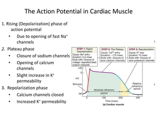
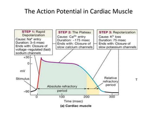
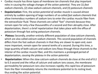
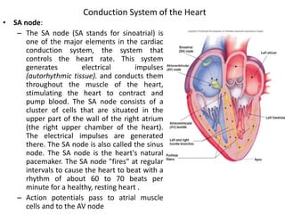
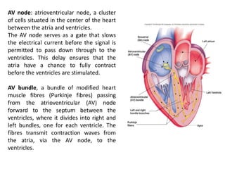
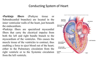
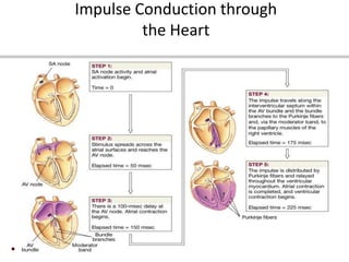
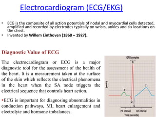
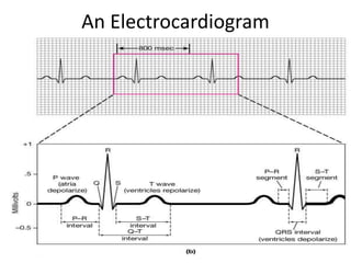
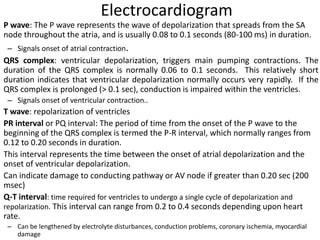
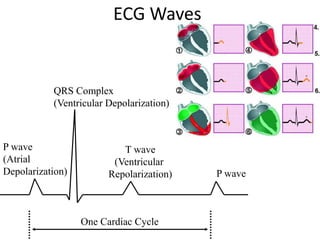
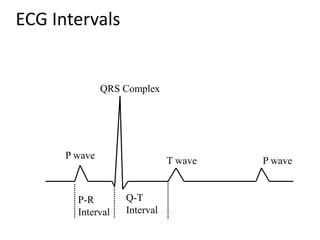
CARDIOVASCULAR
SYSTEM
Md. Saiful Islam
Dept. of Pharmaceutical Sciences
North South University
Facebook Group: Pharmacy Universe
Youtube Channel: Pharmacy Universe
CARDIOVASCULAR SYSTEM
Cardiac Action Potential, ECG
1. Rising (Depolarization) phase of
action potential
• Due to opening of fast Na+
channels
2. Plateau phase
• Closure of sodium channels
• Opening of calcium
channels
• Slight increase in K+
permeability
3. Repolarization phase
• Calcium channels closed
• Increased K+ permeability
The Action Potential in Cardiac Muscle
• Cardiac muscle has three types of membrane ion channels that play important
roles in causing the voltage changes of the action potential. They are (1) fast
sodium channels, (2) slow sodium-calcium channels, and (3) potassium channels
• Depolarization: First, the action potential of cardiac muscle is caused almost
entirely by sudden opening of large numbers of so-called fast sodium channels that
allow tremendous numbers of sodium ions to enter the cardiac muscle fiber from
the extracellular fluid. These channels are called “fast” channels because they
remain open for only a few thousandths of a second and then abruptly close. After
depolarization, there's a brief repolarization that takes place with the efflux of
potassium through fast acting potassium channels.
• Plateau: Secondly, another entirely different population of slow calcium channels,
which are also called calcium-sodium channels. This second population of channels
differs from the fast sodium channels in that they are slower to open and, even
more important, remain open for several tenths of a second. During this time, a
large quantity of both calcium and sodium ions flows through these channels to the
interior of the cardiac muscle fiber, and this maintains a prolonged period of
depolarization, causing the plateau in the action potential.
• Repolarization: When the slow calcium-sodium channels do close at the end of 0.2
to 0.3 second and the influx of calcium and sodium ions ceases, the membrane
permeability for potassium ions also increases rapidly; this rapid loss of potassium
from the fiber immediately returns the membrane potential to its resting level,
thus ending the action potential.
What is Absolute Refractory Period and Relative
refractory period?
The refractory periods are due to the inactivation property of voltage-gated
sodium channels and the lag of potassium channels in closing. Voltage-gated
sodium channels have two gating mechanisms, the activation mechanism that
opens the channel with depolarization and the inactivation mechanism that
closes the channel with repolarization. While the channel is in the inactive
state, it will not open in response to depolarization. The period when the
majority of sodium channels remain in the inactive state is the absolute
refractory period.
After this period, there are enough voltage-activated sodium channels in the
closed (active) state to respond to depolarization. However, voltage-gated
potassium channels that opened in response to repolarization do not close as
quickly as voltage-gated sodium channels; to return to the active closed state.
During this time, the extra potassium conductance means that the membrane
is at a higher threshold and will require a greater stimulus to cause action
potentials to fire. This period is the relative refractory period.
Conduction System of the Heart
• SA node:
– The SA node (SA stands for sinoatrial) is
one of the major elements in the cardiac
conduction system, the system that
controls the heart rate. This system
generates electrical impulses
(autorhythmic tissue). and conducts them
throughout the muscle of the heart,
stimulating the heart to contract and
pump blood. The SA node consists of a
cluster of cells that are situated in the
upper part of the wall of the right atrium
(the right upper chamber of the heart).
The electrical impulses are generated
there. The SA node is also called the sinus
node. The SA node is the heart's natural
pacemaker. The SA node "fires" at regular
intervals to cause the heart to beat with a
rhythm of about 60 to 70 beats per
minute for a healthy, resting heart .
– Action potentials pass to atrial muscle
cells and to the AV node
AV node: atrioventricular node, a cluster
of cells situated in the center of the heart
between the atria and ventricles.
The AV node serves as a gate that slows
the electrical current before the signal is
permitted to pass down through to the
ventricles. This delay ensures that the
atria have a chance to fully contract
before the ventricles are stimulated.
AV bundle, a bundle of modified heart
muscle fibres (Purkinje fibres) passing
from the atrioventricular (AV) node
forward to the septum between the
ventricles, where it divides into right and
left bundles, one for each ventricle. The
fibres transmit contraction waves from
the atria, via the AV node, to the
ventricles.
Conducting System of Heart
•Purkinje fibers (Purkyne tissue or
Subendocardial branches) are located in the
inner ventricular walls of the heart, just beneath
the endocardium.
•Purkinje fibers are specialized myocardial
fibers that carry the electrical impulse from
both the left and right bundle branch to the
myocardium of the ventricles. This causes the
muscle tissue of the ventricles to contract, thus
enabling a force to eject blood out of the heart;
either to the Pulmonary circulation from the
right ventricle or to the Systemic circulation
from the left ventricle.
Impulse Conduction through
the Heart
Electrocardiogram (ECG/EKG)
• ECG is the composite of all action potentials of nodal and myocardial cells detected,
amplified and recorded by electrodes typically on wrists, ankles and six locations on
the chest.
• Invented by Willem Einthoven (1860 – 1927).
Diagnostic Value of ECG
The electrocardiogram or ECG is a major
diagnostic tool for the assessment of the health of
the heart. It is a measurement taken at the surface
of the skin which reflects the electrical phenomena
in the heart when the SA node triggers the
electrical sequence that controls heart action.
ECG is important for diagnosing abnormalities in
conduction pathways, MI, heart enlargement and
electrolyte and hormone imbalances.
An Electrocardiogram
Electrocardiogram
P wave: The P wave represents the wave of depolarization that spreads from the SA
node throughout the atria, and is usually 0.08 to 0.1 seconds (80-100 ms) in duration.
– Signals onset of atrial contraction.
QRS complex: ventricular depolarization, triggers main pumping contractions. The
duration of the QRS complex is normally 0.06 to 0.1 seconds. This relatively short
duration indicates that ventricular depolarization normally occurs very rapidly. If the
QRS complex is prolonged (> 0.1 sec), conduction is impaired within the ventricles.
– Signals onset of ventricular contraction..
T wave: repolarization of ventricles
PR interval or PQ interval: The period of time from the onset of the P wave to the
beginning of the QRS complex is termed the P-R interval, which normally ranges from
0.12 to 0.20 seconds in duration.
This interval represents the time between the onset of atrial depolarization and the
onset of ventricular depolarization.
Can indicate damage to conducting pathway or AV node if greater than 0.20 sec (200
msec)
Q-T interval: time required for ventricles to undergo a single cycle of depolarization and
repolarization. This interval can range from 0.2 to 0.4 seconds depending upon heart
rate.
– Can be lengthened by electrolyte disturbances, conduction problems, coronary ischemia, myocardial
damage
ECG Waves
P wave
(Atrial
Depolarization)
QRS Complex
(Ventricular Depolarization)
T wave
(Ventricular
Repolarization) P wave
One Cardiac Cycle
ECG Intervals
P wave
QRS Complex
T wave P wave
P-R
Interval
Q-T
Interval
THANK YOU

Cardiovascular system (cardiac ap, ECG)

  • 1.
    CARDIOVASCULAR SYSTEM Md. Saiful Islam Dept.of Pharmaceutical Sciences North South University Facebook Group: Pharmacy Universe Youtube Channel: Pharmacy Universe
  • 2.
  • 3.
    1. Rising (Depolarization)phase of action potential • Due to opening of fast Na+ channels 2. Plateau phase • Closure of sodium channels • Opening of calcium channels • Slight increase in K+ permeability 3. Repolarization phase • Calcium channels closed • Increased K+ permeability The Action Potential in Cardiac Muscle
  • 4.
    The Action Potentialin Cardiac Muscle
  • 5.
    • Cardiac musclehas three types of membrane ion channels that play important roles in causing the voltage changes of the action potential. They are (1) fast sodium channels, (2) slow sodium-calcium channels, and (3) potassium channels • Depolarization: First, the action potential of cardiac muscle is caused almost entirely by sudden opening of large numbers of so-called fast sodium channels that allow tremendous numbers of sodium ions to enter the cardiac muscle fiber from the extracellular fluid. These channels are called “fast” channels because they remain open for only a few thousandths of a second and then abruptly close. After depolarization, there's a brief repolarization that takes place with the efflux of potassium through fast acting potassium channels. • Plateau: Secondly, another entirely different population of slow calcium channels, which are also called calcium-sodium channels. This second population of channels differs from the fast sodium channels in that they are slower to open and, even more important, remain open for several tenths of a second. During this time, a large quantity of both calcium and sodium ions flows through these channels to the interior of the cardiac muscle fiber, and this maintains a prolonged period of depolarization, causing the plateau in the action potential. • Repolarization: When the slow calcium-sodium channels do close at the end of 0.2 to 0.3 second and the influx of calcium and sodium ions ceases, the membrane permeability for potassium ions also increases rapidly; this rapid loss of potassium from the fiber immediately returns the membrane potential to its resting level, thus ending the action potential.
  • 6.
    What is AbsoluteRefractory Period and Relative refractory period? The refractory periods are due to the inactivation property of voltage-gated sodium channels and the lag of potassium channels in closing. Voltage-gated sodium channels have two gating mechanisms, the activation mechanism that opens the channel with depolarization and the inactivation mechanism that closes the channel with repolarization. While the channel is in the inactive state, it will not open in response to depolarization. The period when the majority of sodium channels remain in the inactive state is the absolute refractory period. After this period, there are enough voltage-activated sodium channels in the closed (active) state to respond to depolarization. However, voltage-gated potassium channels that opened in response to repolarization do not close as quickly as voltage-gated sodium channels; to return to the active closed state. During this time, the extra potassium conductance means that the membrane is at a higher threshold and will require a greater stimulus to cause action potentials to fire. This period is the relative refractory period.
  • 7.
    Conduction System ofthe Heart • SA node: – The SA node (SA stands for sinoatrial) is one of the major elements in the cardiac conduction system, the system that controls the heart rate. This system generates electrical impulses (autorhythmic tissue). and conducts them throughout the muscle of the heart, stimulating the heart to contract and pump blood. The SA node consists of a cluster of cells that are situated in the upper part of the wall of the right atrium (the right upper chamber of the heart). The electrical impulses are generated there. The SA node is also called the sinus node. The SA node is the heart's natural pacemaker. The SA node "fires" at regular intervals to cause the heart to beat with a rhythm of about 60 to 70 beats per minute for a healthy, resting heart . – Action potentials pass to atrial muscle cells and to the AV node
  • 8.
    AV node: atrioventricularnode, a cluster of cells situated in the center of the heart between the atria and ventricles. The AV node serves as a gate that slows the electrical current before the signal is permitted to pass down through to the ventricles. This delay ensures that the atria have a chance to fully contract before the ventricles are stimulated. AV bundle, a bundle of modified heart muscle fibres (Purkinje fibres) passing from the atrioventricular (AV) node forward to the septum between the ventricles, where it divides into right and left bundles, one for each ventricle. The fibres transmit contraction waves from the atria, via the AV node, to the ventricles.
  • 9.
    Conducting System ofHeart •Purkinje fibers (Purkyne tissue or Subendocardial branches) are located in the inner ventricular walls of the heart, just beneath the endocardium. •Purkinje fibers are specialized myocardial fibers that carry the electrical impulse from both the left and right bundle branch to the myocardium of the ventricles. This causes the muscle tissue of the ventricles to contract, thus enabling a force to eject blood out of the heart; either to the Pulmonary circulation from the right ventricle or to the Systemic circulation from the left ventricle.
  • 10.
  • 11.
    Electrocardiogram (ECG/EKG) • ECGis the composite of all action potentials of nodal and myocardial cells detected, amplified and recorded by electrodes typically on wrists, ankles and six locations on the chest. • Invented by Willem Einthoven (1860 – 1927). Diagnostic Value of ECG The electrocardiogram or ECG is a major diagnostic tool for the assessment of the health of the heart. It is a measurement taken at the surface of the skin which reflects the electrical phenomena in the heart when the SA node triggers the electrical sequence that controls heart action. ECG is important for diagnosing abnormalities in conduction pathways, MI, heart enlargement and electrolyte and hormone imbalances.
  • 12.
  • 13.
    Electrocardiogram P wave: TheP wave represents the wave of depolarization that spreads from the SA node throughout the atria, and is usually 0.08 to 0.1 seconds (80-100 ms) in duration. – Signals onset of atrial contraction. QRS complex: ventricular depolarization, triggers main pumping contractions. The duration of the QRS complex is normally 0.06 to 0.1 seconds. This relatively short duration indicates that ventricular depolarization normally occurs very rapidly. If the QRS complex is prolonged (> 0.1 sec), conduction is impaired within the ventricles. – Signals onset of ventricular contraction.. T wave: repolarization of ventricles PR interval or PQ interval: The period of time from the onset of the P wave to the beginning of the QRS complex is termed the P-R interval, which normally ranges from 0.12 to 0.20 seconds in duration. This interval represents the time between the onset of atrial depolarization and the onset of ventricular depolarization. Can indicate damage to conducting pathway or AV node if greater than 0.20 sec (200 msec) Q-T interval: time required for ventricles to undergo a single cycle of depolarization and repolarization. This interval can range from 0.2 to 0.4 seconds depending upon heart rate. – Can be lengthened by electrolyte disturbances, conduction problems, coronary ischemia, myocardial damage
  • 14.
    ECG Waves P wave (Atrial Depolarization) QRSComplex (Ventricular Depolarization) T wave (Ventricular Repolarization) P wave One Cardiac Cycle
  • 15.
    ECG Intervals P wave QRSComplex T wave P wave P-R Interval Q-T Interval
  • 16.